Download to read offline
![loss of calcium, as well as structural changes, causing the bones to become thinner, more fragile
and more likely to break. DXA is also effective in tracking the effects of treatment for
osteoporosis and other conditions that cause bone loss.
What is Osteoporosis?
Osteoporosis is the weakening of bones in the body. It is caused by lack of calcium deposited in
the bones. This lack of calcium causes the bones to become brittle, so they break easily. Some
side effects are limping. Some symptoms late in the disease include pain in the bones, living in
a wheelchair (as a result of), and lower back pain due to spinal bone fractures.[1] Elderly people
are more likely to develop osteoporosis than younger people. The amount of calcium in the bones
decreases as a person gets older.
Bone density testing is strongly recommended to who?
Those who
are a post-menopausal woman and not taking estrogen.
have a personal or maternal history of hip fracture or smoking.
are a post-menopausal woman who is tall (over 5 feet 7 inches) or thin (less than 125
pounds).
are a man with clinical conditions associated with bone loss.
use medications that are known to cause bone loss, including corticosteroids such as
Prednisone, various anti-seizure medications such as Dilantin and certain barbiturates, or
high-dose thyroid replacement drugs.
have diabetes, liver disease, kidney disease or a family history of osteoporosis.
have high bone turnover, which shows up in the form of excessive collagen in urine samples.
have a thyroid condition, such as hyperthyroidism.
have a parathyroid condition, such as hyperparathyroidism.
have experienced a fracture after only mild trauma.
have had x-ray evidence of vertebral fracture or other signs of osteoporosis.](https://image.slidesharecdn.com/2dc7720a-f9d2-4cda-8506-f8e065e5baa8-141219003130-conversion-gate02/85/BMD-2-320.jpg)
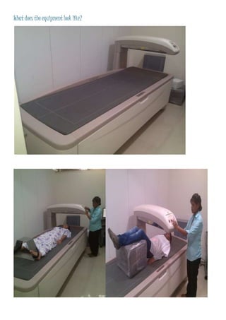

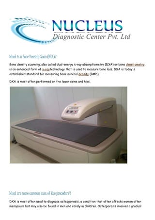
Bone density scanning (DXA) uses x-ray technology to measure bone loss by assessing bone mineral density, most often of the lower spine and hips. DXA is commonly used to diagnose osteoporosis, a condition where decreased bone density causes bones to become brittle and more prone to breaking. It can also be used to monitor osteoporosis treatment effects. Those at high risk for osteoporosis due to factors like post-menopause, family history, smoking or medication use should consider DXA screening. A radiologist will analyze DXA scans and send results to the patient's doctor to discuss.


