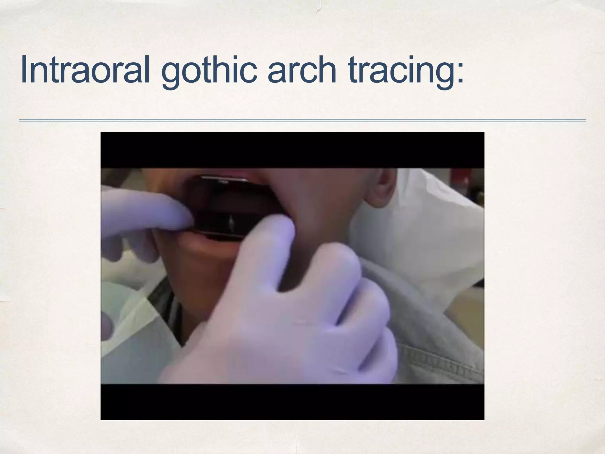
GUIDED BY :
DR SUREKHA GODBOLE
DR SEEMA SATHE
DR ANJALI BORLE
DR TRUPTI DAHANE
“Gothic arch tracing”
PRESENTED BY :
APURVA DESHMUKH
Contents
1. Introduction
2. Terminologies
3. Review of literature
4. Conventional Technique Of Gothic Arch Tracing
5. Extraoral And Intraoral Tracers
6. CLASSIFICATION of ARROW POINT tracings
7. Conclusion
8. References.
Introduction:
✤ Gothic architecture is a style
of architecture which was used in
the Middle Ages. It is most familiar
as the architecture of many of the
great cathedrals, abbeys and
churches of Europe.
✤ The important features of Gothic
architecture is the pointed arches.
Terminologies:
✤ Gothic arch tracing : The pattern obtained on the horizontal
plate used with a central bearing tracing device.
✤ Central bearing point - The contact point of a central bearing
device. - GPT 9.
✤ Central bearing tracing device - A device that provides a
central point of bearing or support between the maxillary and
mandibular dental arches. It consists of a contacting point
that is attached to one dental arch and a plate attached to
the opposing dental arch. The plate provides the surface on
which the bearing point rests or moves and on which the
tracing of the mandibular movement is recorded. It may be
used to distribute occlusal forces evenly during the recording
of maxillo-mandibular relationships and/or for the correction
of disharmonious occlusal contacts. - GPT 9
4
✤ In almost any literature on occlusion and fixed
prosthodontics, centric relation (CR) is the beginning
of the story.
✤ It is the beginning of occlusion, and all treatment
modalities are based on it.
✤ Centric relation: a maxillo-mandibular relationship, independent of tooth
contact, in which the condyles articulate in the anterior-superior position
against the posterior slopes of the articular eminences; in this position, the
mandible is restricted to a purely rotary movement; from this unstrained,
physiologic, maxillo-mandibular relationship, the patient can make vertical,
lateral or protrusive movements; it is a clinically useful, repeatable
reference position.
- GPT 9
6
Methods of recording centric relation :
✤ It is generally agreed that centric relation records can be grouped into
four categories -
1. Direct checkbite (inter-occlusal) recordings - using wax,
compound, plaster & zinc oxide eugenol paste.
2. Graphic recordings (intraoral and extraoral)
3. Functional recordings.
4. Cephalometric
- Myers ML. Centric relation records—historical review. J Prosthet Dent 1982;47:141-5.
Functional methods:
✤ Needle house method
1. Occlusal rims are fabricated in impression compound
2. Four metal beads are embedded in the max. premolar
& molar areas of occlusal rims
3. The rims are inserted into patient’s mouth and close
the occlusal rim & make protrusive, retrusive, right &
left lateral movements
4. Diamond shaped markings on the mandibular
occlusal rim.
Functional methods:
✤ Patterson's method
1. Wax occlusion rims are used.
2. A trench is made in the mandibular rim and a mixture of
half plaster and half carborundum paste is placed in the
trench.
3. The mandibular movements generate compensating
curves in the plaster and carborundum paste.
4. When the paste is reduced to the predetermined vertical
height of occlusion, the patient is instructed to retrude
the mandible and the occlusal rims are joined together
with metal staples.
GRAPHIC METHOD
✤ The graphic methods record a tracing of mandibular
movements in horizontal plane & an arrow point tracing is
achieved.
✤ It indicates the horizontal relation of the mandible to the
maxillae.
✤ The apex of a properly made tracing indicates the most
retruded relation of the mandible to the maxillae from which
lateral movements can take place.
✤ Horizontal relations are those that are established
antero-posteriorly and medio-laterally and so are
classified as :
1. Centric Relations.
2. Eccentric Relations — Protrusive and Lateral
Relation.
Right lateral Left lateral
✤ Graphic methods are either
intraoral or extraoral, depending
upon the placement of the
recording devices.
✤ Extraoral tracing are also referred
to by 2 other names : gothic arch
tracings and arrow point tracings. Intraoral tracers Extraoral tracers
Review of literature:
✤ The earliest graphic recordings were based on studies of mandibular
movements by Balkwill in 1866.
✤ The first known "Needle Point Tracing" was by Hesse in 1897.
✤ The technique was improved and popularised by Gysi around 1910.
The tracer made by Gysi was an extraoral incisal tracer. The tracing
plate coated with wax, was attached to the mandibular rim. A spring-
loaded pin or marker was mounted on the maxillary rim. The rims
were made of modeling compound to maintain the vertical dimension
of occlusion. When a good tracing was recorded, the patient held the
rims in the apex of the tracing while notches were scored in the rims
for orientation. No central bearing point was utilised.
✤ Clapp in 1914 described the use of a Gysi tracer which was
attached directly to the impression trays.
✤ Sears in 1926 used lubricated rims for easier movement. He
placed the needle point tracer on the mandibular rim and the plate
on the maxillary rim. He believed this made the angle of the
tracing more acute and more easily discernible. He would then
cement the rims together for removal.
✤ Phillips in 1927 recognized that any lateral movements of the jaw
would cause interference of the rims which could result in a
distorted record. He developed a plate for the upper rim and a
tripoded ball bearing mounted on a jackscrew for the lower rim.
This innovation was named the "Central Bearing Point".
✤ In 1929, Stansbery introduced a technique which
incorporated a curved plate with a 4-inch radius
(corresponding to Monson's curve) mounted on the
upper rim. A central bearing screw was attached to
the lower plate with a 3-inch radius curve. After the
extraoral tracing was made, plaster was injected
between the rims to form a biconcave centric
registration.
✤ Hall in 1929 used Stansbery's method but
substituted compound for Centric Relation record.
✤ Later graphic recording methods used the central bearing
point to produce the Gothic Arch tracing. Hardy and
Pleasure described the use of Coble Balancer.
✤ Hardy and Porter in 1942 made a depression with a round
bur at the apex of the tracing. The patient would hold the
bearing point in the depression while plaster was injected for
the centric record.
✤ Pleasure in 1955 used a plastic disk which was attached to
the tracing plate with a hole over the apex of the Gothic
Arch. The Centric Relation record could then be made
without a change of vertical dimension.
✤ The Hight Tracer, 1986 — The hight tracer is a
four-component assembly, which consists of
an upper bearing plate, lower bearing plate
with a central screw, a scriber point to be
attached to the upper rim and a tracing
platform which extends in forward and is
attached to the lower rim.
✤ Ballard tracers.
✤ Messerman tracers.
✤ Chandrasekharan Nair developed Chandra Tracer :
1. two component assembly.
1. The upper bearing plate has a pencil holder
2. lower bearing plate has a central bearing screw and tracing platform of
dimensions 35 x 47 mm, which has a facility to hold a drawing sheet.
2. A sharp pencil is attached to the upper component.The tracer is placed in the
mouth and the tracings are recorded on the paper fixed to the table. After many
repetitions, the best tracing is selected, and the centric and protrusive plaster
records are obtained using quick setting plaster. Plastic sheets are used for
stabilisation during the recording.
Extraoral tracing technique:
1. After taking face bow records, it is transferred to the articulator and mounting of the
maxillary cast is done.Then, with tentative centric relation records, mandibular cast to
the mounted.
2. The upper bearing plate is heated and waxed to the maxillary rim, making it flush
with the occlusal plane.
3. The lower occlusal rim is reduced by 3 mm and the lower plate is fixed to the
mandibular rim.
4. The scriber is attached to the maxillary rim and lower tracing platform is
waxed to the mandibular rim.
5. The upper and lower tracers are made parallel.
6. Seat the patient with head upright in a comfortable
position.
7. Intraorally seat the record bases with the attached
devices.
8. Retract the stylus & conduct training exercises by
calmly & quietly instructing the patient to move the jaw
forward, backward, & to the right & left side.
9. When the patient is proficient in executing the
movements, prepare the tracing plate. The tracing plate is
covered with a thin layer of precipitated chalk and
denatured alcohol.
10. The subject is made to perform protrusive and lateral
movements till a Gothic arch tracing with sharp apex is
obtained.
11. A protrusive tracing point is marked, 6mm away from the apex.
12. A plastic sheet with a hole corresponding to the apex and the
protrusive tracing points is mounted over the platform of the tracer.
13. Prior to making records, small nicks are made in the maxillary and mandibular
rims which assist in repositioning the records in their correct positions later.
14. For making the centric and protrusive records, the patient is asked to hold the
pin in the drilled holes to stabilise the position while injecting quick setting plaster
between the rims.
Intraoral gothic arch tracing:
Intra-oral Extra-oral
The intraoral tracings cannot be observed
during the tracing.
Extraoral tracings are visible and can be
observed during the tracing.
Since the intraoral tracings are small, it is
difficult to find the true apex.
Tracings are larger and so the patient can be
directed and guided during the movements.
In extra oral tracing, the stylus cannot be
observed in the apex of the tracing. If the
patient moves the mandible before the
occlusion rims are secured, the accuracy of
the record is destroyed.
While in extra oral tracing, the stylus can be
observed in the apex of the tracing during the
process of injecting plaster between the
occlusion rims.
Comparison of Intraoral vs Extraoral tracing
CLASSIFICATION OF ARROW
POINT TRACING
1. Typical:
- well-defined apex with a symmetrical left and right
lateral component.
- The mean Gothic arch angle is about 120 degrees.
- It reflects a healthy TMJ without interferences in
condylar path and balanced muscle guidance.
- The symmetrical form indicates an uniform movement
of the condyle in fossa and distal slope of eminence
with symmetrical balanced muscle guidance.
CLASSIFICATION OF ARROW
POINT TRACING
2. Flat Form:
- The Gothic arch angle is more than 120°.
- It has more obtuse left and right lateral
tracings.
- This type of arrow point signifies a
marked lateral movement of condyle in the
fossa.
3. Asymmetrical Form:
- The left and right lateral tracings meet in an
arrow point; however their inclination to the
protrusive path is not symmetrical.
- One of the lateral tracing is shorter.
- This form of tracing indicates an inhibition of the
forward movement because of restricted
movement of one joint; either in the left or right
joint.
4. Apex Absent / Round Form:
-Incomplete retrusive movement.
- Movement of record bases.
5. Miniature Arrow Point:
- The extension of tracing is very limited.
- This can be due to restricted mandibular movements improper
seating of record bases, and painfully fitting record bases during
registration.
- It is also an indication of a long period of edentulousness with
an inhibition in condylar movements.
6. Double Arrow Point:
- It is a record of habitual and retruded centric
relation.
- It is also seen when vertical dimension is
altered during registration.
- Allow patient training and repeat till a single
gothic arch is obtained.
7. Dorsally Extended Arrow Point:
- The protrusive path extends beyond the apex of the Gothic
arch.
- Signifies a forced strained retrusive movement of the lower
jaw.
- Sometimes caused by the forward displacement of upper
occlusal rim or backward dislodgement of lower occlusal rim
while removing them from the mouth.
- It can also occur when the head of the patient is tilted too far
posteriorly.
8. Interrupted Gothic Arch:
- Break or loss of continuity of lateral incisal path of
Gothic arch.
- This happens due to posterior interference at the
heels of occlusal rims during lateral movements.
- Check for posterior clearance before recording.
9. Atypical Form:
- Protrusive component does not meet at apex but on
one of the lateral path.
- This may happen in dentulous because of a faulty
muscular pattern due to parafunctional habits.
- It is also seen in very old edentulous patients, who
are using complete denture with incorrect centric
relation.
Why Gothic arch tracings ???
1. Verifies centric relation position most accurately.
2. Store the records for future.
3. To set the horizontal condylar guidance.
Limitations Of Graphic Method
1. It is preferred in good edentulous ridges with normal interarch relation.
2. Arrow point tracing is difficult in excessively resorbed and flabby ridges
as it causes instability of the recording bases and this restricts its use.
3. It is not indicated when there is inadequate inter arch distance, as it is
difficult to accommodate the tracing device without increasing the
vertical dimension.
4. A sharp arrow point cannot be traced in persons with TMJ arthropathy.
In these instances conventional wax closure method is the alternative
choice.
Conclusion :
The accurate determination, recording and transfer of jaw relation
records from the edentulous patient to the articulator are essential for
restoration of function, facial appearance, and the maintenance of the
patient’s health.
Correct recording of centric relation requires the backing of a clear
conceptual understanding.
Various studies have proved the accuracy and reliability of gothic arch
tracings over the check bite records.Therefore, this concept of
verification of centric relation determines the greatest efficiency for
functional occlusion and mouth rehabilitation and should not be
disregarded.
References
1. Glossary of prosthodontic terminologies, 9th edition.
2. Keshvad A, Winstanley RB. An appraisal of the literature on centric relation. Part III. J Oral Rehabil 2001;28:55-63
3. Wilson PHR, Banerjee A. Recording the re- truded contact position: a review of clinical techniques. Br Dent J 2004;196:395-
402.
4. Bansal S, Palaskar J,The Journal of Indian Prosthodontic Society | December 2008 | Vol 8 | Issue 4, 10.4103/0972-
4052.49180
5. Babita Y et al / Int. J. of Allied Med. Sci. and Clin. Research Vol-3(3) 2015 [308-312].
6. Myers M, Dziejma R, Goldberg J, Ross R, Sharry J. Relation of Gothic arch apex to dentist-assisted centric relation. J Prosthet
Dent 1980;44:78-81.
7. Campos AA, Nathanson D, Rose L. Reproducibility and condylar position of a physiologic maxillomandibular centric relation in
upright and supine body position. J Prosthet Dent 1996;76:282-7.
8. Lundeen HC. Centric relation records: the effect of muscle action. J Prosthet Dent 1974;31-3:244-53.
9. Dawson PK. Evaluation, diagnosis, and treatment of occlusal problems.2nd ed. St Louis: CV Mosby; 1989. p. 41-169.
10.Langer A, Michman J. Intraoral technique for recording vertical and horizontal maxillomandibular relations in complete
dentures. J Prosthet Dent. 1969;21:599-606.
THANK YOU !

Gothic arch tracing.

  • 1.
    GUIDED BY : DRSUREKHA GODBOLE DR SEEMA SATHE DR ANJALI BORLE DR TRUPTI DAHANE “Gothic arch tracing” PRESENTED BY : APURVA DESHMUKH
  • 2.
    Contents 1. Introduction 2. Terminologies 3.Review of literature 4. Conventional Technique Of Gothic Arch Tracing 5. Extraoral And Intraoral Tracers 6. CLASSIFICATION of ARROW POINT tracings 7. Conclusion 8. References.
  • 3.
    Introduction: ✤ Gothic architectureis a style of architecture which was used in the Middle Ages. It is most familiar as the architecture of many of the great cathedrals, abbeys and churches of Europe. ✤ The important features of Gothic architecture is the pointed arches.
  • 4.
    Terminologies: ✤ Gothic archtracing : The pattern obtained on the horizontal plate used with a central bearing tracing device. ✤ Central bearing point - The contact point of a central bearing device. - GPT 9. ✤ Central bearing tracing device - A device that provides a central point of bearing or support between the maxillary and mandibular dental arches. It consists of a contacting point that is attached to one dental arch and a plate attached to the opposing dental arch. The plate provides the surface on which the bearing point rests or moves and on which the tracing of the mandibular movement is recorded. It may be used to distribute occlusal forces evenly during the recording of maxillo-mandibular relationships and/or for the correction of disharmonious occlusal contacts. - GPT 9 4
  • 5.
    ✤ In almostany literature on occlusion and fixed prosthodontics, centric relation (CR) is the beginning of the story. ✤ It is the beginning of occlusion, and all treatment modalities are based on it.
  • 6.
    ✤ Centric relation:a maxillo-mandibular relationship, independent of tooth contact, in which the condyles articulate in the anterior-superior position against the posterior slopes of the articular eminences; in this position, the mandible is restricted to a purely rotary movement; from this unstrained, physiologic, maxillo-mandibular relationship, the patient can make vertical, lateral or protrusive movements; it is a clinically useful, repeatable reference position. - GPT 9 6
  • 7.
    Methods of recordingcentric relation : ✤ It is generally agreed that centric relation records can be grouped into four categories - 1. Direct checkbite (inter-occlusal) recordings - using wax, compound, plaster & zinc oxide eugenol paste. 2. Graphic recordings (intraoral and extraoral) 3. Functional recordings. 4. Cephalometric - Myers ML. Centric relation records—historical review. J Prosthet Dent 1982;47:141-5.
  • 8.
    Functional methods: ✤ Needlehouse method 1. Occlusal rims are fabricated in impression compound 2. Four metal beads are embedded in the max. premolar & molar areas of occlusal rims 3. The rims are inserted into patient’s mouth and close the occlusal rim & make protrusive, retrusive, right & left lateral movements 4. Diamond shaped markings on the mandibular occlusal rim.
  • 9.
    Functional methods: ✤ Patterson'smethod 1. Wax occlusion rims are used. 2. A trench is made in the mandibular rim and a mixture of half plaster and half carborundum paste is placed in the trench. 3. The mandibular movements generate compensating curves in the plaster and carborundum paste. 4. When the paste is reduced to the predetermined vertical height of occlusion, the patient is instructed to retrude the mandible and the occlusal rims are joined together with metal staples.
  • 10.
    GRAPHIC METHOD ✤ Thegraphic methods record a tracing of mandibular movements in horizontal plane & an arrow point tracing is achieved. ✤ It indicates the horizontal relation of the mandible to the maxillae. ✤ The apex of a properly made tracing indicates the most retruded relation of the mandible to the maxillae from which lateral movements can take place.
  • 11.
    ✤ Horizontal relationsare those that are established antero-posteriorly and medio-laterally and so are classified as : 1. Centric Relations. 2. Eccentric Relations — Protrusive and Lateral Relation. Right lateral Left lateral
  • 12.
    ✤ Graphic methodsare either intraoral or extraoral, depending upon the placement of the recording devices. ✤ Extraoral tracing are also referred to by 2 other names : gothic arch tracings and arrow point tracings. Intraoral tracers Extraoral tracers
  • 13.
    Review of literature: ✤The earliest graphic recordings were based on studies of mandibular movements by Balkwill in 1866. ✤ The first known "Needle Point Tracing" was by Hesse in 1897. ✤ The technique was improved and popularised by Gysi around 1910. The tracer made by Gysi was an extraoral incisal tracer. The tracing plate coated with wax, was attached to the mandibular rim. A spring- loaded pin or marker was mounted on the maxillary rim. The rims were made of modeling compound to maintain the vertical dimension of occlusion. When a good tracing was recorded, the patient held the rims in the apex of the tracing while notches were scored in the rims for orientation. No central bearing point was utilised.
  • 14.
    ✤ Clapp in1914 described the use of a Gysi tracer which was attached directly to the impression trays. ✤ Sears in 1926 used lubricated rims for easier movement. He placed the needle point tracer on the mandibular rim and the plate on the maxillary rim. He believed this made the angle of the tracing more acute and more easily discernible. He would then cement the rims together for removal. ✤ Phillips in 1927 recognized that any lateral movements of the jaw would cause interference of the rims which could result in a distorted record. He developed a plate for the upper rim and a tripoded ball bearing mounted on a jackscrew for the lower rim. This innovation was named the "Central Bearing Point".
  • 15.
    ✤ In 1929,Stansbery introduced a technique which incorporated a curved plate with a 4-inch radius (corresponding to Monson's curve) mounted on the upper rim. A central bearing screw was attached to the lower plate with a 3-inch radius curve. After the extraoral tracing was made, plaster was injected between the rims to form a biconcave centric registration. ✤ Hall in 1929 used Stansbery's method but substituted compound for Centric Relation record.
  • 16.
    ✤ Later graphicrecording methods used the central bearing point to produce the Gothic Arch tracing. Hardy and Pleasure described the use of Coble Balancer. ✤ Hardy and Porter in 1942 made a depression with a round bur at the apex of the tracing. The patient would hold the bearing point in the depression while plaster was injected for the centric record. ✤ Pleasure in 1955 used a plastic disk which was attached to the tracing plate with a hole over the apex of the Gothic Arch. The Centric Relation record could then be made without a change of vertical dimension.
  • 17.
    ✤ The HightTracer, 1986 — The hight tracer is a four-component assembly, which consists of an upper bearing plate, lower bearing plate with a central screw, a scriber point to be attached to the upper rim and a tracing platform which extends in forward and is attached to the lower rim. ✤ Ballard tracers. ✤ Messerman tracers.
  • 18.
    ✤ Chandrasekharan Nairdeveloped Chandra Tracer : 1. two component assembly. 1. The upper bearing plate has a pencil holder 2. lower bearing plate has a central bearing screw and tracing platform of dimensions 35 x 47 mm, which has a facility to hold a drawing sheet. 2. A sharp pencil is attached to the upper component.The tracer is placed in the mouth and the tracings are recorded on the paper fixed to the table. After many repetitions, the best tracing is selected, and the centric and protrusive plaster records are obtained using quick setting plaster. Plastic sheets are used for stabilisation during the recording.
  • 19.
    Extraoral tracing technique: 1.After taking face bow records, it is transferred to the articulator and mounting of the maxillary cast is done.Then, with tentative centric relation records, mandibular cast to the mounted. 2. The upper bearing plate is heated and waxed to the maxillary rim, making it flush with the occlusal plane. 3. The lower occlusal rim is reduced by 3 mm and the lower plate is fixed to the mandibular rim.
  • 20.
    4. The scriberis attached to the maxillary rim and lower tracing platform is waxed to the mandibular rim. 5. The upper and lower tracers are made parallel.
  • 21.
    6. Seat thepatient with head upright in a comfortable position. 7. Intraorally seat the record bases with the attached devices. 8. Retract the stylus & conduct training exercises by calmly & quietly instructing the patient to move the jaw forward, backward, & to the right & left side.
  • 22.
    9. When thepatient is proficient in executing the movements, prepare the tracing plate. The tracing plate is covered with a thin layer of precipitated chalk and denatured alcohol. 10. The subject is made to perform protrusive and lateral movements till a Gothic arch tracing with sharp apex is obtained.
  • 23.
    11. A protrusivetracing point is marked, 6mm away from the apex. 12. A plastic sheet with a hole corresponding to the apex and the protrusive tracing points is mounted over the platform of the tracer.
  • 24.
    13. Prior tomaking records, small nicks are made in the maxillary and mandibular rims which assist in repositioning the records in their correct positions later. 14. For making the centric and protrusive records, the patient is asked to hold the pin in the drilled holes to stabilise the position while injecting quick setting plaster between the rims.
  • 25.
  • 26.
    Intra-oral Extra-oral The intraoraltracings cannot be observed during the tracing. Extraoral tracings are visible and can be observed during the tracing. Since the intraoral tracings are small, it is difficult to find the true apex. Tracings are larger and so the patient can be directed and guided during the movements. In extra oral tracing, the stylus cannot be observed in the apex of the tracing. If the patient moves the mandible before the occlusion rims are secured, the accuracy of the record is destroyed. While in extra oral tracing, the stylus can be observed in the apex of the tracing during the process of injecting plaster between the occlusion rims. Comparison of Intraoral vs Extraoral tracing
  • 27.
    CLASSIFICATION OF ARROW POINTTRACING 1. Typical: - well-defined apex with a symmetrical left and right lateral component. - The mean Gothic arch angle is about 120 degrees. - It reflects a healthy TMJ without interferences in condylar path and balanced muscle guidance. - The symmetrical form indicates an uniform movement of the condyle in fossa and distal slope of eminence with symmetrical balanced muscle guidance.
  • 28.
    CLASSIFICATION OF ARROW POINTTRACING 2. Flat Form: - The Gothic arch angle is more than 120°. - It has more obtuse left and right lateral tracings. - This type of arrow point signifies a marked lateral movement of condyle in the fossa.
  • 29.
    3. Asymmetrical Form: -The left and right lateral tracings meet in an arrow point; however their inclination to the protrusive path is not symmetrical. - One of the lateral tracing is shorter. - This form of tracing indicates an inhibition of the forward movement because of restricted movement of one joint; either in the left or right joint.
  • 30.
    4. Apex Absent/ Round Form: -Incomplete retrusive movement. - Movement of record bases. 5. Miniature Arrow Point: - The extension of tracing is very limited. - This can be due to restricted mandibular movements improper seating of record bases, and painfully fitting record bases during registration. - It is also an indication of a long period of edentulousness with an inhibition in condylar movements.
  • 31.
    6. Double ArrowPoint: - It is a record of habitual and retruded centric relation. - It is also seen when vertical dimension is altered during registration. - Allow patient training and repeat till a single gothic arch is obtained.
  • 32.
    7. Dorsally ExtendedArrow Point: - The protrusive path extends beyond the apex of the Gothic arch. - Signifies a forced strained retrusive movement of the lower jaw. - Sometimes caused by the forward displacement of upper occlusal rim or backward dislodgement of lower occlusal rim while removing them from the mouth. - It can also occur when the head of the patient is tilted too far posteriorly.
  • 33.
    8. Interrupted GothicArch: - Break or loss of continuity of lateral incisal path of Gothic arch. - This happens due to posterior interference at the heels of occlusal rims during lateral movements. - Check for posterior clearance before recording.
  • 34.
    9. Atypical Form: -Protrusive component does not meet at apex but on one of the lateral path. - This may happen in dentulous because of a faulty muscular pattern due to parafunctional habits. - It is also seen in very old edentulous patients, who are using complete denture with incorrect centric relation.
  • 35.
    Why Gothic archtracings ??? 1. Verifies centric relation position most accurately. 2. Store the records for future. 3. To set the horizontal condylar guidance.
  • 36.
    Limitations Of GraphicMethod 1. It is preferred in good edentulous ridges with normal interarch relation. 2. Arrow point tracing is difficult in excessively resorbed and flabby ridges as it causes instability of the recording bases and this restricts its use. 3. It is not indicated when there is inadequate inter arch distance, as it is difficult to accommodate the tracing device without increasing the vertical dimension. 4. A sharp arrow point cannot be traced in persons with TMJ arthropathy. In these instances conventional wax closure method is the alternative choice.
  • 37.
    Conclusion : The accuratedetermination, recording and transfer of jaw relation records from the edentulous patient to the articulator are essential for restoration of function, facial appearance, and the maintenance of the patient’s health. Correct recording of centric relation requires the backing of a clear conceptual understanding. Various studies have proved the accuracy and reliability of gothic arch tracings over the check bite records.Therefore, this concept of verification of centric relation determines the greatest efficiency for functional occlusion and mouth rehabilitation and should not be disregarded.
  • 38.
    References 1. Glossary ofprosthodontic terminologies, 9th edition. 2. Keshvad A, Winstanley RB. An appraisal of the literature on centric relation. Part III. J Oral Rehabil 2001;28:55-63 3. Wilson PHR, Banerjee A. Recording the re- truded contact position: a review of clinical techniques. Br Dent J 2004;196:395- 402. 4. Bansal S, Palaskar J,The Journal of Indian Prosthodontic Society | December 2008 | Vol 8 | Issue 4, 10.4103/0972- 4052.49180 5. Babita Y et al / Int. J. of Allied Med. Sci. and Clin. Research Vol-3(3) 2015 [308-312]. 6. Myers M, Dziejma R, Goldberg J, Ross R, Sharry J. Relation of Gothic arch apex to dentist-assisted centric relation. J Prosthet Dent 1980;44:78-81. 7. Campos AA, Nathanson D, Rose L. Reproducibility and condylar position of a physiologic maxillomandibular centric relation in upright and supine body position. J Prosthet Dent 1996;76:282-7. 8. Lundeen HC. Centric relation records: the effect of muscle action. J Prosthet Dent 1974;31-3:244-53. 9. Dawson PK. Evaluation, diagnosis, and treatment of occlusal problems.2nd ed. St Louis: CV Mosby; 1989. p. 41-169. 10.Langer A, Michman J. Intraoral technique for recording vertical and horizontal maxillomandibular relations in complete dentures. J Prosthet Dent. 1969;21:599-606.
  • 39.

Editor's Notes

  • #7 The maxillo-mandibular relationship in which the condyles articulate with the thinnest avascular portion of their respective disks with the complex in the anterior-superior position against the shapes of the articular eminencies. This position is independent of tooth contact. This position is clinically discernible when the mandible is directed superior and anteriorly. It is restricted to a purely rotary movement about the transverse horizontal axis (GPT-8)
  • #9 The tracings incorporate the movement in three planes and records are placed on a suitable articulator to receive and duplicate the record.
  • #19 The upper plate is attached flush with the occlusal plane and the lower bearing plate is fixed 3mm below the occlusal plane providing space for the interocclusal record.
  • #20 Positioning Of The Central tracing Point: It can be placed at the midline of the upper arch at the point where it is intersected by a line joining the distal surfaces of the second premolars. Stansberry has suggested placing the central bearing point at the point of intersection of the lines drawn from the cuspid on the side to the second molar on the other side.
  • #21 The upper and lower tracers are made parallel to avoid any interference during jaw movements.
  • #22  7. Make sure that there is no interference between the rims when the mandible is moved in any direction.
  • #25 15. This centric record is used to verify the tentative centric relation and the protrusive record is used to set the horizontal condylar angle.
  • #27 Intraoral tracing is situated closer to the center of movement of TMJ so less errors due to small arch of mandibuar movemnt. Dec 2013 - Dr Sabarigirinathan - International Journal of Advancements in Research & Technology (1) Intraoral graphic tracing (Control group), (2) Extraoral Gothic arch tracing (1mm), (3) Nick and Notch method (3mm) and (4) Mush bite technique (4mm)
  • #36 1. A study was attempted by Dr C Sabari-giri-nathan et al, 2013 to evaluate the efficacy, consistency and relative accuracy of the various methods used to record centric jaw relations. According to the results, among the other three groups, extraoral groups give the closest reading to the control group with the least deviation in the range of 1mm.