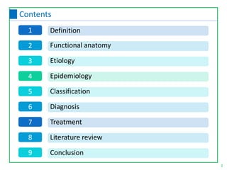
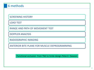
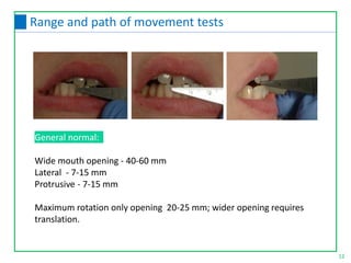
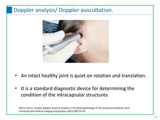
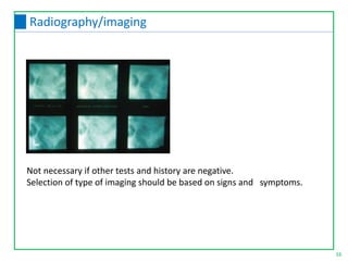
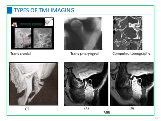
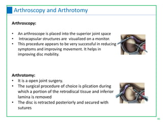
SlideShare a Scribd company logo
TEMPOROMANDIBULAR JOINT DISORDERS
DIAGNOSIS AND MANAGEMENT
Dr.SHARI.S.R
JUNIOR RESIDENT, DEPT. OF PROSTHODONTICS AND CROWN & BRIDGE
GOVT. DENTAL COLLEGE, THIRUVANANTHAPURAM
2
Contents
1
2
3
4
5
6
7
Definition
Functional anatomy
Etiology
Epidemiology
Classification
Diagnosis
Treatment
8 Literature review
9 Conclusion
TMD
Unilateral dull pain in the ear or pre auricular region that is
commonly worse on awakening
Tenderness of one or more muscles of mastication on palpation
Clicking or popping noise in the TMJ
Limitation or deviation of the mandible on opening
4
Signs and symptoms of TMD by Laskin
Diagnosis
5
HOW TO DETERMINE IF THE TMJS ARE HEALTHY !
6
6 ways
Functional occlusion from TMJ to Smile design-Peter.E. Dawson
7
6 methods
SCREENING HISTORY
LOAD TEST
RANGE AND PATH OF MOVEMENT TEST
DOPPLER ANALYSIS
RADIOGRAPHIC IMAGING
ANTERIOR BITE PLANE FOR MUSCLE DEPROGRAMMING
Every patient should be asked key questions about the TMJs
before treating
8
Screening history
9
Sample questionnaire
Diagnostic Criteria for Temporomandibular Disorders Clinical Protocol and Assessment Instruments
International RDC/TMD Consortium Network Version: 20Jan2014
10
Sample questionnaire
11
Load test
For verification of comfort in centric relation or
adapted centric posture. Any sign of tension or
tenderness warrants further evaluation
General normal:
Wide mouth opening - 40-60 mm
Lateral - 7-15 mm
Protrusive - 7-15 mm
Maximum rotation only opening 20-25 mm; wider opening requires
translation.
12
Range and path of movement tests
13
Deviation and Deflection(Path of movement)
Deviation : Movement away from the midline during opening followed
by return to centre during movement. Either “c”or “s” pattern.
Deflection: Movement away from midline during opening without
return to centre during the movement.
On opening, the jaw will normally deviate toward the side of
the displacement
• An intact healthy joint is quiet on rotation and translation.
• It is a standard diagnostic device for determining the
condition of the intracapsular structures
14
Doppler analysis/ Doppler auscultation.
Mario marini. Duplex-doppler spectral analysis in the physiopathology of the temporomandibular joint:
Computerized medical imaging and graphics,18(1)1994 35-43
The coarser the crepitus, the more there is breakdown of the posterior
ligament
Chirping sounds Perforation of the ligament.
Chirping mixed with very coarse crepitus Posterior ligament
severely damaged or lost and bone-to-bone articulation.
Opening and closing clicks at the same protrusive position of the condyle
Ankylosis of the disk
The opening click occuring at a more open relationship than the closing
click Disk is not ankylosed but is still reducible
Crepitus for all jaw movements and no click Disk is not recapturable
15
The character of the amplified sounds
Not necessary if other tests and history are negative.
Selection of type of imaging should be based on signs and symptoms.
16
Radiography/imaging
1. Panoramic radiography
2. Transcranial radiography
3. Trans pharyngeal radiography
4. Anterio - Posterior trans maxillary radiography
5. Computed tomography (CT)
6. Arthrotomography
7. Arthrography with videofluoroscopy
8. Magnetic resonance imaging (MRI)
17
TYPES OF TMJ IMAGING
Management of temporomandibular disorders and occlusion- Jeffrey p. Okeson
Panoramic radiography
• Screening radiograph, they may alert the clinician to a suspected
problem
• Not a dependable modality for assessment of the articular space.
Transcranial radiography
• Readable images can be achieved economically and with minimum
complexity by use of a standard dental x-ray machine.
• Most of the pathologic changes that occur on the articulating
surfaces start at the lateral half of the joint and are visible in the r/g.
• Diagnosis of disk displacement should not be determined solely
from transcranial radiography.
18
TYPES OF TMJ IMAGING
Arthrotomography
• Refers to the injection of a radiopaque contrast medium into the lower
joint space followed by radiography.
• Used to diagnose the position and condition of the meniscus in relation
to the condyle.
• Abnormalities that can be observed include anterior dislocation of the
disk, perforation, degenerative changes, and adhesions
Arthrography with videofluoroscopy
• To observe the movement and contour of the disk in relation to the
condyle as the jaw opens, closes, and translates
Computed tomography (CT)
• Tomography provides a better assessment of the medial pole
area of the condyle
• Improved clarity
19
TYPES OF TMJ IMAGING
Magnetic resonance imaging
Gold standard
Helps in diagnosis of complete disk derangements, or
when unexplainable pain or dysfunction of the TMJs is present
that does not respond to treatment
MRI shows bone marrow changes, disk morphology, mobility, joint effusion
Anterioposterior transmaxillary projection
This offers a good image of superior subarticular bone of the condyle
aswellas medial and lateral poles.
20
TYPES OF TMJ IMAGING
MRI
Trans cranial Trans pharyngeal Computed tomography
21
TYPES OF TMJ IMAGING
CT
Can be used to determine :
- if occlusion is a factor
- if an intracapsular disorder is contributing to the pain.
If a flat, permissive anterior bite plane does not
relieve pain or discomfort at the TMJs
intracapsular disorder as a source of pain.
22
Anterior bite plane for muscle deprogramming
Separating occluso-muscle pain from TMJ pain
23
Functional Occlusion: From TMJ to Smile Design- PETER E DAWSON
Determine whether any masticatory muscle is involved in the
pain. The medial pterygoid muscle is diagnostic – it is almost
always tender to some degree if there is an occluso-muscle
disorder.
24
Separating occluso-muscle pain from TMJ pain
Step 1:
• Ask patient to clench.
• Palpation of the masseter muscle at their superior attachment to the
zygomatic arches.
• Palpation of the superficial masseter muscles near the lower border of
the mandible.
25
Masseter muscle.
26
TEMPORALIS MUSCLE
It has some origin behind the lateral wall of the
orbit of the eye sharp pain behind the eye.
Its aponeurosis extends as an innervated sheath
to the top of the head scalp sore to touch.
Temporal headaches and pain are some of the
most common symptoms related to occluso-
muscle imbalance.
Tendon of the temporalis - Ask patient to open
mouth.
Check for pain. Place finger on anterior ridge of
the coronoid process. Palpate on the superior
aspect of the process.
• Anterior part of insertion can be palpated by placing the finger 45°
in the floor near the base of the relaxed tongue.
• The opposite hand can be used extraorally to palpate posterior
and inferior portions of insertion.
• Body of the muscle can be palpated by rotating the index finger
upwards against the muscle to near its orgin on the tuberosity
27
Medial pterygoid muscle
28
Inferior and Superior lateral pterygoid muscle
Inferior lateral pterygoid muscle
The muscle that pulls the condyle forward every
time the mandible leaves centric relation.
Superior lateral pterygoid muscle.
The muscle that keeps the disk aligned during function.
Ask patient to open the jaw. Ask patient to move his
or her mandible toward the same side, and place
finger on buccal side of alveolar ridge above the
maxillary molars and move finger posteriorly,
medially, and upward as far as possible, and
palpate.
Often involved when deflective occlusal
interferences cause the mandible to be
postured forward to avoid the interferences.
Check for protruded jaw position to achieve
maximum intercuspation.
Muscle involvement If anterior
deprogramming relieves the discomfort
29
The digastric and the hyoid muscles
• If this muscle is tender to palpation, evaluate collateral effects from
head posture and/or cervical misalignments.
• Consider referral to a physical therapist for adjunctive evaluation.
30
Sternocleidomastoid (SCM) muscle.
• Occipital headaches are commonly associated with occlusal interferences.
• May result in combination with head posture and cervical misalignments,
or it may be unrelated to occlusal factors.
• Consider referral to a physical therapist for adjunctive therapy.
31
Occipital area
Occlusal disharmony
Head posture
Muscle pain.
32
Trapezius muscle
Cervical misalignment must always be a consideration.
Consider referral to a physical therapist.
Step 2:
Rule out intracapsular problems.
Verify that centric relation or adapted centric posture can
be achieved.
Load testing must be negative.
33
Separating occluso-muscle pain from TMJ pain
Step 3:
Verify the general acceptability of condyle position and condition
with TMJ radiographs if warranted.
34
Separating occluso-muscle pain from TMJ pain
Step 4:
Rule out pathologic factors as a source of pain.
a. Pulpal
b. Periodontal
c. Soft-tissue
d. Bone
e. Sympathetic and/or referred pain
Step 6:
Correct the cause of the problem.
a. Reversibly with permissive occlusal splint, or
b. Directly with occlusal correction.
Options for treatment:
• Equilibration
• Restorative
• Orthodontics
• Surgery
35
Separating occluso-muscle pain from TMJ pain
Test 1: Clench test
If a patient can clench the teeth together and feel tenderness
in any tooth when the mouth is empty -Positive
Test 2: Anterior deprogramming test
Firmly clench against a cotton roll laid across the arch at
the premolars - If pain is relieved occluso-muscle problem
If clenching on the cotton roll produces discomfort in either
TMJ intracapsular disorder .
Flat anterior deprogramming device [discluder splint] overnight
36
Confirmation of diagnosis of occlusomuscular pain
Treatment
37
Management of temporomandibular disorders and occlusion-
Jeffrey p. Okeson
38
Definitive therapy( for etiologic factors)
• Occlusal appliance(Reversible)
• Selective grinding (irreversible)
• Orthodontic treatment
• Surgical procedures
• Education and cognitive awareness training
• Restrictive use
• Voluntary avoidance
• Relaxation therapy
• Macrotrauma: Mouth guard
• Microtrauma:Occlusal appliance.
For occlusal factors
For emotional stress
For trauma
For deep pain input • Referral to physical therapist.
For parafunctional
activity
• Diurnal activity: Patient education and cognitive
awareness
• Nocturnal activity: Patient education ,Relaxation therapy,
Occlusal appliance therapy ,Clonazepam,Amitryptyline.
PHARMACOLOGIC THERAPY PHYSICAL THERAPY
Analgesics
Anti Inflamatory
Anxiolytics
Muscle Relaxants
Anti-depressants
Anti-convulsants
Injectables
Topicals
MODALITIES
MANUAL
TECHNIQUES
Soft tissue mobilisation
Joint mobilisation
Muscle conditioning
Passive muscle stretching
Active muscle stretching
Resistance exercises
Postural training
39
Supportive therapy
Thermotherapy
Coolant therapy
Ultrasound therapy
Phonophoresis
Iontophoresis
Electro galvanic
stimulation
TENS
Cold laser
Assisted stretching Resisted exercises.
Accupuncture Physical self regulation
40
Supportive therapy
Definitive management is usually based on etiology
Supportive management:
• Restrict the use of the mandible within painless limits
• Soft diet
• Short term analgesics
• Physical self regulation techniques
41
TREATMENT OF MASTICATORY MUSCLE DISORDERS
Myofascial pain
Trigger points management:
• Spray and stretch –Simon and Travel
• Injection and stretch
• Pressure and massage
• Ultrasound and electrogalvanic stimulation
• Muscle relaxants
• Muscle conditioning techniques
42
Disk displacement with reduction:
An Anterior repositioning appliance should be
fabricated to wear at night during sleep and during the
day when needed to reduce symptoms. These adaptive
changes can take 8 to 10 weeks or even longer.
1. Softer foods, slower chewing, smaller bites.
2. If inflammation - NSAID’s , moist heat or ice.
3. Passive jaw movements may be helpful.
4. Distraction manipulation by physical therapist.
Definitive
treatment
01 02 03 04
Supportive therapy:
Patient attempt to reduce the dislocation without
assistance - to move the mandible to the
contralateral side as far as possible. From this
eccentric position the mouth is opened maximally.
Anterior repositioning appliance is contraindicated
43
Disk dislocation without reduction:
Definitive treatment:
Self reduction:
Lateral pterygoid muscle – should be relaxed
If active – Local anesthesia
44
Manual manipulation
3. Joint is distracted, ask the patient to protrude
the mandible.
4. Ask patient to move contra lateral side and ask
to relax, 20-30 sec distractive force is applied to
the joint.
5. Ask the patient to close the mouth to the incisal
end to end position
6. Ask to open wide and return to anterior position
7. Anterior positioning appliance is placed
immediately
1. The thumb - intraorally over the mandibular second molar on the affected side. The
fingers - on the inferior border of the mandible anterior to thumb position.
2. Firm but controlled downward force is then exerted on the molar and at the same
time upward force is placed by the fingers
Surgery should be considered only when conservative therapy fails to resolve
adequately the symptoms and or progression of the disorder.
Arthrocentesis:
• Most conservative surgical procedures.
• Two needles are placed into the joint .
• Sterile saline solution is passed through lavaging the joint. The lavage is thought
to eliminate much of the algogenic substances and breakdown by products that
produce the pain
Pumping the joint:
In cases of disc dislocation without reduction a single needle can be
introduced to the joint and fluid can be forced into the space in an attempt to free
the articular surfaces.
45
Surgical considerations for condyle disc derangement
Arthroscopy:
• An arthroscope is placed into the superior joint space
• Intracapsular structures are visualized on a monitor.
• This procedure appears to be very successful in reducing
symptoms and improving movement. It helps in
improving disc mobility.
Arthrotomy:
• It is a open joint surgery.
• The surgical procedure of choice is plication during
which a portion of the retrodiscal tissue and inferior
lamina is removed
• The disc is retracted posteriorly and secured with
sutures
46
Arthroscopy and Arthrotomy
47
Discectomy
Disc is damaged and can no
longer be maintained for
use. So disc is removed
It leaves a bone to bone
articulation which is likely to
produce some osteoarthritic
changes.
Another choice is to remove the disc
and replace it with a substitute – Discal
implants which include medical silastic,
proplast-Teflon, Dermal and auricular
cartilage grafts.
1
2
3
Adhesions:
• Breaking fibrous attachment using arthroscopic surgery.
• Passive stretching.
• Distraction of the joint.
Subluxations:
• Eminectomy
• Intraoral device that limiting the opening before the point of subluxation
Spontaneous dislocation:
Manual manipulation:
• Thumbs are placed on the mandibular molars & downward pressure is
exerted.
• If not reduced inject LA to inferior pterygoid muscle.
• Chronic spontaneous dislocation ----------- Eminectomy
48
Treatment for structural incompatibility of articular surfaces
Definitive treatment:
Since the etiology is self limiting there is no definitive treatment
indicated. When recurrence of trauma is likely, efforts are made to
protect the joint from any further injury.
Supportive therapy:
•Restrict all mandibular movements within painless limits-soft diet, slow
movements and small bites.
•Constant pain - mild analgesics.
•Moist heat 4-5 times a day for 10-15 minutes.
•Ultrasound therapy – 2-4 times / week.
•Single injection of corticosteriod to the capsular tissues. Repeated
injections are contraindicated.
49
Synovitis, Capsulitis
Definitive treatment:
The mechanical loading should be decreased.
Stabilization appliance- When muscle hyperactivity is suspected.
Supportive therapy
It begins with an explanation of the disease process to the patient.
Analgesics
Anti-inflammatory agents.
Soft diet.
Thermotherapy is usually helpful in reducing symptoms.
Arthritidis:
Retro discitis:
• Along with clenching: Give stabilization appliance.
• Due to micro trauma/Disc dislocation with reduction : Anterior positioning
appliance.
50
Retro discitis and Arthritidis
Definitive treatment:
If function is inadequate or the restriction is intolerable, surgery is the only
definitive treatment available.
Supportive therapy:
Since ankylosis is normally asymptomatic generally no supportive therapy is
indicated. However, if the mandible is forced beyond its restriction, injury
to the tissues can occur. If pain and inflammation result, supportive therapy
is called for and consists of voluantarily restricting movement to either
painless limits. Ankylosis along with deep heat therapy can also be used.
51
Ankylosis
Definition:
An occlusal appliance is a removable device usually made if hard acrylic
that fits over the occlusal and incisal surface of the teeth in one arch,
creating precise occlusal contact with the teeth of the opposing arch.
It is also called as :
1. Bite guard
2. Night guard
3. Interocclusal appliance
4. Orthopedic device
52
Occlusal Splints
• Repositioning of condyles and discs
• Increase of vertical dimension
• Elimination of occlusal interference
• Avoidance of excessive occlusal wear
• Relaxation of jaw and neck muscles
• Stabilization of occlusal and neuromuscular features
• Reduction of headaches
53
Rationale for Splints
Based on Consistency :
• Hard splints e.g.: acrylic resins (heat cure and chemical cure)
• Soft splints e.g.: polyvinyl sheets
Based on the type of fabrication
• Direct – chairside fabrication
• Indirect – laboratory fabrication
54
Occlusal splint classification.
55
Classification of Splints
Dawson classification
Okeson classification
Ash & Ramfjord classification
56
Dawson Classification
 Stabilization appliance
 Anterior positioning appliance
 Other types of occlusal devices:
1. Anterior bite plane
2.Posterior bite plane
3.Pivoting appliance
4.Soft / resilient appliance
Stabilization appliance:
• Primary use is to reduce muscular pain –Muscle relaxation appliance
• Eliminate ORTHOPEDIC INSTABILITY.
Anterior positioning appliance:
• It position the mandible more anteriorly to provide a better condyle
disc relationship.
57
Okeson classification
58
Indications of stabilizing appliances
• Bruxism
• Local muscle soreness
• Centrally mediated myalgia
• Retrodiscitis secondary to microtrauma.
• Osteoarthritis
• Establishment of optimal condylar position in centric relation prior to
definitive occlusal therapy.
• It must accurately fit the maxillary teeth and have
good retention and stability.
• In CR- all mandibular buccal cusps and incisal
edges must contact with even force on flat
surface.
• During protrusive- Mandibular canines must
contact the appliance with even force.
• In lateral movements only canines should exhibit
laterotrusive contact.
• During closure and upright feeding position–
Mandibular posterior teeth must contact the
appliance more heavily than anterior teeth.
59
Final criteria for stabilization appliance
• This helps to position the mandible more anteriorly than the
intercuspal position.
• Provide better condyle-disc relationship
• Provide better oppourtunity for tissue repair.
Indications:
• Disc displacement.
• Dislocation with reduction.
• Retrodiscitis ( due to microtrauma).
60
Anterior repositioning appliance
• Stability and retention
• In established forward position all the mandibular teeth
should contact with even force.
• The forward position is established by asking the patient to
move forward the jaw till the initial clicking starts.
• Lingual retrusive guidance ramp direct the mandible into
the established therapeutic forward position.
61
Final criteria for the anterior positioning appliance
• It provide contact only with mandibular anterior teeth.
• It disengage the posterior teeth & eliminate their influence on
masticatory system.
Indications:
• Muscle disorders related to orthopedic instability
• Accute changes in occlusal condition.
Complications
• Supraeruption of posterior teeth(> 2 weeks)
NTITSS
62
Anterior bite plane
• Fabricated by hard acrylic over the mandibular posterior teeth.
• Indications:
• Severe loss of vertical dimensions
• Mandibular anterior positioning.
• Complication :
• Supra eruption of anterior teeth.
63
Posterior bite plane
64
Hydrostatic Appliances/Aqualizer
65
Pivot appliance
• The pivoting is a hard acrylic
device that covers one arch
and usually provides a single
posterior contact in each
quadrant.
• This appliance lessens
intraarticular pressure and
unload the articular surface of
the joint.
Made using resilient material on maxillary teeth
Indication:
• Protective device who likely to receive trauma
• Clenching & bruxism
• Repeated sinusitis resulting in extremely sensitive teeth
• Complication:
• Difficult to adjust.
Hard splints are better
than soft splints
66
Soft splints / Resilient splints
67
Indication of various splints
68
Occlusal Equilibration
It is a procedure by which the occlusal surfaces of the teeth are precisely
altered to improve the overall contact pattern.
Procedure includes:
• Reduction of all contacting teeth surfaces that interfere with the terminal
hinge axis closure.
• Selective grinding of tooth structures that interfere with lateral excursion.
• Selective grinding of teeth that interferes with protrusive movement.
• Harmonization of anterior guidance
69
Armamentarium for Occlusal correction
Paste ,Spray,Paint on material
70
Rule of Third
Predict success of selective grinding.
71
• INTERFERENCES IN THE ARC OF CLOSURE—Anterior slide
• INTERFERENCES IN THE LINE OF CLOSURE----Right or left slide
(Dawson)
According to Okeson CR slide can be 3 types :
• Anterosuperior
• Antereosuperior and to the right
• Antereosuperior and to the left
Eliminating interferences to centric relation position
• Due to contact between mesial inclines of maxillary cusps and the
distal inclines of the mandibular cusps
Grinding rule MUDL
72
Interferences in the arc of closure
Slide toward cheek:
Due to inner incline of the maxillary lingual cusp against the inner incline
of the mandibular buccal cusp.
Grinding rule: BULL
Slide toward tongue:
Due to lingual incline of the upper or buccal incline of lower.
Grinding rule: LUBL
73
Interferences in the line of closure
Mediotrusive contacts Grinding rule: BUCCAL OF UPPER LINGUAL CUSP AND
LINGUAL OF LOWER BUCCAL CUSP (BUL* LLB).
74
Eliminating the interferences in balancing side
Laterotrusive contacts: LINGUAL OF UPPER BUCCAL CUSP AND BUCCAL OF LOWER
LINGUAL CUSP (LUB* BLL)----Working side interferences
75
Eliminating lateral interferences – BULL’s Law
76
GRINDING BASIC RULES:
• Narrow stamp cusps before reshaping fossae.
• Don’t shorten a stamp cusp.
• Upper teeth are always adjusted on the inclines that face the same
direction as the slide.
• Lower teeth are adjusted by grinding the inclines that face the opposite
direction from path of the slide.
• Adjust centric interferences first
• Then lateral excursive interferences
• Protrusive interferences.
• It can occur between distal inclines of maxillary lingual cusp and
mesial inclines of mandibular buccal cusp.
Grinding rules: DUML
Eliminating interferences in Protrusive contacts
77
Occlusal equilibration in complete denture
• Incorrect registration of RCP
• Irregularities in setting the teeth
• Tooth movement during deflasking and packing.
• Incomplete flask closure.
Causes of occlusal disharmony:
78
Types of occlusal error in centric occlusion and their
correction.: 3
1)Any pair of opposing teeth can be too long and hold the other teeth out of
contact.
Correction: The fossae of the teeth are deepened by grinding so the teeth will in
effect,telescope into each other.The cusp are not shortened.
2)The upper and lower teeth can be too nearly end to end.
Correction: Grind the outer inclines of the functional cusp.
3) The upper teeth can be too far buccally in relation to lower teeth.
Correction: Grind the inner incline of the functional cusp from fossae.so
broadening the fossae near the inner incline.
79
Types of working side occlusal error and their
correction:- 6
1) Both the upper buccal cusp and the lower lingual cusp are too long .
Correction: The length of cusps are reduced by grinding to change the incline
extending from the central fossae to the cusp tip.,Central fossae is not made
deeper,but the cusps made shorter.
2) The buccal cusp make contact but the lingual cusp donot.
Correction: Buccal cusp of the upper teeth are ground from the central fossa to the
cusp tip to shorten the cusp.
3) The lingual cusp make contact but the buccal cusp donot.
Correction: The lower lingual cusps are shortened by changing the buccal incline of
the lower lingual cusp ,so it is not as steep.
80
Types of working side occlusal error and their
correction:- 6
4) Upper buccal or lingual cusps are mesial to their intercuspative postions
Correction: Grinding is done on the mesial inclines of the upper buccal cusps
and distal inclines of the lower cusps.
5)Upper buccal or lingual cusps are distal to their intercusping positions.
Correction: Grinding is done from the distal of the upper cusps and from
the mesial of the lower cusps.
6) Teeth on the working side may not contact.
Due to excessive contact on balancing side.
Correction: Grind lingual incline of the lower buccal cusp.
81
Types of balancing side occlusal error and their
correction:- 2
1) Balancing side contact is so heavy so that working side teeth are held out
of contact.
Correction: Grind the lingual incline of the lower buccal cusp.
2) There is no contact on the balancing side:
Correction: Grind lingual incline of the upper buccal cusps and buccal
incline of lower lingual cusp.
82
LiteratureReview
Ware and Rugh studied a group of bruxism patients without pain and another
group with pain
• Bruxers with pain had a significantly higher number of bruxing events during
REM sleep than did the former.
• Bothgroups, however, bruxed more than a control group.
This study suggests that there might be two types of bruxism patients: one bruxing
more during REM sleep and one bruxing more during the non-REM phases.
• Other studies by these authors showed that the amount of sustained
contraction occurring in bruxism was commonly much higher during the REM
than the non-REM phases of sleep.
• These findings help to explain the conflicting literature on sleep stages and
bruxism and may also explain why some patients awaken with pain but others
with clinical evidence of bruxism report no pain.
83
Literature Review
Study by Rompre et al in 2007 investigated the number of bruxing events per
night in a group of bruxing patients with pain and compared them with
another group of bruxing patients without pain.
The bruxing group without pain actually had more bruxing events per night
than the ones with pain
Logic
• Patients who regularly brux during sleep condition their muscles and adapt
to this activity. Regular exercise leads to stronger, larger, more efficient
muscles.
• This may explain why dentists often observe middle-aged male patients
with major tooth wear secondary to bruxing, yet they have no pain
• Patients who awaken with muscle pain are more likely to be those who do
not frequently brux; therefore their muscles are not conditioned to this
activity. This unconditioned activity is more likely to be associated with pain.
84
Literature Review
Anna Colonna et.al,(2018) Comparative analysis of jaw morphology and
temporomandibular disorders: A three-dimension imaging
study, CRANIO®, DOI: 10.1080/08869634.2018.1507094
• (CT) scans of 20 individuals aged 18 to 40 with (TMD group) or without
TMJ pain (control group) . Three-dimensional reconstructions were
performed to evaluate the gonial angle, condylar volume, and the
distance between the posterior edge of the condyle and the sigmoid
notch.
• There is an association between the presence of TMJ pain and some
features of craniofacial morphology. Individuals with TMJ pain have a
lower condylar volume and a tendency towards hyperdivergent growth.
85
Literature Review
Tissue engineering of the mandibular condyle have been trying to regenerate both
bone and cartilage with distinct structural and functional differences. The
scaffolds fabricated for this purpose must fulfill the biological and mechanical
requirements for cartilage and bone regeneration --- surface chemistry, high
porosity, mechanical compliance, biodegradability and biocompatibility for cell
growth and extracellular matrix deposition
Temporomandibular Joint Replacement—Past, Present and Future: A
Bioengineering Perspective T. Vo Van et al. (eds.), 6th International Conference on
the Development of Biomedical Engineering in Vietnam (BME6), IFMBE
Proceedings 63
Prosthesis Fossa Condyle Ramus
Biomet UHMWPE Cobalt chrome /Ti Cobaltchrome/Titanium
TMJ concepts Titanium Cobalt chrome Titanium
TMJ implants Cobalt chrome Cobalt chrome Cobalt chrome
86
Literature Review
87
LiteratureReview
Biomet Christensen prosthesis
TMJ concepts prosthesis
Temporomandibular disorders (TMD) is an umbrella
term for pain and dysfunction involving the masticatory muscles
and the TMJs. Chronic pain is the overwhelming reason that
patients with TMD seek treatment.
TMD can associate with impaired general health,
depression, and other psychological disabilities, and may affect
the quality of life of the patient The clinician should be able to
differentiate the various clinical symptoms associated with the
disorders and do the treatment accordingly.
88
Conclusion
89
References
1. Functional Occlusion: From TMJ to Smile Design- PETER E DAWSON
2. Management of temporomandibular disorders and occlusion / Jeffrey P. Okeson. --
7th ed.
3. Diagnostic Criteria for Temporomandibular Disorders Clinical Protocol and
Assessment Instruments International RDC/TMD Consortium Network Version:
20Jan2014
4. Ware JC, Rugh JD: Destructive bruxism: sleep stage relationship, Sleep 11(2):172–
181, 1988.
5. Rompre PH, Daigle-Landry D, Guitard F, et al: Identification of a sleep bruxism
subgroup with a higher risk of pain, J Dent Res 86(9):837–842, 2007.
6. Temporomandibular Joint Replacement—Past, Present and Future: A
Bioengineering Perspective T. Vo Van et al. (eds.), 6th International Conference on
the Development of Biomedical Engineering in Vietnam (BME6), IFMBE
Proceedings 63, https://doi.org/10.1007/978-981-10-4361-1_93
7. Anna Colonna et.al,(2018) Comparative analysis of jaw morphology and
temporomandibular disorders: A three-dimension imaging study, CRANIO®, DOI:
10.1080/08869634.2018.1507094
Thank You

More Related Content

What's hot

Temporomandibular joint disorders
Temporomandibular joint disordersTemporomandibular joint disorders
Temporomandibular joint disorders
DR PAAVANA
 
Autogenous bone grafting
Autogenous bone graftingAutogenous bone grafting
Autogenous bone grafting
Dr. Kritika Jangid
 
Genioplasty
GenioplastyGenioplasty
Designing for kennedy class i and class ii
Designing for kennedy class i and class iiDesigning for kennedy class i and class ii
Designing for kennedy class i and class ii
DrLeenaTomer
 
Role of facial muscles in complete denture prosthesis
Role of facial muscles  in complete denture  prosthesisRole of facial muscles  in complete denture  prosthesis
Role of facial muscles in complete denture prosthesis
Ravi banavathu
 
Vestibuloplasty- ridge extension procedures
Vestibuloplasty- ridge extension proceduresVestibuloplasty- ridge extension procedures
Vestibuloplasty- ridge extension procedures
Zeeshan Arif
 
Occlusal splints
Occlusal splintsOcclusal splints
Occlusal splints
padmini rani
 
Muscles surrounding Complete Denture
Muscles surrounding Complete DentureMuscles surrounding Complete Denture
Muscles surrounding Complete Denture
Naveed AnJum
 
Trauma from occlusion
Trauma from occlusionTrauma from occlusion
Trauma from occlusion
Muneeb Muhammed Ali
 
Prosthodontics - realeff relevance in complete denture
Prosthodontics - realeff relevance in complete dentureProsthodontics - realeff relevance in complete denture
Prosthodontics - realeff relevance in complete denture
KIIT ,BHUBANESWAR
 
Examination of tmj &muscles of mastication (2)
Examination of tmj &muscles of mastication (2)Examination of tmj &muscles of mastication (2)
Examination of tmj &muscles of mastication (2)
rachitajainr
 
posterior palatal seal ppt
posterior palatal seal pptposterior palatal seal ppt
posterior palatal seal ppt
Preeti Kalia
 
theories of impression making in complete denture
theories of impression making in complete denturetheories of impression making in complete denture
theories of impression making in complete denture
dipalmawani91
 
Disorders of TMJ
Disorders of TMJDisorders of TMJ
Disorders of TMJ
Dr Nandika Babele
 
Temporomandibular joint /disorders /management / treatment
Temporomandibular joint /disorders /management / treatmentTemporomandibular joint /disorders /management / treatment
Temporomandibular joint /disorders /management / treatment
Cairo University
 
Temporomandibular joint disorders I
Temporomandibular joint disorders ITemporomandibular joint disorders I
Temporomandibular joint disorders I
IAU Dent
 
TMJ DISORDERS - PART 1.ppt
TMJ DISORDERS - PART 1.pptTMJ DISORDERS - PART 1.ppt
TMJ DISORDERS - PART 1.ppt
DentalYoutube
 
Mpds
MpdsMpds
Vestibuloplasty
VestibuloplastyVestibuloplasty
Vestibuloplasty
vrushupatel
 
Horizontal Jaw Relation
Horizontal Jaw RelationHorizontal Jaw Relation
Horizontal Jaw Relation
Dr. Anshul Sahu
 

What's hot (20)

Temporomandibular joint disorders
Temporomandibular joint disordersTemporomandibular joint disorders
Temporomandibular joint disorders
 
Autogenous bone grafting
Autogenous bone graftingAutogenous bone grafting
Autogenous bone grafting
 
Genioplasty
GenioplastyGenioplasty
Genioplasty
 
Designing for kennedy class i and class ii
Designing for kennedy class i and class iiDesigning for kennedy class i and class ii
Designing for kennedy class i and class ii
 
Role of facial muscles in complete denture prosthesis
Role of facial muscles  in complete denture  prosthesisRole of facial muscles  in complete denture  prosthesis
Role of facial muscles in complete denture prosthesis
 
Vestibuloplasty- ridge extension procedures
Vestibuloplasty- ridge extension proceduresVestibuloplasty- ridge extension procedures
Vestibuloplasty- ridge extension procedures
 
Occlusal splints
Occlusal splintsOcclusal splints
Occlusal splints
 
Muscles surrounding Complete Denture
Muscles surrounding Complete DentureMuscles surrounding Complete Denture
Muscles surrounding Complete Denture
 
Trauma from occlusion
Trauma from occlusionTrauma from occlusion
Trauma from occlusion
 
Prosthodontics - realeff relevance in complete denture
Prosthodontics - realeff relevance in complete dentureProsthodontics - realeff relevance in complete denture
Prosthodontics - realeff relevance in complete denture
 
Examination of tmj &muscles of mastication (2)
Examination of tmj &muscles of mastication (2)Examination of tmj &muscles of mastication (2)
Examination of tmj &muscles of mastication (2)
 
posterior palatal seal ppt
posterior palatal seal pptposterior palatal seal ppt
posterior palatal seal ppt
 
theories of impression making in complete denture
theories of impression making in complete denturetheories of impression making in complete denture
theories of impression making in complete denture
 
Disorders of TMJ
Disorders of TMJDisorders of TMJ
Disorders of TMJ
 
Temporomandibular joint /disorders /management / treatment
Temporomandibular joint /disorders /management / treatmentTemporomandibular joint /disorders /management / treatment
Temporomandibular joint /disorders /management / treatment
 
Temporomandibular joint disorders I
Temporomandibular joint disorders ITemporomandibular joint disorders I
Temporomandibular joint disorders I
 
TMJ DISORDERS - PART 1.ppt
TMJ DISORDERS - PART 1.pptTMJ DISORDERS - PART 1.ppt
TMJ DISORDERS - PART 1.ppt
 
Mpds
MpdsMpds
Mpds
 
Vestibuloplasty
VestibuloplastyVestibuloplasty
Vestibuloplasty
 
Horizontal Jaw Relation
Horizontal Jaw RelationHorizontal Jaw Relation
Horizontal Jaw Relation
 

Similar to TEMPOROMANDIBULAR JOINT DISORDERS second part

Tm j examination
Tm j examinationTm j examination
Tm j examination
mortazavimohammad
 
Temporomandibular joint disorders
Temporomandibular joint disorders Temporomandibular joint disorders
Temporomandibular joint disorders
Eman Alsheikh
 
Cervical Hybrid Arthroplasty by Pablo Pazmino MD
Cervical Hybrid Arthroplasty by Pablo Pazmino MDCervical Hybrid Arthroplasty by Pablo Pazmino MD
Cervical Hybrid Arthroplasty by Pablo Pazmino MD
Pablo Pazmino
 
TEMPORO MANDIBULAR JOINT IMAGING PPT
TEMPORO MANDIBULAR JOINT IMAGING PPTTEMPORO MANDIBULAR JOINT IMAGING PPT
TEMPORO MANDIBULAR JOINT IMAGING PPT
Naba Kumar Barman
 
TMJ details and easy to understand it.ppt
TMJ details and easy to understand it.pptTMJ details and easy to understand it.ppt
TMJ details and easy to understand it.ppt
RumelaGhosh5
 
lumbardiscprolapse3-130219095421-phpapp01 (1).pdf
lumbardiscprolapse3-130219095421-phpapp01 (1).pdflumbardiscprolapse3-130219095421-phpapp01 (1).pdf
lumbardiscprolapse3-130219095421-phpapp01 (1).pdf
SriRam071
 
9. TMJ INTERNAL DERANGEMENT.pptx
9. TMJ INTERNAL DERANGEMENT.pptx9. TMJ INTERNAL DERANGEMENT.pptx
9. TMJ INTERNAL DERANGEMENT.pptx
DrChandiniRavikumar
 
Evaluation and management of cervical spine injury
Evaluation and management of cervical spine injuryEvaluation and management of cervical spine injury
Evaluation and management of cervical spine injury
Love2jaipal
 
TMJ 3
TMJ 3TMJ 3
TMJ 3
IAU Dent
 
Makkad tmj disorders/ dental implant courses
Makkad tmj disorders/ dental implant coursesMakkad tmj disorders/ dental implant courses
Makkad tmj disorders/ dental implant courses
Indian dental academy
 
Diagnosis of temporomandibular disorders- Kelly
Diagnosis of temporomandibular disorders- Kelly Diagnosis of temporomandibular disorders- Kelly
Diagnosis of temporomandibular disorders- Kelly
Kelly Norton
 
Temporomandibular joint ankylosis and its management
Temporomandibular joint ankylosis and its managementTemporomandibular joint ankylosis and its management
Temporomandibular joint ankylosis and its management
Dibya Falgoon Sarkar
 
TMD and malocclusion.docx
TMD and malocclusion.docxTMD and malocclusion.docx
TMD and malocclusion.docx
Dr.Mohammed Alruby
 
Radiographic Features of Temporomandibular Joint and Disorders
Radiographic Features of Temporomandibular Joint and DisordersRadiographic Features of Temporomandibular Joint and Disorders
Radiographic Features of Temporomandibular Joint and Disorders
Hadi Munib
 
Cervical degenerative disease and injuries
Cervical degenerative disease and injuriesCervical degenerative disease and injuries
Cervical degenerative disease and injuries
Neurosurgeon Mumtaz Ali Narejo
 
Facial pain due to TMJ, trigminal neuralgia, Sjogren syndrome, Eagles etc..
Facial pain due to TMJ, trigminal neuralgia, Sjogren syndrome, Eagles etc..Facial pain due to TMJ, trigminal neuralgia, Sjogren syndrome, Eagles etc..
Facial pain due to TMJ, trigminal neuralgia, Sjogren syndrome, Eagles etc..
Nelson Hendler
 
Osteochondroma
OsteochondromaOsteochondroma
Osteochondroma
GIRIDHAR BOYAPATI
 
TMJ DISLOCATION AND ITS MANAGEMENT..pptx
TMJ DISLOCATION AND ITS MANAGEMENT..pptxTMJ DISLOCATION AND ITS MANAGEMENT..pptx
TMJ DISLOCATION AND ITS MANAGEMENT..pptx
drash9955
 
Tmj examination & imaging
Tmj examination & imagingTmj examination & imaging
Tmj examination & imaging
Chetan Basnet
 
TMJ overview
TMJ overviewTMJ overview
TMJ overview
Guru Karthik
 

Similar to TEMPOROMANDIBULAR JOINT DISORDERS second part (20)

Tm j examination
Tm j examinationTm j examination
Tm j examination
 
Temporomandibular joint disorders
Temporomandibular joint disorders Temporomandibular joint disorders
Temporomandibular joint disorders
 
Cervical Hybrid Arthroplasty by Pablo Pazmino MD
Cervical Hybrid Arthroplasty by Pablo Pazmino MDCervical Hybrid Arthroplasty by Pablo Pazmino MD
Cervical Hybrid Arthroplasty by Pablo Pazmino MD
 
TEMPORO MANDIBULAR JOINT IMAGING PPT
TEMPORO MANDIBULAR JOINT IMAGING PPTTEMPORO MANDIBULAR JOINT IMAGING PPT
TEMPORO MANDIBULAR JOINT IMAGING PPT
 
TMJ details and easy to understand it.ppt
TMJ details and easy to understand it.pptTMJ details and easy to understand it.ppt
TMJ details and easy to understand it.ppt
 
lumbardiscprolapse3-130219095421-phpapp01 (1).pdf
lumbardiscprolapse3-130219095421-phpapp01 (1).pdflumbardiscprolapse3-130219095421-phpapp01 (1).pdf
lumbardiscprolapse3-130219095421-phpapp01 (1).pdf
 
9. TMJ INTERNAL DERANGEMENT.pptx
9. TMJ INTERNAL DERANGEMENT.pptx9. TMJ INTERNAL DERANGEMENT.pptx
9. TMJ INTERNAL DERANGEMENT.pptx
 
Evaluation and management of cervical spine injury
Evaluation and management of cervical spine injuryEvaluation and management of cervical spine injury
Evaluation and management of cervical spine injury
 
TMJ 3
TMJ 3TMJ 3
TMJ 3
 
Makkad tmj disorders/ dental implant courses
Makkad tmj disorders/ dental implant coursesMakkad tmj disorders/ dental implant courses
Makkad tmj disorders/ dental implant courses
 
Diagnosis of temporomandibular disorders- Kelly
Diagnosis of temporomandibular disorders- Kelly Diagnosis of temporomandibular disorders- Kelly
Diagnosis of temporomandibular disorders- Kelly
 
Temporomandibular joint ankylosis and its management
Temporomandibular joint ankylosis and its managementTemporomandibular joint ankylosis and its management
Temporomandibular joint ankylosis and its management
 
TMD and malocclusion.docx
TMD and malocclusion.docxTMD and malocclusion.docx
TMD and malocclusion.docx
 
Radiographic Features of Temporomandibular Joint and Disorders
Radiographic Features of Temporomandibular Joint and DisordersRadiographic Features of Temporomandibular Joint and Disorders
Radiographic Features of Temporomandibular Joint and Disorders
 
Cervical degenerative disease and injuries
Cervical degenerative disease and injuriesCervical degenerative disease and injuries
Cervical degenerative disease and injuries
 
Facial pain due to TMJ, trigminal neuralgia, Sjogren syndrome, Eagles etc..
Facial pain due to TMJ, trigminal neuralgia, Sjogren syndrome, Eagles etc..Facial pain due to TMJ, trigminal neuralgia, Sjogren syndrome, Eagles etc..
Facial pain due to TMJ, trigminal neuralgia, Sjogren syndrome, Eagles etc..
 
Osteochondroma
OsteochondromaOsteochondroma
Osteochondroma
 
TMJ DISLOCATION AND ITS MANAGEMENT..pptx
TMJ DISLOCATION AND ITS MANAGEMENT..pptxTMJ DISLOCATION AND ITS MANAGEMENT..pptx
TMJ DISLOCATION AND ITS MANAGEMENT..pptx
 
Tmj examination & imaging
Tmj examination & imagingTmj examination & imaging
Tmj examination & imaging
 
TMJ overview
TMJ overviewTMJ overview
TMJ overview
 

More from shari kurup

MAXILLARY MAJOR CONNECTOR.pptx
MAXILLARY MAJOR CONNECTOR.pptxMAXILLARY MAJOR CONNECTOR.pptx
MAXILLARY MAJOR CONNECTOR.pptx
shari kurup
 
1.PLATFORM SWITCHING - Dr Shari S R.pptx
1.PLATFORM SWITCHING - Dr Shari S R.pptx1.PLATFORM SWITCHING - Dr Shari S R.pptx
1.PLATFORM SWITCHING - Dr Shari S R.pptx
shari kurup
 
Pedagogy.pptx
Pedagogy.pptxPedagogy.pptx
Pedagogy.pptx
shari kurup
 
BASICS IN DENTAL IMPLANT
BASICS IN  DENTAL IMPLANT BASICS IN  DENTAL IMPLANT
BASICS IN DENTAL IMPLANT
shari kurup
 
PLATFORM SWITCHING
PLATFORM SWITCHINGPLATFORM SWITCHING
PLATFORM SWITCHING
shari kurup
 
DONNING AND DOFFING
DONNING AND DOFFINGDONNING AND DOFFING
DONNING AND DOFFING
shari kurup
 
SMILE DESIGN
SMILE DESIGNSMILE DESIGN
SMILE DESIGN
shari kurup
 
PROGRESSIVE LOADING IN IMPLANTS
PROGRESSIVE LOADING IN IMPLANTSPROGRESSIVE LOADING IN IMPLANTS
PROGRESSIVE LOADING IN IMPLANTS
shari kurup
 
IMPLANT OCCLUSION
IMPLANT OCCLUSIONIMPLANT OCCLUSION
IMPLANT OCCLUSION
shari kurup
 
MANDIBULAR MOVEMENTS
MANDIBULAR MOVEMENTSMANDIBULAR MOVEMENTS
MANDIBULAR MOVEMENTS
shari kurup
 
Principles and techniques of impresion
Principles and techniques of impresion Principles and techniques of impresion
Principles and techniques of impresion
shari kurup
 
LASERS IN PROSTHODONTICS
LASERS IN PROSTHODONTICSLASERS IN PROSTHODONTICS
LASERS IN PROSTHODONTICS
shari kurup
 
Phonetics
Phonetics Phonetics
Phonetics
shari kurup
 
TOOTH SUPPORTED OVERDENTURE
TOOTH SUPPORTED OVERDENTURETOOTH SUPPORTED OVERDENTURE
TOOTH SUPPORTED OVERDENTURE
shari kurup
 
ARTICULATORS
ARTICULATORSARTICULATORS
ARTICULATORS
shari kurup
 
ETHICS IN DENTISTRY
ETHICS IN DENTISTRYETHICS IN DENTISTRY
ETHICS IN DENTISTRY
shari kurup
 
Selective grinding
Selective grindingSelective grinding
Selective grinding
shari kurup
 
vertical jaw relation
 vertical jaw relation  vertical jaw relation
vertical jaw relation
shari kurup
 
Dental ceramics
Dental ceramicsDental ceramics
Dental ceramics
shari kurup
 
Abrasives and polishing agents of dentistry
Abrasives and polishing agents of dentistryAbrasives and polishing agents of dentistry
Abrasives and polishing agents of dentistry
shari kurup
 

More from shari kurup (20)

MAXILLARY MAJOR CONNECTOR.pptx
MAXILLARY MAJOR CONNECTOR.pptxMAXILLARY MAJOR CONNECTOR.pptx
MAXILLARY MAJOR CONNECTOR.pptx
 
1.PLATFORM SWITCHING - Dr Shari S R.pptx
1.PLATFORM SWITCHING - Dr Shari S R.pptx1.PLATFORM SWITCHING - Dr Shari S R.pptx
1.PLATFORM SWITCHING - Dr Shari S R.pptx
 
Pedagogy.pptx
Pedagogy.pptxPedagogy.pptx
Pedagogy.pptx
 
BASICS IN DENTAL IMPLANT
BASICS IN  DENTAL IMPLANT BASICS IN  DENTAL IMPLANT
BASICS IN DENTAL IMPLANT
 
PLATFORM SWITCHING
PLATFORM SWITCHINGPLATFORM SWITCHING
PLATFORM SWITCHING
 
DONNING AND DOFFING
DONNING AND DOFFINGDONNING AND DOFFING
DONNING AND DOFFING
 
SMILE DESIGN
SMILE DESIGNSMILE DESIGN
SMILE DESIGN
 
PROGRESSIVE LOADING IN IMPLANTS
PROGRESSIVE LOADING IN IMPLANTSPROGRESSIVE LOADING IN IMPLANTS
PROGRESSIVE LOADING IN IMPLANTS
 
IMPLANT OCCLUSION
IMPLANT OCCLUSIONIMPLANT OCCLUSION
IMPLANT OCCLUSION
 
MANDIBULAR MOVEMENTS
MANDIBULAR MOVEMENTSMANDIBULAR MOVEMENTS
MANDIBULAR MOVEMENTS
 
Principles and techniques of impresion
Principles and techniques of impresion Principles and techniques of impresion
Principles and techniques of impresion
 
LASERS IN PROSTHODONTICS
LASERS IN PROSTHODONTICSLASERS IN PROSTHODONTICS
LASERS IN PROSTHODONTICS
 
Phonetics
Phonetics Phonetics
Phonetics
 
TOOTH SUPPORTED OVERDENTURE
TOOTH SUPPORTED OVERDENTURETOOTH SUPPORTED OVERDENTURE
TOOTH SUPPORTED OVERDENTURE
 
ARTICULATORS
ARTICULATORSARTICULATORS
ARTICULATORS
 
ETHICS IN DENTISTRY
ETHICS IN DENTISTRYETHICS IN DENTISTRY
ETHICS IN DENTISTRY
 
Selective grinding
Selective grindingSelective grinding
Selective grinding
 
vertical jaw relation
 vertical jaw relation  vertical jaw relation
vertical jaw relation
 
Dental ceramics
Dental ceramicsDental ceramics
Dental ceramics
 
Abrasives and polishing agents of dentistry
Abrasives and polishing agents of dentistryAbrasives and polishing agents of dentistry
Abrasives and polishing agents of dentistry
 

Recently uploaded

New Directions in Targeted Therapeutic Approaches for Older Adults With Mantl...
New Directions in Targeted Therapeutic Approaches for Older Adults With Mantl...New Directions in Targeted Therapeutic Approaches for Older Adults With Mantl...
New Directions in Targeted Therapeutic Approaches for Older Adults With Mantl...
i3 Health
 
Novas diretrizes da OMS para os cuidados perinatais de mais qualidade
Novas diretrizes da OMS para os cuidados perinatais de mais qualidadeNovas diretrizes da OMS para os cuidados perinatais de mais qualidade
Novas diretrizes da OMS para os cuidados perinatais de mais qualidade
Prof. Marcus Renato de Carvalho
 
Superficial & Deep Fascia of the NECK.pptx
Superficial & Deep Fascia of the NECK.pptxSuperficial & Deep Fascia of the NECK.pptx
Superficial & Deep Fascia of the NECK.pptx
Dr. Rabia Inam Gandapore
 
ARTHROLOGY PPT NCISM SYLLABUS AYURVEDA STUDENTS
ARTHROLOGY PPT NCISM SYLLABUS AYURVEDA STUDENTSARTHROLOGY PPT NCISM SYLLABUS AYURVEDA STUDENTS
ARTHROLOGY PPT NCISM SYLLABUS AYURVEDA STUDENTS
Dr. Vinay Pareek
 
Maxilla, Mandible & Hyoid Bone & Clinical Correlations by Dr. RIG.pptx
Maxilla, Mandible & Hyoid Bone & Clinical Correlations by Dr. RIG.pptxMaxilla, Mandible & Hyoid Bone & Clinical Correlations by Dr. RIG.pptx
Maxilla, Mandible & Hyoid Bone & Clinical Correlations by Dr. RIG.pptx
Dr. Rabia Inam Gandapore
 
NVBDCP.pptx Nation vector borne disease control program
NVBDCP.pptx Nation vector borne disease control programNVBDCP.pptx Nation vector borne disease control program
NVBDCP.pptx Nation vector borne disease control program
Sapna Thakur
 
For Better Surat #ℂall #Girl Service ❤85270-49040❤ Surat #ℂall #Girls
For Better Surat #ℂall #Girl Service ❤85270-49040❤ Surat #ℂall #GirlsFor Better Surat #ℂall #Girl Service ❤85270-49040❤ Surat #ℂall #Girls
For Better Surat #ℂall #Girl Service ❤85270-49040❤ Surat #ℂall #Girls
Savita Shen $i11
 
HOT NEW PRODUCT! BIG SALES FAST SHIPPING NOW FROM CHINA!! EU KU DB BK substit...
HOT NEW PRODUCT! BIG SALES FAST SHIPPING NOW FROM CHINA!! EU KU DB BK substit...HOT NEW PRODUCT! BIG SALES FAST SHIPPING NOW FROM CHINA!! EU KU DB BK substit...
HOT NEW PRODUCT! BIG SALES FAST SHIPPING NOW FROM CHINA!! EU KU DB BK substit...
GL Anaacs
 
Cervical & Brachial Plexus By Dr. RIG.pptx
Cervical & Brachial Plexus By Dr. RIG.pptxCervical & Brachial Plexus By Dr. RIG.pptx
Cervical & Brachial Plexus By Dr. RIG.pptx
Dr. Rabia Inam Gandapore
 
How STIs Influence the Development of Pelvic Inflammatory Disease.pptx
How STIs Influence the Development of Pelvic Inflammatory Disease.pptxHow STIs Influence the Development of Pelvic Inflammatory Disease.pptx
How STIs Influence the Development of Pelvic Inflammatory Disease.pptx
FFragrant
 
Report Back from SGO 2024: What’s the Latest in Cervical Cancer?
Report Back from SGO 2024: What’s the Latest in Cervical Cancer?Report Back from SGO 2024: What’s the Latest in Cervical Cancer?
Report Back from SGO 2024: What’s the Latest in Cervical Cancer?
bkling
 
Pharynx and Clinical Correlations BY Dr.Rabia Inam Gandapore.pptx
Pharynx and Clinical Correlations BY Dr.Rabia Inam Gandapore.pptxPharynx and Clinical Correlations BY Dr.Rabia Inam Gandapore.pptx
Pharynx and Clinical Correlations BY Dr.Rabia Inam Gandapore.pptx
Dr. Rabia Inam Gandapore
 
The Normal Electrocardiogram - Part I of II
The Normal Electrocardiogram - Part I of IIThe Normal Electrocardiogram - Part I of II
The Normal Electrocardiogram - Part I of II
MedicoseAcademics
 
Physiology of Chemical Sensation of smell.pdf
Physiology of Chemical Sensation of smell.pdfPhysiology of Chemical Sensation of smell.pdf
Physiology of Chemical Sensation of smell.pdf
MedicoseAcademics
 
How to Give Better Lectures: Some Tips for Doctors
How to Give Better Lectures: Some Tips for DoctorsHow to Give Better Lectures: Some Tips for Doctors
How to Give Better Lectures: Some Tips for Doctors
LanceCatedral
 
Hemodialysis: Chapter 3, Dialysis Water Unit - Dr.Gawad
Hemodialysis: Chapter 3, Dialysis Water Unit - Dr.GawadHemodialysis: Chapter 3, Dialysis Water Unit - Dr.Gawad
Hemodialysis: Chapter 3, Dialysis Water Unit - Dr.Gawad
NephroTube - Dr.Gawad
 
TEST BANK for Operations Management, 14th Edition by William J. Stevenson, Ve...
TEST BANK for Operations Management, 14th Edition by William J. Stevenson, Ve...TEST BANK for Operations Management, 14th Edition by William J. Stevenson, Ve...
TEST BANK for Operations Management, 14th Edition by William J. Stevenson, Ve...
kevinkariuki227
 
263778731218 Abortion Clinic /Pills In Harare ,
263778731218 Abortion Clinic /Pills In Harare ,263778731218 Abortion Clinic /Pills In Harare ,
263778731218 Abortion Clinic /Pills In Harare ,
sisternakatoto
 
ANATOMY AND PHYSIOLOGY OF URINARY SYSTEM.pptx
ANATOMY AND PHYSIOLOGY OF URINARY SYSTEM.pptxANATOMY AND PHYSIOLOGY OF URINARY SYSTEM.pptx
ANATOMY AND PHYSIOLOGY OF URINARY SYSTEM.pptx
Swetaba Besh
 
Ophthalmology Clinical Tests for OSCE exam
Ophthalmology Clinical Tests for OSCE examOphthalmology Clinical Tests for OSCE exam
Ophthalmology Clinical Tests for OSCE exam
KafrELShiekh University
 

Recently uploaded (20)

New Directions in Targeted Therapeutic Approaches for Older Adults With Mantl...
New Directions in Targeted Therapeutic Approaches for Older Adults With Mantl...New Directions in Targeted Therapeutic Approaches for Older Adults With Mantl...
New Directions in Targeted Therapeutic Approaches for Older Adults With Mantl...
 
Novas diretrizes da OMS para os cuidados perinatais de mais qualidade
Novas diretrizes da OMS para os cuidados perinatais de mais qualidadeNovas diretrizes da OMS para os cuidados perinatais de mais qualidade
Novas diretrizes da OMS para os cuidados perinatais de mais qualidade
 
Superficial & Deep Fascia of the NECK.pptx
Superficial & Deep Fascia of the NECK.pptxSuperficial & Deep Fascia of the NECK.pptx
Superficial & Deep Fascia of the NECK.pptx
 
ARTHROLOGY PPT NCISM SYLLABUS AYURVEDA STUDENTS
ARTHROLOGY PPT NCISM SYLLABUS AYURVEDA STUDENTSARTHROLOGY PPT NCISM SYLLABUS AYURVEDA STUDENTS
ARTHROLOGY PPT NCISM SYLLABUS AYURVEDA STUDENTS
 
Maxilla, Mandible & Hyoid Bone & Clinical Correlations by Dr. RIG.pptx
Maxilla, Mandible & Hyoid Bone & Clinical Correlations by Dr. RIG.pptxMaxilla, Mandible & Hyoid Bone & Clinical Correlations by Dr. RIG.pptx
Maxilla, Mandible & Hyoid Bone & Clinical Correlations by Dr. RIG.pptx
 
NVBDCP.pptx Nation vector borne disease control program
NVBDCP.pptx Nation vector borne disease control programNVBDCP.pptx Nation vector borne disease control program
NVBDCP.pptx Nation vector borne disease control program
 
For Better Surat #ℂall #Girl Service ❤85270-49040❤ Surat #ℂall #Girls
For Better Surat #ℂall #Girl Service ❤85270-49040❤ Surat #ℂall #GirlsFor Better Surat #ℂall #Girl Service ❤85270-49040❤ Surat #ℂall #Girls
For Better Surat #ℂall #Girl Service ❤85270-49040❤ Surat #ℂall #Girls
 
HOT NEW PRODUCT! BIG SALES FAST SHIPPING NOW FROM CHINA!! EU KU DB BK substit...
HOT NEW PRODUCT! BIG SALES FAST SHIPPING NOW FROM CHINA!! EU KU DB BK substit...HOT NEW PRODUCT! BIG SALES FAST SHIPPING NOW FROM CHINA!! EU KU DB BK substit...
HOT NEW PRODUCT! BIG SALES FAST SHIPPING NOW FROM CHINA!! EU KU DB BK substit...
 
Cervical & Brachial Plexus By Dr. RIG.pptx
Cervical & Brachial Plexus By Dr. RIG.pptxCervical & Brachial Plexus By Dr. RIG.pptx
Cervical & Brachial Plexus By Dr. RIG.pptx
 
How STIs Influence the Development of Pelvic Inflammatory Disease.pptx
How STIs Influence the Development of Pelvic Inflammatory Disease.pptxHow STIs Influence the Development of Pelvic Inflammatory Disease.pptx
How STIs Influence the Development of Pelvic Inflammatory Disease.pptx
 
Report Back from SGO 2024: What’s the Latest in Cervical Cancer?
Report Back from SGO 2024: What’s the Latest in Cervical Cancer?Report Back from SGO 2024: What’s the Latest in Cervical Cancer?
Report Back from SGO 2024: What’s the Latest in Cervical Cancer?
 
Pharynx and Clinical Correlations BY Dr.Rabia Inam Gandapore.pptx
Pharynx and Clinical Correlations BY Dr.Rabia Inam Gandapore.pptxPharynx and Clinical Correlations BY Dr.Rabia Inam Gandapore.pptx
Pharynx and Clinical Correlations BY Dr.Rabia Inam Gandapore.pptx
 
The Normal Electrocardiogram - Part I of II
The Normal Electrocardiogram - Part I of IIThe Normal Electrocardiogram - Part I of II
The Normal Electrocardiogram - Part I of II
 
Physiology of Chemical Sensation of smell.pdf
Physiology of Chemical Sensation of smell.pdfPhysiology of Chemical Sensation of smell.pdf
Physiology of Chemical Sensation of smell.pdf
 
How to Give Better Lectures: Some Tips for Doctors
How to Give Better Lectures: Some Tips for DoctorsHow to Give Better Lectures: Some Tips for Doctors
How to Give Better Lectures: Some Tips for Doctors
 
Hemodialysis: Chapter 3, Dialysis Water Unit - Dr.Gawad
Hemodialysis: Chapter 3, Dialysis Water Unit - Dr.GawadHemodialysis: Chapter 3, Dialysis Water Unit - Dr.Gawad
Hemodialysis: Chapter 3, Dialysis Water Unit - Dr.Gawad
 
TEST BANK for Operations Management, 14th Edition by William J. Stevenson, Ve...
TEST BANK for Operations Management, 14th Edition by William J. Stevenson, Ve...TEST BANK for Operations Management, 14th Edition by William J. Stevenson, Ve...
TEST BANK for Operations Management, 14th Edition by William J. Stevenson, Ve...
 
263778731218 Abortion Clinic /Pills In Harare ,
263778731218 Abortion Clinic /Pills In Harare ,263778731218 Abortion Clinic /Pills In Harare ,
263778731218 Abortion Clinic /Pills In Harare ,
 
ANATOMY AND PHYSIOLOGY OF URINARY SYSTEM.pptx
ANATOMY AND PHYSIOLOGY OF URINARY SYSTEM.pptxANATOMY AND PHYSIOLOGY OF URINARY SYSTEM.pptx
ANATOMY AND PHYSIOLOGY OF URINARY SYSTEM.pptx
 
Ophthalmology Clinical Tests for OSCE exam
Ophthalmology Clinical Tests for OSCE examOphthalmology Clinical Tests for OSCE exam
Ophthalmology Clinical Tests for OSCE exam
 

TEMPOROMANDIBULAR JOINT DISORDERS second part

  • 1. TEMPOROMANDIBULAR JOINT DISORDERS DIAGNOSIS AND MANAGEMENT Dr.SHARI.S.R JUNIOR RESIDENT, DEPT. OF PROSTHODONTICS AND CROWN & BRIDGE GOVT. DENTAL COLLEGE, THIRUVANANTHAPURAM
  • 3. TMD
  • 4. Unilateral dull pain in the ear or pre auricular region that is commonly worse on awakening Tenderness of one or more muscles of mastication on palpation Clicking or popping noise in the TMJ Limitation or deviation of the mandible on opening 4 Signs and symptoms of TMD by Laskin
  • 6. HOW TO DETERMINE IF THE TMJS ARE HEALTHY ! 6 6 ways
  • 7. Functional occlusion from TMJ to Smile design-Peter.E. Dawson 7 6 methods SCREENING HISTORY LOAD TEST RANGE AND PATH OF MOVEMENT TEST DOPPLER ANALYSIS RADIOGRAPHIC IMAGING ANTERIOR BITE PLANE FOR MUSCLE DEPROGRAMMING
  • 8. Every patient should be asked key questions about the TMJs before treating 8 Screening history
  • 9. 9 Sample questionnaire Diagnostic Criteria for Temporomandibular Disorders Clinical Protocol and Assessment Instruments International RDC/TMD Consortium Network Version: 20Jan2014
  • 11. 11 Load test For verification of comfort in centric relation or adapted centric posture. Any sign of tension or tenderness warrants further evaluation
  • 12. General normal: Wide mouth opening - 40-60 mm Lateral - 7-15 mm Protrusive - 7-15 mm Maximum rotation only opening 20-25 mm; wider opening requires translation. 12 Range and path of movement tests
  • 13. 13 Deviation and Deflection(Path of movement) Deviation : Movement away from the midline during opening followed by return to centre during movement. Either “c”or “s” pattern. Deflection: Movement away from midline during opening without return to centre during the movement. On opening, the jaw will normally deviate toward the side of the displacement
  • 14. • An intact healthy joint is quiet on rotation and translation. • It is a standard diagnostic device for determining the condition of the intracapsular structures 14 Doppler analysis/ Doppler auscultation. Mario marini. Duplex-doppler spectral analysis in the physiopathology of the temporomandibular joint: Computerized medical imaging and graphics,18(1)1994 35-43
  • 15. The coarser the crepitus, the more there is breakdown of the posterior ligament Chirping sounds Perforation of the ligament. Chirping mixed with very coarse crepitus Posterior ligament severely damaged or lost and bone-to-bone articulation. Opening and closing clicks at the same protrusive position of the condyle Ankylosis of the disk The opening click occuring at a more open relationship than the closing click Disk is not ankylosed but is still reducible Crepitus for all jaw movements and no click Disk is not recapturable 15 The character of the amplified sounds
  • 16. Not necessary if other tests and history are negative. Selection of type of imaging should be based on signs and symptoms. 16 Radiography/imaging
  • 17. 1. Panoramic radiography 2. Transcranial radiography 3. Trans pharyngeal radiography 4. Anterio - Posterior trans maxillary radiography 5. Computed tomography (CT) 6. Arthrotomography 7. Arthrography with videofluoroscopy 8. Magnetic resonance imaging (MRI) 17 TYPES OF TMJ IMAGING Management of temporomandibular disorders and occlusion- Jeffrey p. Okeson
  • 18. Panoramic radiography • Screening radiograph, they may alert the clinician to a suspected problem • Not a dependable modality for assessment of the articular space. Transcranial radiography • Readable images can be achieved economically and with minimum complexity by use of a standard dental x-ray machine. • Most of the pathologic changes that occur on the articulating surfaces start at the lateral half of the joint and are visible in the r/g. • Diagnosis of disk displacement should not be determined solely from transcranial radiography. 18 TYPES OF TMJ IMAGING
  • 19. Arthrotomography • Refers to the injection of a radiopaque contrast medium into the lower joint space followed by radiography. • Used to diagnose the position and condition of the meniscus in relation to the condyle. • Abnormalities that can be observed include anterior dislocation of the disk, perforation, degenerative changes, and adhesions Arthrography with videofluoroscopy • To observe the movement and contour of the disk in relation to the condyle as the jaw opens, closes, and translates Computed tomography (CT) • Tomography provides a better assessment of the medial pole area of the condyle • Improved clarity 19 TYPES OF TMJ IMAGING
  • 20. Magnetic resonance imaging Gold standard Helps in diagnosis of complete disk derangements, or when unexplainable pain or dysfunction of the TMJs is present that does not respond to treatment MRI shows bone marrow changes, disk morphology, mobility, joint effusion Anterioposterior transmaxillary projection This offers a good image of superior subarticular bone of the condyle aswellas medial and lateral poles. 20 TYPES OF TMJ IMAGING
  • 21. MRI Trans cranial Trans pharyngeal Computed tomography 21 TYPES OF TMJ IMAGING CT
  • 22. Can be used to determine : - if occlusion is a factor - if an intracapsular disorder is contributing to the pain. If a flat, permissive anterior bite plane does not relieve pain or discomfort at the TMJs intracapsular disorder as a source of pain. 22 Anterior bite plane for muscle deprogramming
  • 23. Separating occluso-muscle pain from TMJ pain 23 Functional Occlusion: From TMJ to Smile Design- PETER E DAWSON
  • 24. Determine whether any masticatory muscle is involved in the pain. The medial pterygoid muscle is diagnostic – it is almost always tender to some degree if there is an occluso-muscle disorder. 24 Separating occluso-muscle pain from TMJ pain Step 1:
  • 25. • Ask patient to clench. • Palpation of the masseter muscle at their superior attachment to the zygomatic arches. • Palpation of the superficial masseter muscles near the lower border of the mandible. 25 Masseter muscle.
  • 26. 26 TEMPORALIS MUSCLE It has some origin behind the lateral wall of the orbit of the eye sharp pain behind the eye. Its aponeurosis extends as an innervated sheath to the top of the head scalp sore to touch. Temporal headaches and pain are some of the most common symptoms related to occluso- muscle imbalance. Tendon of the temporalis - Ask patient to open mouth. Check for pain. Place finger on anterior ridge of the coronoid process. Palpate on the superior aspect of the process.
  • 27. • Anterior part of insertion can be palpated by placing the finger 45° in the floor near the base of the relaxed tongue. • The opposite hand can be used extraorally to palpate posterior and inferior portions of insertion. • Body of the muscle can be palpated by rotating the index finger upwards against the muscle to near its orgin on the tuberosity 27 Medial pterygoid muscle
  • 28. 28 Inferior and Superior lateral pterygoid muscle Inferior lateral pterygoid muscle The muscle that pulls the condyle forward every time the mandible leaves centric relation. Superior lateral pterygoid muscle. The muscle that keeps the disk aligned during function. Ask patient to open the jaw. Ask patient to move his or her mandible toward the same side, and place finger on buccal side of alveolar ridge above the maxillary molars and move finger posteriorly, medially, and upward as far as possible, and palpate.
  • 29. Often involved when deflective occlusal interferences cause the mandible to be postured forward to avoid the interferences. Check for protruded jaw position to achieve maximum intercuspation. Muscle involvement If anterior deprogramming relieves the discomfort 29 The digastric and the hyoid muscles
  • 30. • If this muscle is tender to palpation, evaluate collateral effects from head posture and/or cervical misalignments. • Consider referral to a physical therapist for adjunctive evaluation. 30 Sternocleidomastoid (SCM) muscle.
  • 31. • Occipital headaches are commonly associated with occlusal interferences. • May result in combination with head posture and cervical misalignments, or it may be unrelated to occlusal factors. • Consider referral to a physical therapist for adjunctive therapy. 31 Occipital area
  • 32. Occlusal disharmony Head posture Muscle pain. 32 Trapezius muscle Cervical misalignment must always be a consideration. Consider referral to a physical therapist.
  • 33. Step 2: Rule out intracapsular problems. Verify that centric relation or adapted centric posture can be achieved. Load testing must be negative. 33 Separating occluso-muscle pain from TMJ pain
  • 34. Step 3: Verify the general acceptability of condyle position and condition with TMJ radiographs if warranted. 34 Separating occluso-muscle pain from TMJ pain Step 4: Rule out pathologic factors as a source of pain. a. Pulpal b. Periodontal c. Soft-tissue d. Bone e. Sympathetic and/or referred pain
  • 35. Step 6: Correct the cause of the problem. a. Reversibly with permissive occlusal splint, or b. Directly with occlusal correction. Options for treatment: • Equilibration • Restorative • Orthodontics • Surgery 35 Separating occluso-muscle pain from TMJ pain
  • 36. Test 1: Clench test If a patient can clench the teeth together and feel tenderness in any tooth when the mouth is empty -Positive Test 2: Anterior deprogramming test Firmly clench against a cotton roll laid across the arch at the premolars - If pain is relieved occluso-muscle problem If clenching on the cotton roll produces discomfort in either TMJ intracapsular disorder . Flat anterior deprogramming device [discluder splint] overnight 36 Confirmation of diagnosis of occlusomuscular pain
  • 37. Treatment 37 Management of temporomandibular disorders and occlusion- Jeffrey p. Okeson
  • 38. 38 Definitive therapy( for etiologic factors) • Occlusal appliance(Reversible) • Selective grinding (irreversible) • Orthodontic treatment • Surgical procedures • Education and cognitive awareness training • Restrictive use • Voluntary avoidance • Relaxation therapy • Macrotrauma: Mouth guard • Microtrauma:Occlusal appliance. For occlusal factors For emotional stress For trauma For deep pain input • Referral to physical therapist. For parafunctional activity • Diurnal activity: Patient education and cognitive awareness • Nocturnal activity: Patient education ,Relaxation therapy, Occlusal appliance therapy ,Clonazepam,Amitryptyline.
  • 39. PHARMACOLOGIC THERAPY PHYSICAL THERAPY Analgesics Anti Inflamatory Anxiolytics Muscle Relaxants Anti-depressants Anti-convulsants Injectables Topicals MODALITIES MANUAL TECHNIQUES Soft tissue mobilisation Joint mobilisation Muscle conditioning Passive muscle stretching Active muscle stretching Resistance exercises Postural training 39 Supportive therapy Thermotherapy Coolant therapy Ultrasound therapy Phonophoresis Iontophoresis Electro galvanic stimulation TENS Cold laser
  • 40. Assisted stretching Resisted exercises. Accupuncture Physical self regulation 40 Supportive therapy
  • 41. Definitive management is usually based on etiology Supportive management: • Restrict the use of the mandible within painless limits • Soft diet • Short term analgesics • Physical self regulation techniques 41 TREATMENT OF MASTICATORY MUSCLE DISORDERS Myofascial pain Trigger points management: • Spray and stretch –Simon and Travel • Injection and stretch • Pressure and massage • Ultrasound and electrogalvanic stimulation • Muscle relaxants • Muscle conditioning techniques
  • 42. 42 Disk displacement with reduction: An Anterior repositioning appliance should be fabricated to wear at night during sleep and during the day when needed to reduce symptoms. These adaptive changes can take 8 to 10 weeks or even longer. 1. Softer foods, slower chewing, smaller bites. 2. If inflammation - NSAID’s , moist heat or ice. 3. Passive jaw movements may be helpful. 4. Distraction manipulation by physical therapist. Definitive treatment 01 02 03 04 Supportive therapy:
  • 43. Patient attempt to reduce the dislocation without assistance - to move the mandible to the contralateral side as far as possible. From this eccentric position the mouth is opened maximally. Anterior repositioning appliance is contraindicated 43 Disk dislocation without reduction: Definitive treatment: Self reduction: Lateral pterygoid muscle – should be relaxed If active – Local anesthesia
  • 44. 44 Manual manipulation 3. Joint is distracted, ask the patient to protrude the mandible. 4. Ask patient to move contra lateral side and ask to relax, 20-30 sec distractive force is applied to the joint. 5. Ask the patient to close the mouth to the incisal end to end position 6. Ask to open wide and return to anterior position 7. Anterior positioning appliance is placed immediately 1. The thumb - intraorally over the mandibular second molar on the affected side. The fingers - on the inferior border of the mandible anterior to thumb position. 2. Firm but controlled downward force is then exerted on the molar and at the same time upward force is placed by the fingers
  • 45. Surgery should be considered only when conservative therapy fails to resolve adequately the symptoms and or progression of the disorder. Arthrocentesis: • Most conservative surgical procedures. • Two needles are placed into the joint . • Sterile saline solution is passed through lavaging the joint. The lavage is thought to eliminate much of the algogenic substances and breakdown by products that produce the pain Pumping the joint: In cases of disc dislocation without reduction a single needle can be introduced to the joint and fluid can be forced into the space in an attempt to free the articular surfaces. 45 Surgical considerations for condyle disc derangement
  • 46. Arthroscopy: • An arthroscope is placed into the superior joint space • Intracapsular structures are visualized on a monitor. • This procedure appears to be very successful in reducing symptoms and improving movement. It helps in improving disc mobility. Arthrotomy: • It is a open joint surgery. • The surgical procedure of choice is plication during which a portion of the retrodiscal tissue and inferior lamina is removed • The disc is retracted posteriorly and secured with sutures 46 Arthroscopy and Arthrotomy
  • 47. 47 Discectomy Disc is damaged and can no longer be maintained for use. So disc is removed It leaves a bone to bone articulation which is likely to produce some osteoarthritic changes. Another choice is to remove the disc and replace it with a substitute – Discal implants which include medical silastic, proplast-Teflon, Dermal and auricular cartilage grafts. 1 2 3
  • 48. Adhesions: • Breaking fibrous attachment using arthroscopic surgery. • Passive stretching. • Distraction of the joint. Subluxations: • Eminectomy • Intraoral device that limiting the opening before the point of subluxation Spontaneous dislocation: Manual manipulation: • Thumbs are placed on the mandibular molars & downward pressure is exerted. • If not reduced inject LA to inferior pterygoid muscle. • Chronic spontaneous dislocation ----------- Eminectomy 48 Treatment for structural incompatibility of articular surfaces
  • 49. Definitive treatment: Since the etiology is self limiting there is no definitive treatment indicated. When recurrence of trauma is likely, efforts are made to protect the joint from any further injury. Supportive therapy: •Restrict all mandibular movements within painless limits-soft diet, slow movements and small bites. •Constant pain - mild analgesics. •Moist heat 4-5 times a day for 10-15 minutes. •Ultrasound therapy – 2-4 times / week. •Single injection of corticosteriod to the capsular tissues. Repeated injections are contraindicated. 49 Synovitis, Capsulitis
  • 50. Definitive treatment: The mechanical loading should be decreased. Stabilization appliance- When muscle hyperactivity is suspected. Supportive therapy It begins with an explanation of the disease process to the patient. Analgesics Anti-inflammatory agents. Soft diet. Thermotherapy is usually helpful in reducing symptoms. Arthritidis: Retro discitis: • Along with clenching: Give stabilization appliance. • Due to micro trauma/Disc dislocation with reduction : Anterior positioning appliance. 50 Retro discitis and Arthritidis
  • 51. Definitive treatment: If function is inadequate or the restriction is intolerable, surgery is the only definitive treatment available. Supportive therapy: Since ankylosis is normally asymptomatic generally no supportive therapy is indicated. However, if the mandible is forced beyond its restriction, injury to the tissues can occur. If pain and inflammation result, supportive therapy is called for and consists of voluantarily restricting movement to either painless limits. Ankylosis along with deep heat therapy can also be used. 51 Ankylosis
  • 52. Definition: An occlusal appliance is a removable device usually made if hard acrylic that fits over the occlusal and incisal surface of the teeth in one arch, creating precise occlusal contact with the teeth of the opposing arch. It is also called as : 1. Bite guard 2. Night guard 3. Interocclusal appliance 4. Orthopedic device 52 Occlusal Splints
  • 53. • Repositioning of condyles and discs • Increase of vertical dimension • Elimination of occlusal interference • Avoidance of excessive occlusal wear • Relaxation of jaw and neck muscles • Stabilization of occlusal and neuromuscular features • Reduction of headaches 53 Rationale for Splints
  • 54. Based on Consistency : • Hard splints e.g.: acrylic resins (heat cure and chemical cure) • Soft splints e.g.: polyvinyl sheets Based on the type of fabrication • Direct – chairside fabrication • Indirect – laboratory fabrication 54 Occlusal splint classification.
  • 55. 55 Classification of Splints Dawson classification Okeson classification Ash & Ramfjord classification
  • 57.  Stabilization appliance  Anterior positioning appliance  Other types of occlusal devices: 1. Anterior bite plane 2.Posterior bite plane 3.Pivoting appliance 4.Soft / resilient appliance Stabilization appliance: • Primary use is to reduce muscular pain –Muscle relaxation appliance • Eliminate ORTHOPEDIC INSTABILITY. Anterior positioning appliance: • It position the mandible more anteriorly to provide a better condyle disc relationship. 57 Okeson classification
  • 58. 58 Indications of stabilizing appliances • Bruxism • Local muscle soreness • Centrally mediated myalgia • Retrodiscitis secondary to microtrauma. • Osteoarthritis • Establishment of optimal condylar position in centric relation prior to definitive occlusal therapy.
  • 59. • It must accurately fit the maxillary teeth and have good retention and stability. • In CR- all mandibular buccal cusps and incisal edges must contact with even force on flat surface. • During protrusive- Mandibular canines must contact the appliance with even force. • In lateral movements only canines should exhibit laterotrusive contact. • During closure and upright feeding position– Mandibular posterior teeth must contact the appliance more heavily than anterior teeth. 59 Final criteria for stabilization appliance
  • 60. • This helps to position the mandible more anteriorly than the intercuspal position. • Provide better condyle-disc relationship • Provide better oppourtunity for tissue repair. Indications: • Disc displacement. • Dislocation with reduction. • Retrodiscitis ( due to microtrauma). 60 Anterior repositioning appliance
  • 61. • Stability and retention • In established forward position all the mandibular teeth should contact with even force. • The forward position is established by asking the patient to move forward the jaw till the initial clicking starts. • Lingual retrusive guidance ramp direct the mandible into the established therapeutic forward position. 61 Final criteria for the anterior positioning appliance
  • 62. • It provide contact only with mandibular anterior teeth. • It disengage the posterior teeth & eliminate their influence on masticatory system. Indications: • Muscle disorders related to orthopedic instability • Accute changes in occlusal condition. Complications • Supraeruption of posterior teeth(> 2 weeks) NTITSS 62 Anterior bite plane
  • 63. • Fabricated by hard acrylic over the mandibular posterior teeth. • Indications: • Severe loss of vertical dimensions • Mandibular anterior positioning. • Complication : • Supra eruption of anterior teeth. 63 Posterior bite plane
  • 65. 65 Pivot appliance • The pivoting is a hard acrylic device that covers one arch and usually provides a single posterior contact in each quadrant. • This appliance lessens intraarticular pressure and unload the articular surface of the joint.
  • 66. Made using resilient material on maxillary teeth Indication: • Protective device who likely to receive trauma • Clenching & bruxism • Repeated sinusitis resulting in extremely sensitive teeth • Complication: • Difficult to adjust. Hard splints are better than soft splints 66 Soft splints / Resilient splints
  • 68. 68 Occlusal Equilibration It is a procedure by which the occlusal surfaces of the teeth are precisely altered to improve the overall contact pattern. Procedure includes: • Reduction of all contacting teeth surfaces that interfere with the terminal hinge axis closure. • Selective grinding of tooth structures that interfere with lateral excursion. • Selective grinding of teeth that interferes with protrusive movement. • Harmonization of anterior guidance
  • 69. 69 Armamentarium for Occlusal correction Paste ,Spray,Paint on material
  • 70. 70 Rule of Third Predict success of selective grinding.
  • 71. 71 • INTERFERENCES IN THE ARC OF CLOSURE—Anterior slide • INTERFERENCES IN THE LINE OF CLOSURE----Right or left slide (Dawson) According to Okeson CR slide can be 3 types : • Anterosuperior • Antereosuperior and to the right • Antereosuperior and to the left Eliminating interferences to centric relation position
  • 72. • Due to contact between mesial inclines of maxillary cusps and the distal inclines of the mandibular cusps Grinding rule MUDL 72 Interferences in the arc of closure
  • 73. Slide toward cheek: Due to inner incline of the maxillary lingual cusp against the inner incline of the mandibular buccal cusp. Grinding rule: BULL Slide toward tongue: Due to lingual incline of the upper or buccal incline of lower. Grinding rule: LUBL 73 Interferences in the line of closure
  • 74. Mediotrusive contacts Grinding rule: BUCCAL OF UPPER LINGUAL CUSP AND LINGUAL OF LOWER BUCCAL CUSP (BUL* LLB). 74 Eliminating the interferences in balancing side
  • 75. Laterotrusive contacts: LINGUAL OF UPPER BUCCAL CUSP AND BUCCAL OF LOWER LINGUAL CUSP (LUB* BLL)----Working side interferences 75 Eliminating lateral interferences – BULL’s Law
  • 76. 76 GRINDING BASIC RULES: • Narrow stamp cusps before reshaping fossae. • Don’t shorten a stamp cusp. • Upper teeth are always adjusted on the inclines that face the same direction as the slide. • Lower teeth are adjusted by grinding the inclines that face the opposite direction from path of the slide. • Adjust centric interferences first • Then lateral excursive interferences • Protrusive interferences. • It can occur between distal inclines of maxillary lingual cusp and mesial inclines of mandibular buccal cusp. Grinding rules: DUML Eliminating interferences in Protrusive contacts
  • 77. 77 Occlusal equilibration in complete denture • Incorrect registration of RCP • Irregularities in setting the teeth • Tooth movement during deflasking and packing. • Incomplete flask closure. Causes of occlusal disharmony:
  • 78. 78 Types of occlusal error in centric occlusion and their correction.: 3 1)Any pair of opposing teeth can be too long and hold the other teeth out of contact. Correction: The fossae of the teeth are deepened by grinding so the teeth will in effect,telescope into each other.The cusp are not shortened. 2)The upper and lower teeth can be too nearly end to end. Correction: Grind the outer inclines of the functional cusp. 3) The upper teeth can be too far buccally in relation to lower teeth. Correction: Grind the inner incline of the functional cusp from fossae.so broadening the fossae near the inner incline.
  • 79. 79 Types of working side occlusal error and their correction:- 6 1) Both the upper buccal cusp and the lower lingual cusp are too long . Correction: The length of cusps are reduced by grinding to change the incline extending from the central fossae to the cusp tip.,Central fossae is not made deeper,but the cusps made shorter. 2) The buccal cusp make contact but the lingual cusp donot. Correction: Buccal cusp of the upper teeth are ground from the central fossa to the cusp tip to shorten the cusp. 3) The lingual cusp make contact but the buccal cusp donot. Correction: The lower lingual cusps are shortened by changing the buccal incline of the lower lingual cusp ,so it is not as steep.
  • 80. 80 Types of working side occlusal error and their correction:- 6 4) Upper buccal or lingual cusps are mesial to their intercuspative postions Correction: Grinding is done on the mesial inclines of the upper buccal cusps and distal inclines of the lower cusps. 5)Upper buccal or lingual cusps are distal to their intercusping positions. Correction: Grinding is done from the distal of the upper cusps and from the mesial of the lower cusps. 6) Teeth on the working side may not contact. Due to excessive contact on balancing side. Correction: Grind lingual incline of the lower buccal cusp.
  • 81. 81 Types of balancing side occlusal error and their correction:- 2 1) Balancing side contact is so heavy so that working side teeth are held out of contact. Correction: Grind the lingual incline of the lower buccal cusp. 2) There is no contact on the balancing side: Correction: Grind lingual incline of the upper buccal cusps and buccal incline of lower lingual cusp.
  • 83. Ware and Rugh studied a group of bruxism patients without pain and another group with pain • Bruxers with pain had a significantly higher number of bruxing events during REM sleep than did the former. • Bothgroups, however, bruxed more than a control group. This study suggests that there might be two types of bruxism patients: one bruxing more during REM sleep and one bruxing more during the non-REM phases. • Other studies by these authors showed that the amount of sustained contraction occurring in bruxism was commonly much higher during the REM than the non-REM phases of sleep. • These findings help to explain the conflicting literature on sleep stages and bruxism and may also explain why some patients awaken with pain but others with clinical evidence of bruxism report no pain. 83 Literature Review
  • 84. Study by Rompre et al in 2007 investigated the number of bruxing events per night in a group of bruxing patients with pain and compared them with another group of bruxing patients without pain. The bruxing group without pain actually had more bruxing events per night than the ones with pain Logic • Patients who regularly brux during sleep condition their muscles and adapt to this activity. Regular exercise leads to stronger, larger, more efficient muscles. • This may explain why dentists often observe middle-aged male patients with major tooth wear secondary to bruxing, yet they have no pain • Patients who awaken with muscle pain are more likely to be those who do not frequently brux; therefore their muscles are not conditioned to this activity. This unconditioned activity is more likely to be associated with pain. 84 Literature Review
  • 85. Anna Colonna et.al,(2018) Comparative analysis of jaw morphology and temporomandibular disorders: A three-dimension imaging study, CRANIO®, DOI: 10.1080/08869634.2018.1507094 • (CT) scans of 20 individuals aged 18 to 40 with (TMD group) or without TMJ pain (control group) . Three-dimensional reconstructions were performed to evaluate the gonial angle, condylar volume, and the distance between the posterior edge of the condyle and the sigmoid notch. • There is an association between the presence of TMJ pain and some features of craniofacial morphology. Individuals with TMJ pain have a lower condylar volume and a tendency towards hyperdivergent growth. 85 Literature Review
  • 86. Tissue engineering of the mandibular condyle have been trying to regenerate both bone and cartilage with distinct structural and functional differences. The scaffolds fabricated for this purpose must fulfill the biological and mechanical requirements for cartilage and bone regeneration --- surface chemistry, high porosity, mechanical compliance, biodegradability and biocompatibility for cell growth and extracellular matrix deposition Temporomandibular Joint Replacement—Past, Present and Future: A Bioengineering Perspective T. Vo Van et al. (eds.), 6th International Conference on the Development of Biomedical Engineering in Vietnam (BME6), IFMBE Proceedings 63 Prosthesis Fossa Condyle Ramus Biomet UHMWPE Cobalt chrome /Ti Cobaltchrome/Titanium TMJ concepts Titanium Cobalt chrome Titanium TMJ implants Cobalt chrome Cobalt chrome Cobalt chrome 86 Literature Review
  • 88. Temporomandibular disorders (TMD) is an umbrella term for pain and dysfunction involving the masticatory muscles and the TMJs. Chronic pain is the overwhelming reason that patients with TMD seek treatment. TMD can associate with impaired general health, depression, and other psychological disabilities, and may affect the quality of life of the patient The clinician should be able to differentiate the various clinical symptoms associated with the disorders and do the treatment accordingly. 88 Conclusion
  • 89. 89 References 1. Functional Occlusion: From TMJ to Smile Design- PETER E DAWSON 2. Management of temporomandibular disorders and occlusion / Jeffrey P. Okeson. -- 7th ed. 3. Diagnostic Criteria for Temporomandibular Disorders Clinical Protocol and Assessment Instruments International RDC/TMD Consortium Network Version: 20Jan2014 4. Ware JC, Rugh JD: Destructive bruxism: sleep stage relationship, Sleep 11(2):172– 181, 1988. 5. Rompre PH, Daigle-Landry D, Guitard F, et al: Identification of a sleep bruxism subgroup with a higher risk of pain, J Dent Res 86(9):837–842, 2007. 6. Temporomandibular Joint Replacement—Past, Present and Future: A Bioengineering Perspective T. Vo Van et al. (eds.), 6th International Conference on the Development of Biomedical Engineering in Vietnam (BME6), IFMBE Proceedings 63, https://doi.org/10.1007/978-981-10-4361-1_93 7. Anna Colonna et.al,(2018) Comparative analysis of jaw morphology and temporomandibular disorders: A three-dimension imaging study, CRANIO®, DOI: 10.1080/08869634.2018.1507094

Editor's Notes

  1. It is not necessary to use all six methods if no problem is suspected. Specific methods should be selected in response to the patient’s history or suspected problems. A negative history, normal range and path of motion, and negative response to load testing (i.e., zero tension or tenderness) typically indicates no intracapsular TMDs. Complete release of discomfort when an anterior deprogramming splint is in place indicates a probable occluso-muscle disorder.
  2. It is not necessary to use all six methods if no problem is suspected. Specific methods should be selected in response to the patient’s history or suspected problems. A negative history, normal range and path of motion, and negative response to load testing (i.e., zero tension or tenderness) typically indicates no intracapsular TMDs. Complete release of discomfort when an anterior deprogramming splint is in place indicates a probable occluso-muscle disorder.
  3. It begins with a complete medical questionnaire because major medical problems can play an important role in functional disturbances. The history should include the initial onset, history of previous treatment, other associated symptoms and emotional stress.
  4. This is a grating or scraping noise that occurs on jaw movement which can be noticed by the patient and often can be palpated by the clinician. It is said by the patient to feel like sand paper rubbing together. It is caused by roughened, irregular articular surfaces of the osteoarthritic joint
  5. Panoramic radiography Screening radiograph, they may alert the clinician to a suspected problem Not a dependable modality for assessment of the articular space Transcranial radiography economically – with standard dental x-ray machine. Most of the pathologic changes that occur on the articulating surfaces start at the lateral half of the joint and are visible in the r/g. Diagnosis of disk displacement should not be determined solely from transcranial radiography.
  6. Perforations of the disk or the retrodiskal ligament are easily recognized on arthrograms because the contrast medium injected into the lower compartment leaks into the upper compartment.
  7. A logical diagnostic process requires a structure by structure analysis to determine which tissues are a source of pain There are exceptions to the source of pain being at the site of pain. It is important to recognize the role of sympathetic sources of pain such as referred pain or complex regional pain syndrome (CRPS)
  8. Tenderness to palpation almost always indicates some degree of occlusal interference that requires displacement of the same side condyle to achieve maximum intercuspation. The muscle may feel quite enlarged/hypertrophied in strong clenchers and bruxers. Tenderness and restricted opening in the morning are almost certain indications of nighttime bruxing. Occlusal correction may or may not reduce the bruxing, but it almost always relieves the soreness in the muscle, and it most certainly reduces the damage that strong bruxers inflict on the dentition.
  9. Testing - Have the patient slightly protrude the mandible. Then apply distalization pressure on the jaw to provoke a muscle response. A sore muscle will respond to this test. Any sign of tension or tenderness on loading indicates either muscle bracing or an intracapsular disorder. This is the perfect indication for anterior deprogramming to differentiate. If there is no intracapsular disorder, the muscle will release. Superior lateral pterygoid -if there is a disk misalignment because the disk is held forward if it fails to release contraction when the condyle goes to centric position. A reciprocal click is indicative of hyperactivity or spasm of this muscle. Deprogramming often causes release of contraction and spontaneous reduction of the disk displacement.
  10. Hyoid area. The digastric and the hyoid muscles are often involved when deflective occlusal interferences cause the mandible to be postured forward to avoid the interferences. Look for protruded jaw position to achieve maximum intercuspation. Many patients develop a forward head posture in response to occlusal deflections and use these muscles as an aid to relieve the lateral pterygoid muscles. You can test this involvement by ascertaining if anterior deprogramming relieves the discomfort. It frequently does.
  11. Sternocleidomastoid (SCM) muscle. If this muscle is tender to palpation, evaluate collateral effects from head posture and/or cervical misalignments. Consider referral to a physical therapist for adjunctive evaluation. Be aware that occlusal disharmony is not the only cause for head and neck muscle problems.
  12. Occipital area. Occipital headaches are commonly associated with occlusal interferences. If tender, look for occlusal interferences to centric relation or excursions. Recognize that this problem may result in combination with head posture and cervical misalignments, or it may be unrelated to occlusal factors. Consider referral to a physical therapist for adjunctive therapy.
  13. Trapezius muscle. In spite of claims that a form of TMD includes shoulder and back pain, clinical experience indicates that while some pain in this area does disappear when the occlusion is corrected, the result is probably more related to the improvement of head posture that is common when occlusal disharmony is corrected. Cervical misalignment must always be a consideration. Consider referral to a physical therapist.
  14. Note: Precise positioning for centric is not achievable by radiographs. Comparative transcranial films show the condyle fully seated in centric relation versus the condyle down and forward during maximum intercuspation (right). This relates to comfort in the centric relation position versus muscle pain in the displaced position when the teeth are clenched. See also Chapter 27 for other imaging options.
  15. Note: The cotton-roll clench test may require a few minutes of tooth separation to allow release of the lateral pterygoid contraction before firm clenching pressure is applied. Usually 5 to 30 minutes is sufficient. The effectiveness of anterior deprogramming is usually determinable within minutes or hours. If relief is not noticeable with overnight use, either the splint was improperly made (a far too common problem) or there are other causes of the pain that should be evaluated and diagnosed with Specificity If neither an occluso-muscle disorder nor an intracapsular disorder can be affirmed as the source of the pain, it is imperative that other potential causes for pain be explored, including sympathetic pain sources. A last resort is to blame the primary cause of pain on a psychological or emotional etiology. While psychological factors may influence patient responses to pain, a diagnosis of psychosocial factors as the primary source of pain is almost invariably a missed diagnosis.
  16. Have to re-establish a normal condyle-disc relationship. The treatment goal is to reduce intracapsular pain and not to recapture the disc.
  17. Technique for manual manipulation: The lateral pterygoid muscle must be relaxed. If it remains active by pain or dysfunction it should be injected with local anesthetic prior to any attempt to reduce the disc. Definitive treatment begins by having the patient attempt to reduce the dislocation without assistance. The patient is asked to move the mandible to the contralateral side as far as possible. From this eccentric position the mouth is opened maximally. If it fails, assistance with manipulating is needed. The thumb is placed intraorally over the mandibular second molar on the affected side. The fingers are placed on the inferior border of the mandible anterior to thumb position. Firm but controlled downward force is then exerted on the molar and at the same time upward force is placed by the fingers. The opposite hand helps stabilize the cranium above the joint that is being distracted. While the joint is thus being distracted, the condyle is brought downward and forward which translates it out of the fossa. It may be helpful also to bring the mandible to the contralateral side during the distraction procedure since the disk is likely to be dislocated anteriorly and medially and a contralateral movement will move the condyle onto it better. Once the full range of laterotrusive excursion has been reached, the patients is asked to relax while 20-30 seconds of constant destructive force is applied to the joint. The patient then lightly closes to the incisal end to end position on the anterior teeth and after relaxing for few seconds open wide and returns to this anterior position. An anterior repositioning appliance is immediately placed to prevent any clenching on the posterior teeth which would likely redislocate the disc. If the disc is not successfully reduced, a second and possibly a third attempt will be needed.
  18. the most effective treatment for the effects of bruxism is perfection of the occlusion. This can be accomplished in two ways: Directly: By equilibration, occlusal restorations, or orthodontics Indirectly: By occlusal splints Direct
  19. the most effective treatment for the effects of bruxism is perfection of the occlusion. This can be accomplished in two ways: Directly: By equilibration, occlusal restorations, or orthodontics Indirectly: By occlusal splints Direct
  20. the most effective treatment for the effects of bruxism is perfection of the occlusion. This can be accomplished in two ways: Directly: By equilibration, occlusal restorations, or orthodontics Indirectly: By occlusal splints Direct
  21. the most effective treatment for the effects of bruxism is perfection of the occlusion. This can be accomplished in two ways: Directly: By equilibration, occlusal restorations, or orthodontics Indirectly: By occlusal splints Direct
  22. Study by Rompre et al198 investigated the number of bruxing events per night in a group of bruxing patients with pain and compared them with another group of bruxing patients without pain. The investigators noted that the bruxing group without pain actually had more bruxing events per night than the ones with pain. Intuitively this does not make any sense; however, as one appreciates muscle function, it is very logical. Patients who regularly brux during sleep condition their muscles and adapt to this activity. This is precisely what bodybuilders do. Regular exercise leads to stronger, larger, more efficient muscles. This may explain why dentists often observe middle-aged male patients with major tooth wear secondary to bruxing, yet they have no pain. These individuals have conditioned their muscles much as weightlifters do. Patients who awaken with muscle pain are more likely to be those who do not frequently brux; therefore their muscles are not conditioned to this activity. This unconditioned activity is more likely to be associated with pain