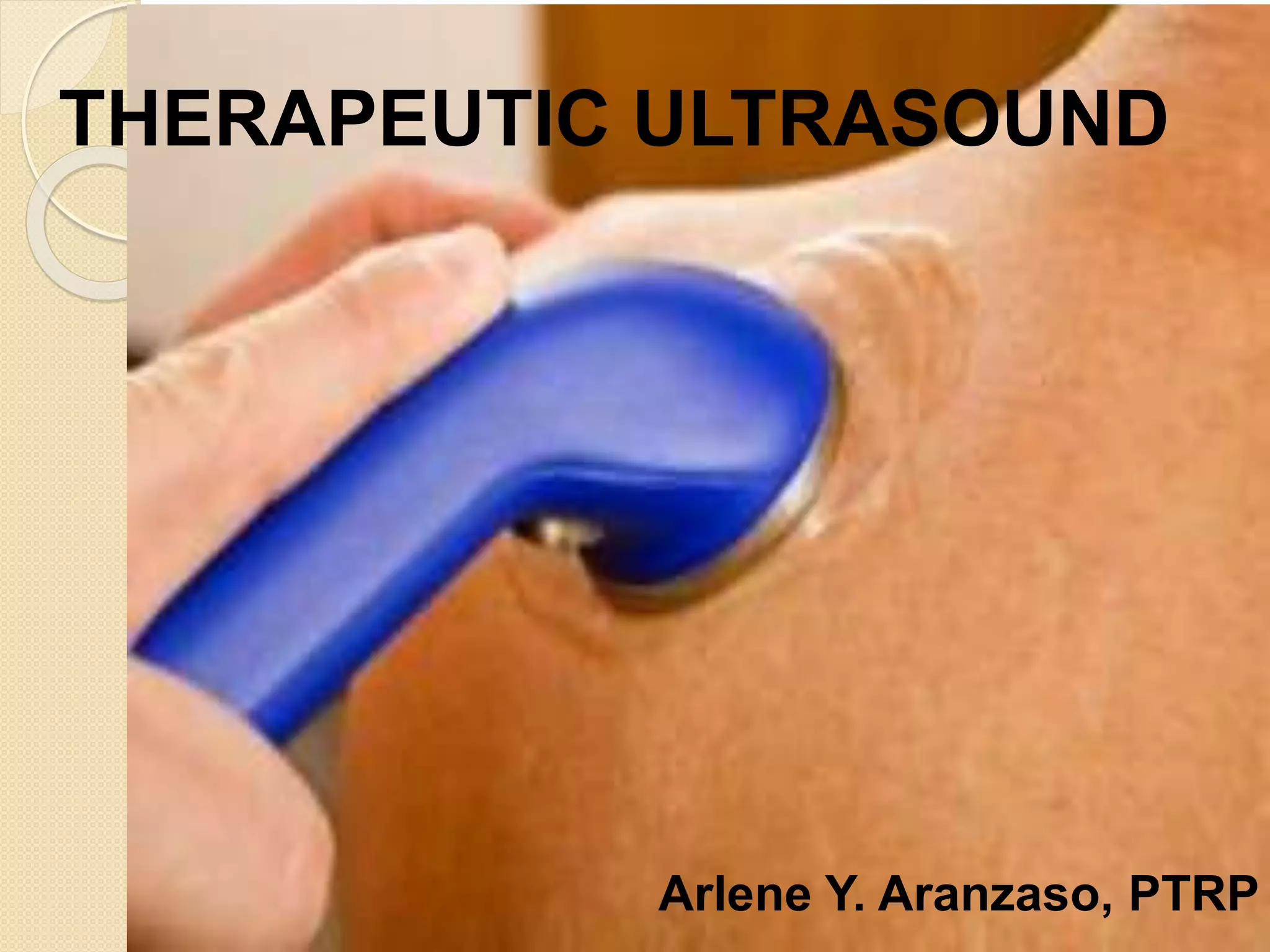
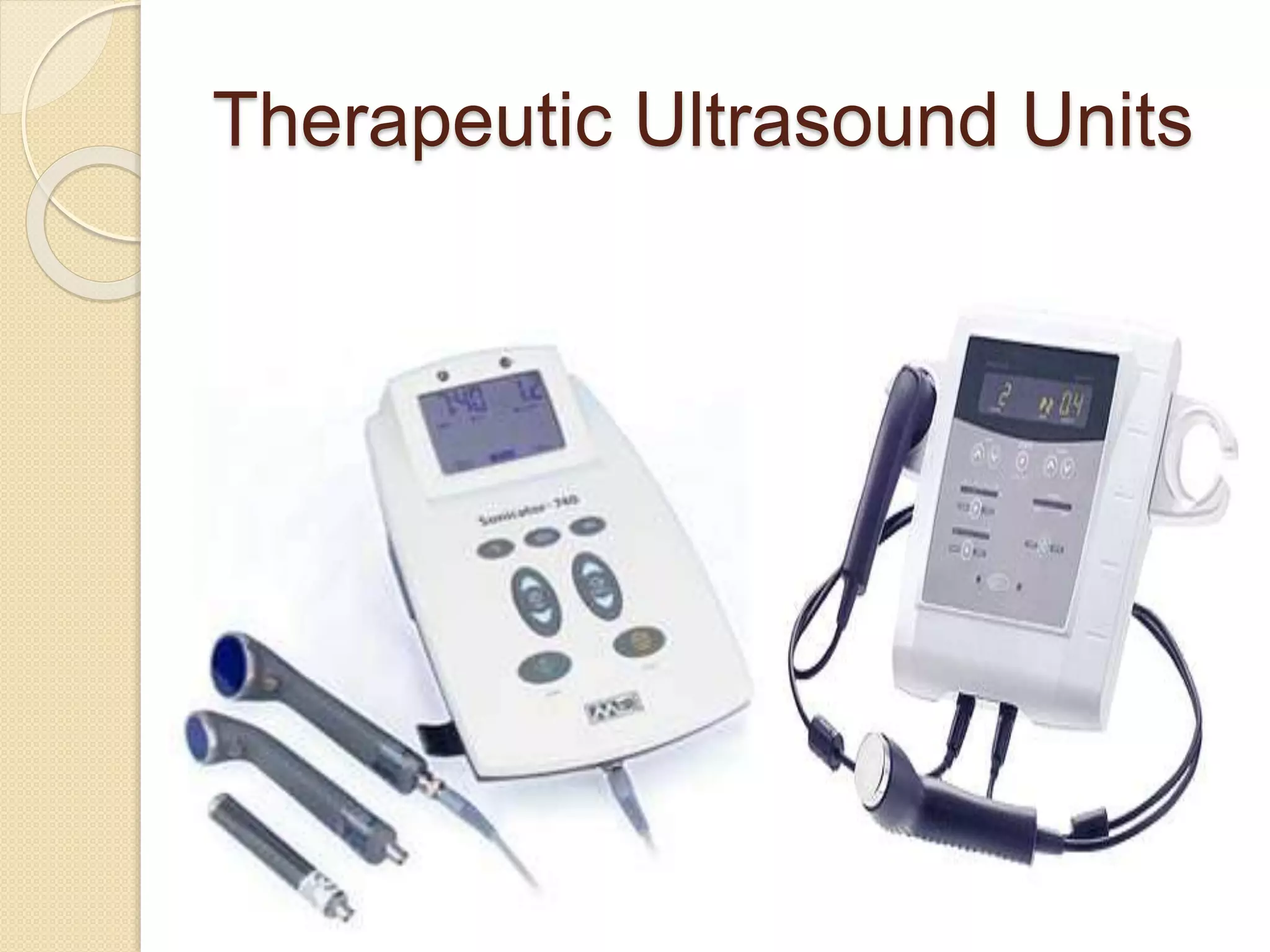
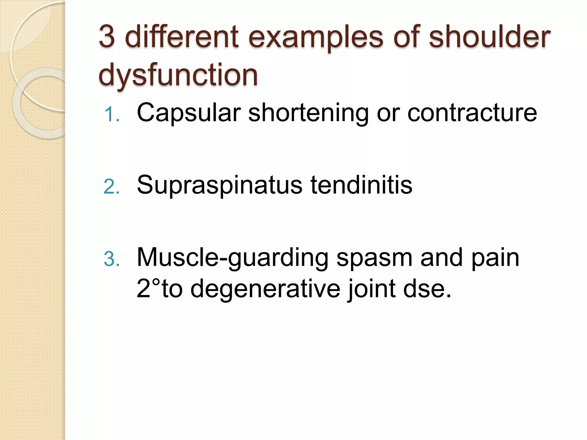
THERAPEUTIC ULTRASOUND
Arlene Y. Aranzaso, PTRP
Objectives:
1. To present the physical principles and
biophysical effects of Ultrasound
2. Discuss the clinical conditions for which
ultrasound is effective
3. Discuss the clinical procedures for the
application of ultrasound
4. Present guidelines for the safe use of
ultrasound, including a discussion of the
contraindications and precautions for
treatment with this agent
 Ultrasound
◦ Is used in medicine for diagnosis (
imaging of internal structures)
◦ Physical therapy (functional restoration
and healing of soft tissue ailments)
◦ Tissue destruction
Physical Principles
 Nature of Sound
◦ Sound is a non-ionizing radiation, it is
propagation of the vibratory motion.
 Frequency
◦ Number of oscillations a molecule
undergoes in 1 second
◦ Hz
◦ Human ear – 16 Hz and 20, 000 Hz
◦ Greater than 20,000 Hz is Ultrasound
◦ Ultrasound beams is collimated
◦ 85 KHz and 3 MHz
 Attenuation – reduction of acoustical
energy as it passes through soft
tissue.
◦ Scattering
◦ Absorption - attenuation
frequency
 Sound Velocity
◦ Is the speed at
which the vibratory
motion is
propagated through
a material
◦ 1540 m/s –soft
tissue
◦ 4000 m/s –
compact bone
 Wavelength
◦ Distance between 2
successive peaks in the
pressure wave
◦ Phase shift – time delay
◦ Condensations – the
concentration of
molecules increase in
the regions
◦ Rarefactions –
decrease in alternating
regions
Types of Waves
 Longitudinal waves
 Transverse waves
 Continuous wave
 Pulsed wave
Duty cycle = duration of pulse (time on)
pulse period (time on + time off)
< 50% is Pulsed US
Intensity
 It determined the strength of an US
beam
 It is the rate at which energy is delivered
per unit
 Watts/square centimeter
 W/cm²
 The greater intensity result greater
Temperature elevation
 PT – 0.25 to 2.0 W/cm² (therapeutic
application)
- 1 to 3 W/cm² (Sullivan&Siegelman)
 Spatial Average Intensity
Total Power Output (watts)
area (cm²)
 Spatial Peak Intensity – the greatest
intensity anywhere within the beam.
 BNR (beam non-uniformity ratio)-
defines the maximum point intensity
on the transducer to the average
intensity value across the transducer
surface.
 2:1 and 6:1
 Low BNR – more even energy
distribution and less risk of tissue
 Example:
 CW
◦ 3.0 MHz/0.5 W/cm²/CW/5min
 PW
◦ 3.0 MHz/0.5 W/cm²/p 20%/5min
Generation of
Ultrasound
The Piezoelectric Effect
 2 forms
A. Direct Piezoelectric effect
B. Reverse Piezoelectric effect (indirect)
Direct Piezoelectric Effect
 Is the generation of an electric voltage
across a crystal when the crystal is
compressed.
Reverse Piezoelectric Effect
 Is the contraction or expansion of a
crystal in response to a voltage
applied across it face.
The Transducer
Is any device that converts one form of energy into
another
Piezoelectric crystal is a transducer that converts
electrical energy into sound energy, and vice versa.
1 cm² to 10 cm²
5 cm² most commonly used
ERA (effective radiating area) –the area of the faceplate
(crystal size), which is smaller than the sound head.
Biophysical Effects
1. Thermal
 Those effects produced by the ability of
ultrasound to elevate tissue temperature.
2. Non- Thermal
 Those effects that must be attributed to
mechanism other than an increase in tissue
temperature.
Thermal Effects
 Increase collagen tissue extensibility
 Alterations in blood flow
 Changes in nerve conduction velocity
 Increased pain threshold
 Increase enzymatic activity
 Changes in contractile activity of
skeletal muscle
 3 MHz – most of the energy is
absorbed within a depth of 1 to 2 cm
 1 MHz – absorption in deeper tissue
- deeper than 2 cm from the
skin surface
Tissue Attenuation (%/cm)
Blood
Fat
Muscle
Blood vessel
Skin
Tendon
Cartilage
Bone
3
13
24
32
39
59
68
96
Attenuation of a 1 MHz Ultrasound Beam
Non-Thermal effects
 Cavitation
 Mechanical alterations
 Chemical alterations
 Cavitation
◦ is the vibrational effect on gas bubbles by
an US beam.
◦ Stable cavitation – result diffusional
changes along cell membrane and alter
cell function.
◦ Unstable or transient cavitation – the
violent collapse of bubbles within the
sound field result tissue destruction.
 Mechanical
◦ Acoustical streaming – refer to the
movement of fluids along the boundaries of
cell membranes
 Increase fibroblastic activity
 Increase calcium fluxes
 Alteration of cell membrane activity
 Increased cell wall permeability
 Increased protein synthesis
Clinical Application of
Ultrasound at Therapeutic
Intensities
1. Joint contracture and scar tissue
2. Reduction of pain and muscle spasm
3. Bursitis and tendinitis
4. Calcium deposits
5. Phonophoresis
6. Wound healing
7. Chronic wounds
Therapeutic Ultrasound Units
Basic components
 Power supply
 Oscillator circuit
 Transformer
 Coaxial cable
 Ultrasound applicator
Guidelines for Clinical
Administration
 Coupling Techniques
 Moving vs. Stationary Applicator
◦ Stationary
◦ Moving – slowly ~ 4 cm/s
- longitudinal stroking or circular
movements can be used
- total area covered – 2-3x the size of
the irradiating crystal for every
5 minutes exposure.
 Exposure Factors
◦ 5 minutes – increase tissue extensibility
◦ General use – it should be:
 an appropriate frequency for the depth of the
tissue to be treated
 A continuous wave or pulsed wave – according
to treatment aims
 The lowest intensity and duration that achieved
desired result
 Indirect contact
◦ Water immersion
◦ Using thin walled bag
Technique of Phonophoresis
 Medication is rubbed directly onto the
surface of the skin
 Coupling gel spread over the
medication
 Then sonation is initiated
Patient Positioning and Field
Selection
3 different examples of shoulder
dysfunction
1. Capsular shortening or contracture
2. Supraspinatus tendinitis
3. Muscle-guarding spasm and pain
2°to degenerative joint dse.
Treatment Precautions
1. Should not be applied over the eye
2. Irradiation over the heart should be avoided
3. Over pregnant uterus
4. Over testes
5. Over malignant tissue
6. Impaired sensation
7. Impaired circulation
8. Impaired cognitive function
9. Over thrombophlebitis
10. Over epiphyseal area in children
11. Over exposed or unprotected spinal cord
Clinical Decision Making
1. Stage of inflammation and repair
2. Site of pathology
3. Total amount of tissue to be heated
4. Presence or absence of orthopedic
implants
CASE STUDY
Joint Contracture and Scar
Tissue
 A 28 year old man sustained partial
lacerations to the extensor tendons to
the ring and small fingers over the
metacarpophalangeal joints when he
cut his fingers on a knife in dish water.
The tendons were surgically repaired
and immobilized for 2 weeks 2° to
wound infection. You are now seeing
the patient 4 wks.postop.
 US
◦ 3 MHz/0.5 W/cm²/CW/5 min.
◦ Small head
Reduction of Pain and Muscle
Spasm
 The patient, a 45 year old woman, has
had pain in the right cervical and
interscapular area for ~ 2 wks. The
onset of pain was caused by repetitive
activity during spring cleaning.
 US
◦ 1.0 MHz/0.5 W/cm²/p 20%/5 min.
◦
◦ Lying in prone or
◦ Seated with UE well supported
Bursitis and Tendinitis
 A 26 year old male patient has a
patellar tendinitis. The insidious onset
of pain occurred ~ 3 wks. ago. The
patient was told to place ice on the
area, rest, and do range of motion
exercises.
 US
◦ 3.0 MHz/0.8 W/cm²/p 50%/5 min.
Phonophoresis
 The patient, a 38 year old man, has a
lateral epicondylitis. His symptoms,
which first began after prolonged
hammering, have continued for ~ 6
weeks. He was instructed to use ice
on the affected area and to rest. In
addition, anti-inflammatory medication
was prescribed.
 Phonophoresis
◦ 3.0 MHz/1.0 W/cm²/CW/3 min.
Chronic Wounds
 A 40 year old male factory worker
sustained a sprain of the lateral ankle.
This occurred when he overturned his
foot 1 day ago. Rest, ice,
compression, and elevation were
recommended to the patient by the
company nurse.
 US
◦ Days 1-5 = 3.0 MHz/0.5 W/cm²/p 20%/5
min
◦ Days 6-14 = 3.0 MHz/1.0 W/cm²/p 20%/5
min
References :
 Thermal Agents in Rehabilitation by
Susan L. Michlovitz 3rd edition
 Physical Agents in Rehabilitation
From Research to Practice
by Michelle H. Cameron 2nd edition
 NPTE Review & Study Guide
by O’ Sullivan & Siegelman
Therapeutic ultrasound

Therapeutic ultrasound

  • 1.
  • 2.
    Objectives: 1. To presentthe physical principles and biophysical effects of Ultrasound 2. Discuss the clinical conditions for which ultrasound is effective 3. Discuss the clinical procedures for the application of ultrasound 4. Present guidelines for the safe use of ultrasound, including a discussion of the contraindications and precautions for treatment with this agent
  • 3.
     Ultrasound ◦ Isused in medicine for diagnosis ( imaging of internal structures) ◦ Physical therapy (functional restoration and healing of soft tissue ailments) ◦ Tissue destruction
  • 4.
    Physical Principles  Natureof Sound ◦ Sound is a non-ionizing radiation, it is propagation of the vibratory motion.
  • 5.
     Frequency ◦ Numberof oscillations a molecule undergoes in 1 second ◦ Hz ◦ Human ear – 16 Hz and 20, 000 Hz ◦ Greater than 20,000 Hz is Ultrasound ◦ Ultrasound beams is collimated ◦ 85 KHz and 3 MHz
  • 6.
     Attenuation –reduction of acoustical energy as it passes through soft tissue. ◦ Scattering ◦ Absorption - attenuation frequency
  • 7.
     Sound Velocity ◦Is the speed at which the vibratory motion is propagated through a material ◦ 1540 m/s –soft tissue ◦ 4000 m/s – compact bone
  • 8.
     Wavelength ◦ Distancebetween 2 successive peaks in the pressure wave ◦ Phase shift – time delay ◦ Condensations – the concentration of molecules increase in the regions ◦ Rarefactions – decrease in alternating regions
  • 9.
    Types of Waves Longitudinal waves  Transverse waves
  • 11.
  • 12.
    Duty cycle =duration of pulse (time on) pulse period (time on + time off) < 50% is Pulsed US
  • 13.
    Intensity  It determinedthe strength of an US beam  It is the rate at which energy is delivered per unit  Watts/square centimeter  W/cm²  The greater intensity result greater Temperature elevation  PT – 0.25 to 2.0 W/cm² (therapeutic application) - 1 to 3 W/cm² (Sullivan&Siegelman)
  • 14.
     Spatial AverageIntensity Total Power Output (watts) area (cm²)  Spatial Peak Intensity – the greatest intensity anywhere within the beam.
  • 16.
     BNR (beamnon-uniformity ratio)- defines the maximum point intensity on the transducer to the average intensity value across the transducer surface.  2:1 and 6:1  Low BNR – more even energy distribution and less risk of tissue
  • 18.
     Example:  CW ◦3.0 MHz/0.5 W/cm²/CW/5min  PW ◦ 3.0 MHz/0.5 W/cm²/p 20%/5min
  • 19.
  • 20.
    The Piezoelectric Effect 2 forms A. Direct Piezoelectric effect B. Reverse Piezoelectric effect (indirect)
  • 21.
    Direct Piezoelectric Effect Is the generation of an electric voltage across a crystal when the crystal is compressed.
  • 22.
    Reverse Piezoelectric Effect Is the contraction or expansion of a crystal in response to a voltage applied across it face.
  • 24.
    The Transducer Is anydevice that converts one form of energy into another Piezoelectric crystal is a transducer that converts electrical energy into sound energy, and vice versa. 1 cm² to 10 cm² 5 cm² most commonly used ERA (effective radiating area) –the area of the faceplate (crystal size), which is smaller than the sound head.
  • 26.
    Biophysical Effects 1. Thermal Those effects produced by the ability of ultrasound to elevate tissue temperature. 2. Non- Thermal  Those effects that must be attributed to mechanism other than an increase in tissue temperature.
  • 27.
    Thermal Effects  Increasecollagen tissue extensibility  Alterations in blood flow  Changes in nerve conduction velocity  Increased pain threshold  Increase enzymatic activity  Changes in contractile activity of skeletal muscle
  • 28.
     3 MHz– most of the energy is absorbed within a depth of 1 to 2 cm  1 MHz – absorption in deeper tissue - deeper than 2 cm from the skin surface
  • 29.
    Tissue Attenuation (%/cm) Blood Fat Muscle Bloodvessel Skin Tendon Cartilage Bone 3 13 24 32 39 59 68 96 Attenuation of a 1 MHz Ultrasound Beam
  • 30.
    Non-Thermal effects  Cavitation Mechanical alterations  Chemical alterations
  • 31.
     Cavitation ◦ isthe vibrational effect on gas bubbles by an US beam. ◦ Stable cavitation – result diffusional changes along cell membrane and alter cell function. ◦ Unstable or transient cavitation – the violent collapse of bubbles within the sound field result tissue destruction.
  • 34.
     Mechanical ◦ Acousticalstreaming – refer to the movement of fluids along the boundaries of cell membranes  Increase fibroblastic activity  Increase calcium fluxes  Alteration of cell membrane activity  Increased cell wall permeability  Increased protein synthesis
  • 35.
    Clinical Application of Ultrasoundat Therapeutic Intensities
  • 36.
    1. Joint contractureand scar tissue 2. Reduction of pain and muscle spasm 3. Bursitis and tendinitis 4. Calcium deposits 5. Phonophoresis 6. Wound healing 7. Chronic wounds
  • 37.
  • 38.
    Basic components  Powersupply  Oscillator circuit  Transformer  Coaxial cable  Ultrasound applicator
  • 39.
  • 40.
     Moving vs.Stationary Applicator ◦ Stationary ◦ Moving – slowly ~ 4 cm/s - longitudinal stroking or circular movements can be used - total area covered – 2-3x the size of the irradiating crystal for every 5 minutes exposure.
  • 41.
     Exposure Factors ◦5 minutes – increase tissue extensibility ◦ General use – it should be:  an appropriate frequency for the depth of the tissue to be treated  A continuous wave or pulsed wave – according to treatment aims  The lowest intensity and duration that achieved desired result
  • 42.
     Indirect contact ◦Water immersion ◦ Using thin walled bag
  • 43.
    Technique of Phonophoresis Medication is rubbed directly onto the surface of the skin  Coupling gel spread over the medication  Then sonation is initiated
  • 45.
    Patient Positioning andField Selection
  • 46.
    3 different examplesof shoulder dysfunction 1. Capsular shortening or contracture 2. Supraspinatus tendinitis 3. Muscle-guarding spasm and pain 2°to degenerative joint dse.
  • 47.
    Treatment Precautions 1. Shouldnot be applied over the eye 2. Irradiation over the heart should be avoided 3. Over pregnant uterus 4. Over testes 5. Over malignant tissue 6. Impaired sensation 7. Impaired circulation 8. Impaired cognitive function 9. Over thrombophlebitis 10. Over epiphyseal area in children 11. Over exposed or unprotected spinal cord
  • 48.
    Clinical Decision Making 1.Stage of inflammation and repair 2. Site of pathology 3. Total amount of tissue to be heated 4. Presence or absence of orthopedic implants
  • 49.
  • 50.
    Joint Contracture andScar Tissue  A 28 year old man sustained partial lacerations to the extensor tendons to the ring and small fingers over the metacarpophalangeal joints when he cut his fingers on a knife in dish water. The tendons were surgically repaired and immobilized for 2 weeks 2° to wound infection. You are now seeing the patient 4 wks.postop.
  • 51.
     US ◦ 3MHz/0.5 W/cm²/CW/5 min. ◦ Small head
  • 52.
    Reduction of Painand Muscle Spasm  The patient, a 45 year old woman, has had pain in the right cervical and interscapular area for ~ 2 wks. The onset of pain was caused by repetitive activity during spring cleaning.
  • 53.
     US ◦ 1.0MHz/0.5 W/cm²/p 20%/5 min. ◦ ◦ Lying in prone or ◦ Seated with UE well supported
  • 54.
    Bursitis and Tendinitis A 26 year old male patient has a patellar tendinitis. The insidious onset of pain occurred ~ 3 wks. ago. The patient was told to place ice on the area, rest, and do range of motion exercises.
  • 55.
     US ◦ 3.0MHz/0.8 W/cm²/p 50%/5 min.
  • 56.
    Phonophoresis  The patient,a 38 year old man, has a lateral epicondylitis. His symptoms, which first began after prolonged hammering, have continued for ~ 6 weeks. He was instructed to use ice on the affected area and to rest. In addition, anti-inflammatory medication was prescribed.
  • 57.
     Phonophoresis ◦ 3.0MHz/1.0 W/cm²/CW/3 min.
  • 58.
    Chronic Wounds  A40 year old male factory worker sustained a sprain of the lateral ankle. This occurred when he overturned his foot 1 day ago. Rest, ice, compression, and elevation were recommended to the patient by the company nurse.
  • 59.
     US ◦ Days1-5 = 3.0 MHz/0.5 W/cm²/p 20%/5 min ◦ Days 6-14 = 3.0 MHz/1.0 W/cm²/p 20%/5 min
  • 60.
    References :  ThermalAgents in Rehabilitation by Susan L. Michlovitz 3rd edition  Physical Agents in Rehabilitation From Research to Practice by Michelle H. Cameron 2nd edition  NPTE Review & Study Guide by O’ Sullivan & Siegelman

Editor's Notes

  • #4 A deep heating agent Employed in medicine for over 50 years Wood and loomis in 1927-biologic effect in tissue to US 1930 germany –application of US 1940 – Unuted states Lowest intensity – use for diagnostic High intensity – use for tissue destruction
  • #5 Ionizing radiation – cause cancer production and chromosome breakage Acoustic radiation= US
  • #6 1 Hz = 1 cycle/sec. 1 kHz = 1000 cycles/sec 1 MHz = 1 million cycles/sec. Collimated beam that oscillating crystal produces sound waves with little dispersion of energy
  • #7 1.0 MHz most use for deep penetration 3.0 MHZ widely used for superficial Absorption highest muscle, tendons, ligaments and capsule Scattering – reflection and refraction
  • #8 The more rigid the material= the greater the velocity of sound passing through it
  • #9 Wavelength is inversely related to frequency V= FxW
  • #10 LW- the direction of motion of the molecules is parallel to the direction of wave propagation TW-is perpendicular No TW in gases and liquids Solids= LW & TW- is found in bone
  • #12 CW- sound intensity is constant – use to achieve thermal effects – chronic conditions PW – intermittently interrupted – used when non thermal effects are desired – acute soft tissue injuries Duty cycle- fractions of time in pulsed wave
  • #13 Pulsed mode -0.05 (5%) to 0.5 (50%) 20% is most commonly used – 2msec. On time and 8 msec.off time 51%-99%- it produces less acoustic energy and less heat Pulsed mode –duty cycle is important
  • #15 Continuous US – spatial characteristic is predominant SPI – hot spot – using moving technique and pulsed lower the SPI
  • #18 Temporal peak intensity or pulse average intensity- maximum intensity in pulsed Temporal average intensity –US power average over 1 pulsed period Tgemporal average intensity- on period+off period Pulsed –minimize heating needed- like stasis ulcers and acute coft tissue injuries Hot spots =can cause damage to the insonated tissue Moving applicator technique
  • #19 SAI – 0.25 to 2.0 W/cm used therapeutic application 10.0 W/cm used to destroy tissue surgically 0.1 W/cm used for diagnostic purposes Parasitic radiation –therapist result acute pain in hand fingers
  • #25 Is any device that converts one form of energy into another Piezoelectric crystal is a transducer that converts electrical energy into sound energy, and vice versa. Has a variety of sizes
  • #27 Conversion – mechanical energy produces by sound waves
  • #28 Increase temp-5 cm or more High dose = retard long bone growth, damage spinal cord tissue, destroy other tissue Thermal using Continuous mode Tissue with high collagen absorb a large amt.of US beam
  • #30 Attenuation of a 1 MHz Ultrasound Beam US as deep heating – elevate temp 40 °C to 45 °C
  • #31 Using pulsed mode
  • #32 Rarefactions- small bubbles expand Condensations- bubbles compressed SC – resonate without tissue damage USC- high temp.
  • #35 Result mechanical pressure wave Ion fluxes- streaming Protein systhesis
  • #37 Heat and stretch 45 degrees C- elevate tissue temp
  • #39 Ratio of the acoustic power to the intensity is constant ERA –effective radiating area Quality control – calibrated and electrically safe
  • #40 Coupling medium to transmit US energy from transducer to irradiated surface Water soluble gel Eliminates the air to minimize the amt of sound entering the body Immersion technique -0.5 to 3.0 cm from the body
  • #41 Direct contact Used stationary if the area is too small with low intensity Hot spots= tissue damage Rapid- decreased the amt.of enrgy the tissue absorb Distribute the energy even
  • #42 Heat – inadequate coupling medium Not moving Choice of transducer Tx.time 3-10 mins
  • #43 Irregular body parts Plastic container-less reflection 1 cm from skin surface – ryt angle Wipe off bubbles Nor widely used but can used as alternative for immersion
  • #44 Then the part placed in water bath 80% Anti inflammatory drugs – cortisol, dexamethasone, salicylates, lidocaine Pulsed mode 20% 0.5 -0.75 W/cm²
  • #47 1- glenohumeral jt-affected abduction and ER-position of the pt.shpoulder 2- supraspinatus insertion – abducted and ER –acromion process 3- comfortable and relaxed position
  • #48 Can produce cataract Retina’s destruction 2- study ECG changes happened Cardiac pacemaker 3- abnormality sich as low birth wt., brain size reduction, orthopedic abnormalities (study) 10 days after the onset of menses – ok 4- temporary sterility 5- metatasis- increase tumor growth Not over healing fracture
  • #49 Factors need to be considered 2- depth , location Adjunct with HMP But cold should not be given prior to US 9-12 US should be given then discontinued for 2 weeks
  • #51 Problems: decreased gliding of the tendon Wound infection and slow healing Limited functional activities Assessment –ROM, wound/scar eval, functional daily activities Goals – Increase tendon gliding, ROM, FA
  • #52 Superficial Subacute Measure ROM after tx. HP= AROM Scar- fiction massage and ROM
  • #53 Problems: pain, limited ROM Assessment: pain quality, quantity, location Functional activity Cervical ROM Goals: Reduce pain, Increase FA, ROM
  • #54 Deep tissue Subacute Pulse mode- pain relief
  • #55 Problem: persistent pain, limited FA Assessment: Pain, FA Goals: reduce pain, increase FA
  • #56 Deep Subacute Pulse- healing, pain
  • #57 Problem: temporary relief, aggravation with activity, limited FA Assessment: pain, FA Gials: reduce pain, incA use of involved side
  • #58 In order to induce dexamethasone 3MHz- less than 2cm deep Subacute to chronic Thermal effect to increase diffusion of the drugs
  • #59 Problems: swelling, bruising of the ankle, pain, decrease ROM, limited FA Assessment: volumetrics of ankle, pain, ROM, FA, gait Goals: decrease swelling,reduce pain, increase ROM foot and ankle, strength, improve gait, FA
  • #60 Low dose US to reduce edema and increase mast cell degranulation Superficial healing