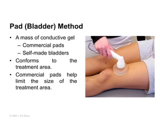
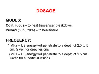
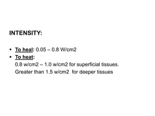
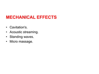
THERAPEUTIC ULTRASOUND
Dr. Dheeraj Lamba
Associate Professor/ Head Department of
Physiotherapy
At the end of this lesson the student will be able to:
• State the definition of Ultrasound.
• Describe the Parameters of Ultrasound.
• Demonstrate the methods of application of ultrasound.
• Determine the dosage of ultrasound.
• Demonstrate an understanding about the Uses,
Physiological and therapeutic effects.
• Identify the contraindications and dangers.
• Demonstrate the method of Phonophoresis.
OBJECTIVES:
DEFINITION:
• A form of acoustic vibrations occurring at a frequency
that is too high to be perceived by the human ear.
• The therapeutic frequency is in the range of 0.5 to 5
MHz.
• Commonly used frequency in physiotherapy is 1MHz
and 3MHz.
PIEZOELECTRIC EFFECT :
• It is the ability of certain materials to generate an electric
charge in response to applied mechanical stress.
• Reverse piezoelectric effect –electrical energy is used to
produce mechanical stress and deformation
PARAMETERS
• Intensity / output.
• Duration
• Mark- Space ratio.
TYPES OF ULTRASOUND WAVES
• Continuous Wave - no interruption of beam: best for maximum
buildup, sometimes heat.
• Pulsed Wave - intermittent “on-off” beam modulation. It ensures that
the energy available in each pulse (pulsed averaged intensity) is
high enough for mechanical rather than thermal effects.
• It is represented as
• The mark : space ratio
It is the ratio of the pulse length to the interval.
Ultrasound Machine & Coupling Agent
Dispensers
METHODS OF APPLICATION
• Direct method / contact cream method.
• Bath method or immersion technique.
• Bag method.
Direct method
• Gel or Creams should be
applied over the
treatment part
• Only use approved
coupling agents.
• Remove air bubbles by
passing sound head over
area (before power is
increased)
© 2005 – FA Davis
Immersion Technique
• Used to treat irregularly
shaped areas.
• The limb is immersed in a
tub of degassed water.
• Transducer is held appx. 1”
from the body part.
• Avoid the formation of air
bubbles.
© 2005 – FA Davis
Pad (Bladder) Method
• A mass of conductive gel
– Commercial pads
– Self-made bladders
• Conforms to the
treatment area.
• Commercial pads help
limit the size of the
treatment area.
© 2005 – FA Davis
DOSAGE
MODES:
Continuous – to heat tissue/scar breakdown.
Pulsed (50%, 20%) – to heal tissue.
FREQUENCY:
1 MHz – US energy will penetrate to a depth of 2.5 to 5
cm. Given for deep lesions.
3 MHz – US energy will penetrate to a depth of 1.5 cm.
Given for superficial lesions.
INTENSITY:
 To heal: 0.05 – 0.8 W/cm2
 To heat:
0.8 w/cm2 – 1.0 w/cm2 for superficial tissues.
Greater than 1.5 w/cm2 for deeper tissues
MECHANICAL EFFECTS
• Cavitation's.
• Acoustic streaming.
• Standing waves.
• Micro massage.
Cavitations:
Formation of gas bubbles that expand & compress due to
pressure changes in tissue fluids.
• Stable cavitation
Occurs when the bubbles oscillate to and fro within the
ultrasound pressure waves but remain intact.
• Transient (or collapse) cavitation
Occurs when the volume of the bubble changes rapidly
and then collapses
Causes high pressure and temperature changes .
Results in gross damage to tissues.
Acoustic Streaming
• Unidirectional flow currents in a fluid due to the presence
of sound waves.
• Exerts viscous stress on the cell membrane and
increase membrane permeability.
Standing waves
• Formed due to reflected waves being superimposed on
the incident waves.
• Possibility of marked local heating where the amplitude
of the combined waves is high.
Micromassage
The micromassage effect of ultrasound occurs at a
cellular level where the cells are alternately compressed
and then pulled further apart.
The waves of compression and rarefaction may produce
a form of micromassage, which could reduce oedema.
PHYSIOLOGICAL EFFECTS
• Increased peripheral arterial blood flow.
• Increased tissue metabolism.
• Increased permeability of membrane.
• Increased pain threshold.
• Relief of muscle spasm.
THERAPEUTIC EFFECTS
• Pain relief.
• Resolution of inflammation.
• Effect on healing.
Physiological and therapeutic effects
INDICATIONS
• Bursitis.
• Tendonitis.
• Pressure sores.
• TMJ syndrome.
• Sports injuries.
• Capsulitis.
• Contractures
• Plantar fascitis.
• Calcaneal spur.
• Tennis / Golfer's elbow.
• Coccydynia.
• OA / RA.
• Scars.
.
CONTRAINDICATIONS
• Rapid dividing tissues -Neoplasm's
• Pregnant Uterus.
• Epiphyseal Plates
• Infections.
• Vascular Problems
Hemorrhage.
Hematoma.
Haemarthrosis.
Thrombosis.
Thrombophlebitis.
Ischemia.
• Radiotherapy.
• Metal / plastic / acrylic implant.
• Pace maker.
• Avoid specialized tissues – eyes, ears, ovaries and
testes.
• Central nervous system tissue.
• Anesthetic areas.
DANGERS
• Burns could occur if the heat generated exceeded the
physiological ability to dissipate it.
• Tissue destruction would result from transient cavitation.
• Blood cell stasis and endothelial damage may occur if
there is standing wave formation.
These dangers are more likely with high-intensity
continuous output with a stationary head or over bony
prominences.
PRECAUTIONS
• Check thermal sensation.
• Use the right dosage.
• Move the transducer.
• Avoid bony prominences.
• Avoid excess heat sensation.
• Don’t keep the ultrasound head in air when the machine
is ON.
• Maintain good contact between ultrasound head and
tissues.
• Test the equipment.
PHONOPHORESIS
It is the movement of drugs through skin into the
subcutaneous tissues under the influence of ultrasound.
• Ultrasound facilitates the passage of some drugs into
and through the skin.
• The effects are due both to absorption of the drug and to
the ultrasound.
• Lower ultrasonic frequencies lead to deeper drug
penetration.
• Pulsing ultrasound lead to better drug penetration.
ULTRASOUND.ppt

ULTRASOUND.ppt

  • 1.
    THERAPEUTIC ULTRASOUND Dr. DheerajLamba Associate Professor/ Head Department of Physiotherapy
  • 2.
    At the endof this lesson the student will be able to: • State the definition of Ultrasound. • Describe the Parameters of Ultrasound. • Demonstrate the methods of application of ultrasound. • Determine the dosage of ultrasound. • Demonstrate an understanding about the Uses, Physiological and therapeutic effects. • Identify the contraindications and dangers. • Demonstrate the method of Phonophoresis. OBJECTIVES:
  • 3.
    DEFINITION: • A formof acoustic vibrations occurring at a frequency that is too high to be perceived by the human ear. • The therapeutic frequency is in the range of 0.5 to 5 MHz. • Commonly used frequency in physiotherapy is 1MHz and 3MHz.
  • 4.
    PIEZOELECTRIC EFFECT : •It is the ability of certain materials to generate an electric charge in response to applied mechanical stress. • Reverse piezoelectric effect –electrical energy is used to produce mechanical stress and deformation
  • 6.
    PARAMETERS • Intensity /output. • Duration • Mark- Space ratio.
  • 7.
    TYPES OF ULTRASOUNDWAVES • Continuous Wave - no interruption of beam: best for maximum buildup, sometimes heat. • Pulsed Wave - intermittent “on-off” beam modulation. It ensures that the energy available in each pulse (pulsed averaged intensity) is high enough for mechanical rather than thermal effects. • It is represented as • The mark : space ratio It is the ratio of the pulse length to the interval.
  • 9.
    Ultrasound Machine &Coupling Agent Dispensers
  • 10.
    METHODS OF APPLICATION •Direct method / contact cream method. • Bath method or immersion technique. • Bag method.
  • 11.
    Direct method • Gelor Creams should be applied over the treatment part • Only use approved coupling agents. • Remove air bubbles by passing sound head over area (before power is increased) © 2005 – FA Davis
  • 12.
    Immersion Technique • Usedto treat irregularly shaped areas. • The limb is immersed in a tub of degassed water. • Transducer is held appx. 1” from the body part. • Avoid the formation of air bubbles. © 2005 – FA Davis
  • 13.
    Pad (Bladder) Method •A mass of conductive gel – Commercial pads – Self-made bladders • Conforms to the treatment area. • Commercial pads help limit the size of the treatment area. © 2005 – FA Davis
  • 14.
    DOSAGE MODES: Continuous – toheat tissue/scar breakdown. Pulsed (50%, 20%) – to heal tissue. FREQUENCY: 1 MHz – US energy will penetrate to a depth of 2.5 to 5 cm. Given for deep lesions. 3 MHz – US energy will penetrate to a depth of 1.5 cm. Given for superficial lesions.
  • 15.
    INTENSITY:  To heal:0.05 – 0.8 W/cm2  To heat: 0.8 w/cm2 – 1.0 w/cm2 for superficial tissues. Greater than 1.5 w/cm2 for deeper tissues
  • 16.
    MECHANICAL EFFECTS • Cavitation's. •Acoustic streaming. • Standing waves. • Micro massage.
  • 17.
    Cavitations: Formation of gasbubbles that expand & compress due to pressure changes in tissue fluids. • Stable cavitation Occurs when the bubbles oscillate to and fro within the ultrasound pressure waves but remain intact. • Transient (or collapse) cavitation Occurs when the volume of the bubble changes rapidly and then collapses Causes high pressure and temperature changes . Results in gross damage to tissues.
  • 18.
    Acoustic Streaming • Unidirectionalflow currents in a fluid due to the presence of sound waves. • Exerts viscous stress on the cell membrane and increase membrane permeability. Standing waves • Formed due to reflected waves being superimposed on the incident waves. • Possibility of marked local heating where the amplitude of the combined waves is high.
  • 19.
    Micromassage The micromassage effectof ultrasound occurs at a cellular level where the cells are alternately compressed and then pulled further apart. The waves of compression and rarefaction may produce a form of micromassage, which could reduce oedema.
  • 21.
    PHYSIOLOGICAL EFFECTS • Increasedperipheral arterial blood flow. • Increased tissue metabolism. • Increased permeability of membrane. • Increased pain threshold. • Relief of muscle spasm.
  • 22.
    THERAPEUTIC EFFECTS • Painrelief. • Resolution of inflammation. • Effect on healing.
  • 23.
  • 24.
    INDICATIONS • Bursitis. • Tendonitis. •Pressure sores. • TMJ syndrome. • Sports injuries. • Capsulitis. • Contractures • Plantar fascitis. • Calcaneal spur. • Tennis / Golfer's elbow. • Coccydynia. • OA / RA. • Scars. .
  • 25.
    CONTRAINDICATIONS • Rapid dividingtissues -Neoplasm's • Pregnant Uterus. • Epiphyseal Plates • Infections. • Vascular Problems Hemorrhage. Hematoma. Haemarthrosis. Thrombosis. Thrombophlebitis. Ischemia.
  • 26.
    • Radiotherapy. • Metal/ plastic / acrylic implant. • Pace maker. • Avoid specialized tissues – eyes, ears, ovaries and testes. • Central nervous system tissue. • Anesthetic areas.
  • 27.
    DANGERS • Burns couldoccur if the heat generated exceeded the physiological ability to dissipate it. • Tissue destruction would result from transient cavitation. • Blood cell stasis and endothelial damage may occur if there is standing wave formation. These dangers are more likely with high-intensity continuous output with a stationary head or over bony prominences.
  • 28.
    PRECAUTIONS • Check thermalsensation. • Use the right dosage. • Move the transducer. • Avoid bony prominences. • Avoid excess heat sensation. • Don’t keep the ultrasound head in air when the machine is ON. • Maintain good contact between ultrasound head and tissues. • Test the equipment.
  • 29.
    PHONOPHORESIS It is themovement of drugs through skin into the subcutaneous tissues under the influence of ultrasound. • Ultrasound facilitates the passage of some drugs into and through the skin. • The effects are due both to absorption of the drug and to the ultrasound. • Lower ultrasonic frequencies lead to deeper drug penetration. • Pulsing ultrasound lead to better drug penetration.