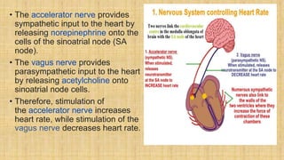
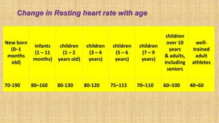
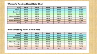
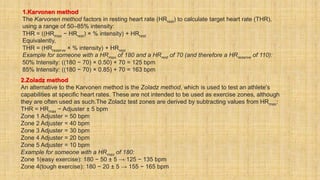
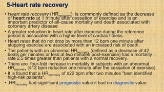
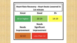
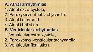
SlideShare a Scribd company logo
Heart Rate
Dr. Ajay Kumar Singh
Department of Physiology
MLN Medical College Prayagraj
Contents
A-Definition of heart rate
B-Physiology of heart rate
C-Factors influencing heart rate
1.Epinephrine and norepinephrine
2.Thyroid hormones
3.Calcium
4.Caffeine and nicotine
5.Effects of stress
6.Factors decreasing heart rate
7.Physiological control over heart rate
D. Different circumstances of heart rate
1.Resting heart rate
2.Maximum heart rate
3.Heart rate reserve
4.Target heart rate
5.Heart rate recovery
E. Development of heart rate
F. Measurement of heart rate
1-Manual measurement
2-Electronic measurement
3-Optical measurements
G. Clinical significance of heart
rate
1-Arrhythmia
2-Tachycardia
3-Bradycardia
(A) Definition of Heart Rate
Heart rate is the number of heartbeats per
unit of time, usually per minute.
The heart rate is based on the number of
contractions of the ventricles (the lower
chambers of the heart).
Normal Heart Rate- The normal resting adult
human heart rate is 60–100 beat per minute.
(B) Physiology of Heart Rate
•Heart rhythm is regulated
by the sinoatrial node (SA
node ) under normal
conditions.
•Heart rate is regulated by
sympathetic and
parasympathetic input to
the sinoatrial node.
• The accelerator nerve provides
sympathetic input to the heart by
releasing norepinephrine onto the
cells of the sinoatrial node (SA
node).
• The vagus nerve provides
parasympathetic input to the heart
by releasing acetylcholine onto
sinoatrial node cells.
• Therefore, stimulation of
the accelerator nerve increases
heart rate, while stimulation of the
vagus nerve decreases heart rate.
Neural control of heart rate
• Neural control over the heart rate is
centralized within the two
paired cardiovascular canters of
the medulla oblongata.
• The cardioaccelerator regions stimulate
heart activity via sympathetic stimulation
as one component of cardioaccelerator
nerves.
• The cardioinhibitory centers decrease
heart activity via parasympathetic
stimulation as one component of
the vagus nerve.
• Both sympathetic and parasympathetic
stimuli flow through the paired cardiac
plexus near the base of the heart.
•The cardioaccelerator
center also sends
additional fibers, forming
the cardiac nerves via
sympathetic ganglia (the
cervical ganglia plus
superior thoracic ganglia
T1–T4) to both the SA
and AV nodes, plus
additional fibers to the
atria and ventricles.
• Sympathetic stimulation causes the release of the
neurotransmitter norepinephrine at the neuromuscular
junction of the cardiac nerves.
• Norepinephrine opens chemical or ligand-gated
sodium and calcium ion channels, allowing an influx of
positively charged ions.
• Norepinephrine shortens the repolarization period, and
speeding the rate of depolarization and contraction,
which results in an increased heartrate.
• Norepinephrine binds to the beta–1 receptor and
increases the heart rate.
•Parasympathetic
stimulation originates
from the cardioinhibitory
region of the brain and
impulses traveling via
the vagus nerve (X
cranial nerve ).
•The vagus nerve sends
branches to both the SA
and AV nodes, and to
portions of both the atria
and ventricles.
• Parasympathetic stimulation releases the neurotransmitter
acetylcholine (ACh) at the neuromuscular junction.
• ACh slows HR by opening chemical- or ligand-gated potassium ion
channels to slow the rate of spontaneous depolarization, which
extends repolarization and increases the time before the next
spontaneous depolarization occurs.
• Without any nervous stimulation, the SA node would establish a
sinus rhythm of approximately 100 bpm. Since resting rates are
considerably less than this, it becomes evident that parasympathetic
stimulation normally slows HR.
• Decreasing parasympathetic stimulation decreases the release of
ACh, which allows HR to increase up to approximately 100 bpm. Any
increases beyond this rate would require sympathetic stimulation.
Effects of Parasympathetic and Sympathetic Stimulation on Normal Sinus Rhythm
• The wave of depolarization in a normal
sinus rhythm shows a stable resting HR.
• Parasympathetic stimulation releases the
neurotransmitter acetylcholine (ACh) at the
neuromuscular junction. ACh slows the rate
of spontaneous depolarization, which
extends repolarization and increases the
time before the next spontaneous
depolarization occurs.
• Sympathetic stimulation causes the release
of the neurotransmitter norepinephrine at
the neuromuscular junction of the cardiac
nerves. This shortens the repolarization
period, thus speeding the rate of
depolarization and contraction, which
results in an increased heartrate.
Input to the cardiovascular centres
• The cardiovascular centres
receive sensory input from a
series of visceral receptors.
• Sensory impulses traveling from
visceral receptors to
cardiovascular centers through
visceral sensory fibers within the
vagus and sympathetic nerves
via the cardiac plexus.
• Among these receptors are
various proprioceptors,
baroreceptors, chemoreceptors,
and stimuli from the limbic
system which normally enable
the precise regulation of heart
function, via cardiac reflexes.
Proprioreceptors-
•Various proprioreceptors located in muscles,
joint capsules, and tendons.
•Increased physical activity results in increased
rates of firing by the different proprioreceptors.
•The cardiovascular centres monitor these
increased rates of firing, suppressing
parasympathetic stimulation or increasing
sympathetic stimulation as needed in order to
increase blood flow.
Baroreceptors -
• Baroreceptors are stretch receptors located in the aortic sinus, carotid
bodies, the venae cava, and other locations, including pulmonary
vessels and the right side of the heart itself.
• Rates of firing from the baroreceptors represent blood pressure, level
of physical activity, and the relative distribution of blood.
• The cardiac centers monitor baroreceptor firing to maintain cardiac
homeostasis, a mechanism called the baroreceptor reflex.
• With increased pressure and stretch, the rate of baroreceptor firing
increases, and the cardiac centers decrease sympathetic stimulation
and increase parasympathetic stimulation.
• As pressure and stretch decrease, the rate of baroreceptor firing
decreases, and the cardiac centers increase sympathetic stimulation
and decrease parasympathetic stimulation.
• There is a similar reflex, called the atrial reflex
or Bainbridge reflex, associated with varying rates
of blood flow to the atria.
• Increased venous return stretches the walls of the
atria where specialized baroreceptors are located.
However, as the atrial baroreceptors increase their
rate of firing and as they stretch due to the
increased blood pressure, the cardiac center
responds by increasing sympathetic stimulation
and inhibiting parasympathetic stimulation to
increase HR. The opposite is also true.
Chemoreceptors-
•Increased metabolic byproducts associated with
increased activity, such as carbon dioxide, hydrogen
ions, and lactic acid, plus falling oxygen levels, are
detected by a suite of chemoreceptors innervated by
the glossopharyngeal and vagus nerves.
• These chemoreceptors provide feedback to the
cardiovascular centers about the need for increased or
decreased blood flow, based on the relative levels of
these substances.
• The limbic system significantly impact HR related to
emotional state.
• During periods of stress, it is not unusual to identify higher
than normal HRs, often accompanied by a surge in the stress
hormone cortisol.
• Individuals experiencing extreme anxiety may manifest panic
attacks with symptoms that resemble those of heart attacks.
These events are typically transient and treatable.
• Meditation techniques have been developed to ease anxiety
and have been shown to lower HR effectively.
• Doing simple deep and slow breathing exercises with one's
eyes closed can also significantly reduce this anxiety and
HR.
(C) Factors influencing heart rate
1.Epinephrine and norepinephrine
2.Thyroid hormones
3.Calcium
4.Caffeine and Nicotine
5.Effects of stress
6.Factors decreasing heart rate
7.Physiological control over heart rate
• Using a combination of autorhythmicity and innervation, the
cardiovascular center is able to provide relatively precise control
over the heart rate, but other factors can impact on this.
• These include hormones, notably epinephrine, norepinephrine, and
thyroid hormones; levels of various ions including calcium,
potassium, and sodium; body temperature, hypoxia,and pH balance.
Factors increasing heart rate and force of contraction
Factor Effect
Cardioaccelerator nerves Release of norepinephrine
Proprioceptors Increased rates of firing during exercise
Chemoreceptors Decreased levels of O2; increased levels of H+, CO2, and lactic acid
Baroreceptors Decreased rates of firing, indicating falling blood volume/pressure
Limbic system Anticipation of physical exercise or strong emotions
Catecholamines Increased epinephrine and norepinephrine
Thyroid hormones Increased T3 and T4
Calcium Increased Ca2+
Potassium Decreased K+
Sodium Decreased Na+
Body temperature Increased body temperature
Nicotine and caffeine Stimulants, increasing heart rate
Factors decreasing heart rate and force of contraction
Factor Effect
Cardioinhibitor nerves (vagus) Release of acetylcholine
Proprioreceptors Decreased rates of firing following exercise
Chemoreceptors Increased levels of O2; decreased levels of H+ and CO2
Baroreceptors Increased rates of firing, indicating higher blood volume/pressure
Limbic system Anticipation of relaxation
Catecholamines Decreased epinephrine and norepinephrine
Thyroid hormones Decreased T3 and T4
Calcium Decreased Ca2+
Potassium Increased K+
Sodium Increased Na+
Body temperature Decrease in body temperature
1-Epinephrine and norepinephrine
• The catecholamines (epinephrine and norepinephrine)
secreted by the adrenal medulla have similar effects: binding to
the beta-1 adrenergic receptors, and open sodium and calcium
ion chemical- or ligand-gated channels.
• The rate of depolarization is increased by this additional influx
of positively charged ions, so the threshold is reached more
quickly and the period of repolarization is shortened.
• However, massive releases of these hormones coupled with
sympathetic stimulation may actually lead to arrhythmias.
• There is no parasympathetic stimulation to the adrenal
medulla.
2-Thyroid hormones
• Increased levels of the thyroid hormones increase the heart
rate.
• Excessive levels can trigger tachycardia.
• The impact of thyroid hormones is typically of a much longer
duration than that of the catecholamines.
• The physiologically active form of triiodothyronine, has been
shown to directly enter cardiomyocytes and alter activity at the
level of the genome.
• It also impacts the beta adrenergic response similar to
epinephrine and norepinephrine
3-Calcium
•Calcium ion levels have a great impact on heart rate
and contractility: increased calcium levels cause an
increase in both.
•Excessive levels of calcium can induce cardiac arrest.
•Drugs known as calcium channel blockers slow HR by
binding to these channels and blocking or slowing the
inward movement of calcium ions.
4-Caffeine and Nicotine
• Caffeine and nicotine are both stimulants of the nervous
system and of the cardiac centres causing an increased
heart rate.
• Caffeine works by increasing the rates of depolarization
at the SA node, whereas nicotine stimulates the activity
of the sympathetic neurons that deliver impulses to the
heart.
• Both stimulants are legal and unregulated, and nicotine
is very addictive.
5-Effects of stress
•Both surprise and stress induce physiological
response: elevate heart rate substantially.
•Negative emotion/stimulus has a prolonged effect
on heart rate in individuals who are directly
impacted.
6-Factors decreasing heart rate
I-Altered sodium and potassium levels
• The relationship between electrolytes and HR is complex, but maintaining
electrolyte balance is critical to the normal wave of depolarization.
• Of the two ions, potassium has the greater clinical significance.
• Both hyponatremia (low sodium levels) and hypernatremia (high sodium
levels) may lead to tachycardia.
• Severely hypernatremia may lead to fibrillation, which may cause CO to
cease.
• Severe hyponatremia leads to both bradycardia and arrhythmias.
• Hypokalemia (low potassium levels) leads to arrhythmias,
whereas hyperkalemia (high potassium levels) causes the heart to become
weak and flaccid, and ultimately to fail.
II-Hypoxia
• Heart muscle relies exclusively on aerobic metabolism for energy. Severe
hypoxia (an insufficient supply of oxygen) leads to decreasing HRs, since
metabolic reactions fueling heart contraction are restricted.
III-Acidosis, and Alkalosis
• Acidosis is a condition in which excess hydrogen ions are
present, and the patient's blood expresses a low pH value.
• Alkalosis is a condition in which there are too few hydrogen
ions, and the patient's blood has an elevated pH.
• Normal blood pH falls in the range of 7.35–7.45, so a number
lower than this range represents acidosis and a higher number
represents alkalosis.
• Enzymes, being the regulators or catalysts of virtually all
biochemical reactions - are sensitive to pH and will change
shape slightly with values outside their normal range. These
variations in pH and accompanying slight physical changes to
the active site on the enzyme decrease the rate of formation of
the enzyme-substrate complex, subsequently decreasing the
rate of many enzymatic reactions, which can have complex
effects on HR. Severe changes in pH will lead to denaturation of
the enzyme.
IV-Body temperature
• Elevated body temperature is called hyperthermia, and suppressed
body temperature is called hypothermia.
• Slight hyperthermia results in increasing HR and strength of
contraction.
• Hypothermia slows the rate and strength of heart contractions. This
distinct slowing of the heart is one component of the larger diving
reflex that diverts blood to essential organs while submerged.
• If sufficiently chilled, the heart will stop beating, a technique that may
be employed during open heart surgery. In this case, the patient's
blood is normally diverted to an artificial heart-lung machine to
maintain the body's blood supply and gas exchange until the surgery
is complete, and sinus rhythm can be restored.
7-Physiological control over heart rate
•A study shows that bottlenose dolphins can learn –
apparently via instrumental conditioning – to rapidly
and selectively slow down their heart rate during
diving for conserving oxygen depending on external
signals.
•In humans regulating heart rate by methods such as
listening to music, meditation or a vagal
maneuver takes longer and only lowers the rate to a
much smaller extent
D. Different circumstances of heart rate
1.Resting heart rate
2.Maximum heart rate
3.Heart rate reserve
4.Target heart rate
5.Heart rate recovery
1-Resting heart rate
• The basal or resting heart rate (HRrest) is defined as the heart
rate when a person is awake, in a normal environment , and
has not been subject to any recent exertion or stimulation,
such as stress or surprise.
• The normal range for resting heart rate is 50-90 beats per
minute.
• This resting heart rate is often correlated with mortality. For
example, all-cause mortality is increased by 1.22 (hazard
ratio) when heart rate exceeds 90 beats per minute.
• The mortality rate of patients with myocardial infarction
increased from 15% to 41% if their admission heart rate was
greater than 90 beats per minute.
• Cardiologists suggested that as a desirable target range,
50 to 90 beats per minute is more appropriate than 60 to
100.
• The normal resting heart rate is based on the at-rest firing
rate of the heart's sinoatrial node.
• For endurance athletes at the elite level, it is not unusual
to have a resting heart rate between 33 and 50 bpm.
• The lowest resting heart rate ever recorded was 27bpm,
achieved by Martin Brady.
Change in Resting heart rate with age
New born
(0–1
months
old)
infants
(1 – 11
months)
children
(1 – 2
years old)
children
(3 – 4
years)
children
(5 – 6
years)
children
(7 – 9
years)
children
over 10
years
& adults,
including
seniors
well-
trained
adult
athletes
70-190 80–160 80-130 80-120 75–115 70–110 60–100 40–60
2-Maximum heart rate
• The maximum heart rate (HRmax) is the highest heart rate an
individual can achieve without severe problems through exercise
stress.
• Maximum heart rate are generally decreases with age.
• Since HRmax varies by individual, the most accurate way of
measuring any single person's HRmax is via a cardiac stress test.
• In cardiac stress test, a person is subjected to controlled physiologic
stress (generally by treadmill) while being monitored by an ECG. The
intensity of exercise is periodically increased until certain changes in
heart function are detected on the ECG monitor, at which point the
subject is directed to stop. Typical duration of the test ranges ten to
twenty minutes.
• The theoretical maximum heart rate of a human is 300bpm, however
there have been multiple cases where this theoretical upper limit has
been exceeded.
• The fastest human ventricular conduction rate recorded to this day is
a conducted tachyarrhythmia with ventricular rate of 480 beats per
minute, which is comparable to the heart rate of a mouse.
• Adults who are beginning a new exercise regimen are often advised
to perform this test only in the presence of medical staff due to risks
associated with high heart rates.
• For general purposes, a formula is often employed to estimate a
person's maximum heart rate.
Limitations of maximum heart rate
• Maximum heart rates vary significantly between individuals.
• Even within a single elite sports team, such as Olympic rowers in
their 20s, maximum heart rates have been reported as varying from
160 to 220.
• Figures are generally considered averages, and depend greatly on
individual physiology and fitness. For example, an endurance
runner's rates will typically be lower due to the increased size of the
heart required to support the exercise, while a sprinter's rates will be
higher due to the improved response time and short duration. While
each may have predicted heart rates of 180 (= 220 − age), these two
people could have actual HRmax 20 beats apart (e.g., 170–190).
• Individuals of the same age, the same training, in the same sport, on
the same team, can have actual HRmax 60 bpm apart (160–220): the
range is extremely broad, and some say "The heart rate is probably
the least important variable in comparing athletes."
3-Heart rate reserve
• Heart rate reserve (HRreserve) is the difference between a person's
measured or predicted maximum heart rate and resting heart rate.
• Some methods of measurement of exercise intensity measure
percentage of heart rate reserve.
• Additionally, as a person increases their cardiovascular fitness, their
HRrest will drop, and the heart rate reserve will increase.
• Percentage of HRreserve is equivalent to percentage of VO2 reserve
HRreserve = HRmax − HRrest
4-Target heart rate
• For healthy people, the Target Heart Rate (THR) or Training Heart
Rate Range (THRR) is a desired range of heart rate reached
during aerobic exercise which enables one's heart and lungs to
receive the most benefit from a workout.
• Target heart rate depends on many factors like age, person's
physical condition, sex, and previous training also are used in the
calculation.
• There are two method to calculate Target Heart Rate.
• 1. Karvonen method
• 2. Zoladz method
• In each of these methods, there is an element called "intensity"
which is expressed as a percentage. The THR can be calculated as
a range of 65–85% intensity.
1.Karvonen method
The Karvonen method factors in resting heart rate (HRrest) to calculate target heart rate (THR),
using a range of 50–85% intensity:
THR = ((HRmax − HRrest) × % intensity) + HRrest
Equivalently,
THR = (HRreserve × % intensity) + HRrest
Example for someone with a HRmax of 180 and a HRrest of 70 (and therefore a HRreserve of 110):
50% Intensity: ((180 − 70) × 0.50) + 70 = 125 bpm
85% Intensity: ((180 − 70) × 0.85) + 70 = 163 bpm
2.Zoladz method
An alternative to the Karvonen method is the Zoladz method, which is used to test an athlete's
capabilities at specific heart rates. These are not intended to be used as exercise zones, although
they are often used as such.The Zoladz test zones are derived by subtracting values from HRmax:
THR = HRmax − Adjuster ± 5 bpm
Zone 1 Adjuster = 50 bpm
Zone 2 Adjuster = 40 bpm
Zone 3 Adjuster = 30 bpm
Zone 4 Adjuster = 20 bpm
Zone 5 Adjuster = 10 bpm
Example for someone with a HRmax of 180:
Zone 1(easy exercise): 180 − 50 ± 5 → 125 − 135 bpm
Zone 4(tough exercise): 180 − 20 ± 5 → 155 − 165 bpm
5-Heart rate recovery
• Heart rate recovery (HRrecovery) is commonly defined as the decrease
of heart rate at 1 minute after cessation of exercise and is an
important predictor of all‐cause mortality and death associated with
coronary artery disease
• A greater reduction in heart rate after exercise during the reference
period is associated with a higher level of cardiac fitness.
• Heart rates that do not drop by more than 12 bpm one minute after
stopping exercise are associated with an increased risk of death.
• The patients with an abnormal HRrecovery (defined as a decrease of 42
beats per minutes or less at two minutes post-exercise) had a mortality
rate 2.5 times greater than patients with a normal recovery.
• There are four-fold increase in mortality in subjects with an abnormal
HRrecovery (≤12 bpm reduction one minute after the cessation of exercise).
• It is found that a HRrecovery of ≤22 bpm after two minutes "best identified
high-risk patients".
• HRrecovery had significant prognostic value it had no diagnostic value.
E- Development of Heart rate
• The human heart beats more than 3.5 billion times in an average
lifetime.
• The heartbeat of a human embryo begins at approximately 21 days
after conception, or five weeks after the last normal menstrual
period (LMP), which is the date normally used to date pregnancy in
the medical community.
• The electrical depolarizations that trigger cardiac myocytes to
contract arise spontaneously within the myocyte itself. The
heartbeat is initiated in the pacemaker regions and spreads to the
rest of the heart through a conduction pathway. Pacemaker cells
develop in the primitive atrium and the sinus venosus to form
the sinoatrial node and the atrioventricular node respectively.
Conductive cells develop the bundle of His and carry
the depolarization into the lower heart.
• The human heart begins beating at a rate near the mother's,
about 75–80 beats per minute (bpm). The embryonic heart rate
then accelerates linearly for the first month of beating, peaking at
165–185 bpm during the early 7th week, (early 9th week after the
LMP). This acceleration is approximately 3.3 bpm per day, or
about 10 bpm every three days, an increase of 100 bpm in the
first month.
• After peaking at about 9.2 weeks after the LMP, it decelerates to
about 150 bpm (+/-25 bpm) during the 15th week after the LMP.
After the 15th week the deceleration slows reaching an average
rate of about 145 (+/-25 bpm) bpm at term. The regression
formula which describes this acceleration before the embryo
reaches 25 mm in crown-rump length or 9.2 LMP weeks is:
• There is no difference in male and female heart rates before birth.
F-Measurement of heart rate
1-Manual measurement of heart rate
2-Electronic measurement of heart rate
3-Optical measurements of heart rate
1-Manual measurement of heart rate
• Heart rate is measured by finding the pulse of the heart.
• This pulse rate can be found at any point on the body
where the artery's pulsation is transmitted to the surface
by pressuring it with the index and middle fingers; often
it is compressed against an underlying structure like
bone.
• The thumb should not be used for measuring another
person's heart rate, as its strong pulse may interfere
with the correct perception of the target pulse.
Pulse examination with the index and middle fingers
• The radial artery is the easiest to use to check the heart
rate.
• In emergency situations the most reliable arteries to measure
heart rate are carotid arteries. This is important mainly in
patients with atrial fibrillation, in whom heart beats are
irregular and stroke volume is largely different from one beat
to another.
• In those beats following a shorter diastolic interval left
ventricle does not fill properly, stroke volume is lower and
pulse wave is not strong enough to be detected by palpation
on a distal artery like the radial artery. It can be detected,
however, by doppler
Possible points for measuring the heart rate are
1.The ventral aspect of the wrist on the side of the thumb (radial artery).
2.The ulnar artery.
3.The inside of the elbow, or under the biceps muscle (brachial artery).
4.The groin (femoral artery).
5.Behind the medial malleolus on the feet (posterior tibial artery).
6.Middle of dorsum of the foot (dorsalis pedis).
7.Behind the knee (popliteal artery).
8.Over the abdomen (abdominal aorta).
9.The chest (apex of the heart), which can be felt with one's hand or
fingers. It is also possible to auscultate the heart using a stethoscope.
10.In the neck, lateral of the larynx (carotid artery)
11.The temple (superficial temporal artery).
12.The lateral edge of the mandible (facial artery).
13.The side of the head near the ear (posterior auricular artery).
2-Electronic measurement of heart rate
• A more precise method of determining heart rate involves the use of
an electrocardiograph.
• An ECG generates a pattern based on electrical activity of the heart,
which closely follows heart function.
• On the ECG, instantaneous heart rate is calculated using the R wave-to-
R wave (RR) interval and multiplying/dividing in order to derive heart rate
in heartbeats/min. Multiple methods exist:
• HR = 300/number of "large" squares between successive R waves.
• HR= 1,500 number of small square between successive R wave
• Heart rate monitors allow measurements to be taken continuously and
can be used during exercise when manual measurement would be
difficult or impossible .
• Various commercial heart rate monitors are available. Some monitors,
used during sport, consist of a chest strap with electrodes. The signal is
transmitted to a wrist receiver for display.
• Alternative methods of measurement include seismocardiography.
Seismocardiogram (SCG)
•Seismocardiogram (SCG) is the recording of
body vibrations induced by the heart beat. SCG
contains information on cardiac mechanics, in
particular heart sounds and cardiac output.
3-Optical measurements of heart rate
Pulse oximetry of the finger and laser Doppler imaging of the
eye fundus are often used in the clinics. Those techniques
can assess the heart rate by measuring the delay
between pulses.
Pulse oximetry is a noninvasive and painless test that
measures your oxygen saturation level, or the oxygen levels
in your blood. It can rapidly detect even small changes in
how efficiently oxygen is being carried to the extremities
furthest from the heart, including the legs and the arms.
G. Clinical significance of heart rate
1.Arrhythmia
2.Tachycardia
3.bradycardia
1-Arrhythmia
•Arrhythmia, also known as cardiac
arrhythmia or heart arrhythmia, refers to disruption
of the normal cardiac rhythm.
•The normal cardiac rhythm implies a regular sinus
rhythm with a normal cardiac rate, between 60 and 100
beats/min (average 72 beats/min).
Cardiac arrhythmias may be grouped as:
1.Abnormal sinus rhythm,
2.Conduction disturbances (heart blocks) and
3.Ectopic cardiac rhythm.
1.Abnormal sinus rhythm-
I-Sinus Arrhythmia
• Sinus arrhythmia is characterized by a normal sinus rhythm
except for the R–R interval (cardiac rate) which varies in a set
pattern.
• Sinus arrhythmia is alternate period of tachycardia and
bradycardia which occur due to an irregular discharge of SA
node associated with phase of respiration.
• Sinus arrhythmia is usually, but not always synchronized with
respiration.
• Heart rate increases during inspiration and
decreases during expiration, as a result of
variations in vagal tone that affect the SA node.
• During inspiration, impulses from lung stretch
receptors carried by vagii inhibit cardioinhibitory
area (vagal centre) in the medulla, resulting
decrease in tonic vagal discharge (vagal tone)
and rise in heart rate.
• Sinus arrhythmia is common in children and in
endurance athletes with slow heart rates.
II-Sinus Tachycardia
•A resting heart rate over 100 beats per minute is
accepted as tachycardia in adults.
•Sinus tachycardia is characterized by a normal sinus
rhythm except for increased heart rate (i.e.
decreased but regular R–R interval).
•Heart rates above the resting rate may be normal
(such as with exercise) or abnormal (such as with
electrical problems within the heart).
Sinus tachycardia
• The body has several feedback mechanisms to
maintain adequate blood flow and blood pressure.
• If blood pressure decreases, the heart beats faster in
an attempt to raise it. This is called reflex tachycardia.
• This can happen in response to a decrease in blood
volume (through dehydration or bleeding), or an
unexpected change in blood flow.
• The most common cause of the change in blood flow
is orthostatic hypotension (also called postural
hypotension).
•Fever, hyperventilation, diarrhea and
severe infections can also cause tachycardia, primarily
due to increase in metabolic demands.
Physiological cause of sinus tachycardia
1.Pregnancy
2.Emotional conditions such as anxiety or stress.
3.Exercise
4.High altitude
5.Excitement
6.Pain
7.Post prandial
Pathological cause of sinus tachycardia
1.Anxiety state
2.Sepsis
3.Fever
4.Anaemia
5.Hypoxia
6.Hyperthyroidism
7.Cardiac failure
8.Hypersecretion of catecholamines
9.Haemorrhage and shock
Diagnosis of tachycardia-
The upper threshold of a normal human resting heart rate is based on age.
Cutoff values for tachycardia in different age groups are fairly well
standardized; typical cutoffs are listed below:
•1–2 days: Tachycardia >159 beats per minute (bpm)
•3–6 days: Tachycardia >166 bpm
•1–3 weeks: Tachycardia >182 bpm
•1–2 months: Tachycardia >179 bpm
•3–5 months: Tachycardia >186 bpm
•6–11 months: Tachycardia >169 bpm
•1–2 years: Tachycardia >151 bpm
•3–4 years: Tachycardia >137 bpm
•5–7 years: Tachycardia >133 bpm
•8–11 years: Tachycardia >130 bpm
•12–15 years: Tachycardia >119 bpm
•>15 years – adult: Tachycardia >100 bpm
Diagnosis of sinus tachycardia-The R-R interval is
less than 15 small square with normal PQRST
complex occurring at regular intervals.
III-Sinus Bradycardia
•Bradycardia was defined as a heart rate
less than 60 beats per minute.
•Sinus bradycardia occur when SA node
discharged less than 60 times per minute
•The normal heart rate number can vary as
children and adolescents tend to have
faster heart rates than average adults.
Physiological cause of sinus bradycardia
1.Sleep and Rest
2.Cold
3.Fright
4.Starvation
5.Athletes
6.Convalescence from infectious disease
Pathological cause of sinus bradycardia
1. Acute MI
2. Hypothyroidism
3. Raised intracranial pressure
4. Obstructive jaundice
5. Glaucoma
6. Drugs: Propranolol
Diagnosis of sinus bradycardia-
The R-R interval is more than 25 small squares with
normal PQRST complexes occurring at regular intervals
2-Cardiac arrhythmia due to conduction
disturbances (heart block)
•Heart blocks refer to slowing down or blockage of
cardiac impulse (generated from SA node) along the
cardiac conductive pathway.
•Conduction blockage may occur as:
1. SA nodal block,
2. AV nodal block and
3. Bundle branch block.
III-Arrhythmia due to Ectopic cardiac rhythm
•Ectopic cardiac rhythm refers to abnormal
cardiac excitation produced either by an ectopic
focus or a re-entry phenomenon.
•Ectopic cardiac rhythm includes the following
conditions:
A. Atrial arrhythmias
B. Ventricular arrhythmias
A. Atrial arrhythmias
1. Atrial extra systole,
2. Paroxysmal atrial tachycardia,
3. Atrial flutter and
4. Atrial fibrillation.
B. Ventricular arrhythmias
1. Ventricular extra systole,
2. Paroxysmal ventricular tachycardia
3. Ventricular fibrillation.

More Related Content

What's hot

Physiology of cardiovascular system dr toufiqur rahman
Physiology of cardiovascular system   dr toufiqur rahmanPhysiology of cardiovascular system   dr toufiqur rahman
Physiology of cardiovascular system dr toufiqur rahman
PROFESSOR DR. MD. TOUFIQUR RAHMAN
 
Physiology of Heart
Physiology of HeartPhysiology of Heart
Physiology of Heart
Nosheen Almas
 
Regulation of arterial blood pressure (The Guyton and Hall Physiology)
Regulation of arterial blood pressure (The Guyton and Hall Physiology)Regulation of arterial blood pressure (The Guyton and Hall Physiology)
Regulation of arterial blood pressure (The Guyton and Hall Physiology)
Maryam Fida
 
(heart beat) regulation of cardiac activity
(heart beat) regulation of cardiac activity(heart beat) regulation of cardiac activity
(heart beat) regulation of cardiac activity
Kuvempu university
 
Arterial blood pressure regulation
Arterial blood pressure regulationArterial blood pressure regulation
Arterial blood pressure regulation
Lubna Abu Alrub,DDS
 
Nervous control of blood vessels regulation of arterial pressure
Nervous control of blood vessels regulation of arterial pressureNervous control of blood vessels regulation of arterial pressure
Nervous control of blood vessels regulation of arterial pressure
Amen Ullah
 
Cardiac Output and its Regulation.pptx
Cardiac Output and its Regulation.pptxCardiac Output and its Regulation.pptx
Cardiac Output and its Regulation.pptx
sakshiupadhyay88
 
Cerebral circulation
Cerebral circulationCerebral circulation
Cerebral circulation
Dr Nilesh Kate
 
Cardiac muscle (The Guyton and Hall Physiology)
Cardiac muscle (The Guyton and Hall Physiology)Cardiac muscle (The Guyton and Hall Physiology)
Cardiac muscle (The Guyton and Hall Physiology)
Maryam Fida
 
Cutaneous circulation & splanchnic circulation
Cutaneous circulation & splanchnic circulationCutaneous circulation & splanchnic circulation
Cutaneous circulation & splanchnic circulationYogesh Ramasamy
 
Cardiac cycle by dr ambareesha
Cardiac cycle by dr ambareeshaCardiac cycle by dr ambareesha
Cardiac cycle by dr ambareesha
Dr K Ambareesha Goud PhD
 
Cardiac cycle
Cardiac cycle   Cardiac cycle
Cardiac cycle
Faez Toushiro
 
Local control of blood flow
Local control of blood flow Local control of blood flow
Local control of blood flow
mediciti institute of medical sciences
 
Cardio regulatory mechanism
Cardio regulatory mechanismCardio regulatory mechanism
Cardio regulatory mechanism
Dr K Ambareesha Goud PhD
 
Arterial blood pressure
Arterial blood pressureArterial blood pressure
Arterial blood pressure
Jamilah AlQahtani
 
Cardiac cycle made easy
Cardiac cycle made easy Cardiac cycle made easy
Cardiac cycle made easy
Md Rahman
 
Cardiac innervation seminar by Dr Manish Ruhela, SMS Medical College,jaipur
Cardiac innervation seminar by Dr Manish Ruhela, SMS Medical College,jaipurCardiac innervation seminar by Dr Manish Ruhela, SMS Medical College,jaipur
Cardiac innervation seminar by Dr Manish Ruhela, SMS Medical College,jaipur
manishdmcardio
 
Cardiovascular physiology
Cardiovascular physiologyCardiovascular physiology
Cardiovascular physiology
Jonathan Downham
 
Cardiac output 2
Cardiac output 2Cardiac output 2
Cardiac output 2
Dr Nilesh Kate
 

What's hot (20)

Physiology of cardiovascular system dr toufiqur rahman
Physiology of cardiovascular system   dr toufiqur rahmanPhysiology of cardiovascular system   dr toufiqur rahman
Physiology of cardiovascular system dr toufiqur rahman
 
Physiology of Heart
Physiology of HeartPhysiology of Heart
Physiology of Heart
 
Regulation of arterial blood pressure (The Guyton and Hall Physiology)
Regulation of arterial blood pressure (The Guyton and Hall Physiology)Regulation of arterial blood pressure (The Guyton and Hall Physiology)
Regulation of arterial blood pressure (The Guyton and Hall Physiology)
 
(heart beat) regulation of cardiac activity
(heart beat) regulation of cardiac activity(heart beat) regulation of cardiac activity
(heart beat) regulation of cardiac activity
 
Cardiac output
Cardiac outputCardiac output
Cardiac output
 
Arterial blood pressure regulation
Arterial blood pressure regulationArterial blood pressure regulation
Arterial blood pressure regulation
 
Nervous control of blood vessels regulation of arterial pressure
Nervous control of blood vessels regulation of arterial pressureNervous control of blood vessels regulation of arterial pressure
Nervous control of blood vessels regulation of arterial pressure
 
Cardiac Output and its Regulation.pptx
Cardiac Output and its Regulation.pptxCardiac Output and its Regulation.pptx
Cardiac Output and its Regulation.pptx
 
Cerebral circulation
Cerebral circulationCerebral circulation
Cerebral circulation
 
Cardiac muscle (The Guyton and Hall Physiology)
Cardiac muscle (The Guyton and Hall Physiology)Cardiac muscle (The Guyton and Hall Physiology)
Cardiac muscle (The Guyton and Hall Physiology)
 
Cutaneous circulation & splanchnic circulation
Cutaneous circulation & splanchnic circulationCutaneous circulation & splanchnic circulation
Cutaneous circulation & splanchnic circulation
 
Cardiac cycle by dr ambareesha
Cardiac cycle by dr ambareeshaCardiac cycle by dr ambareesha
Cardiac cycle by dr ambareesha
 
Cardiac cycle
Cardiac cycle   Cardiac cycle
Cardiac cycle
 
Local control of blood flow
Local control of blood flow Local control of blood flow
Local control of blood flow
 
Cardio regulatory mechanism
Cardio regulatory mechanismCardio regulatory mechanism
Cardio regulatory mechanism
 
Arterial blood pressure
Arterial blood pressureArterial blood pressure
Arterial blood pressure
 
Cardiac cycle made easy
Cardiac cycle made easy Cardiac cycle made easy
Cardiac cycle made easy
 
Cardiac innervation seminar by Dr Manish Ruhela, SMS Medical College,jaipur
Cardiac innervation seminar by Dr Manish Ruhela, SMS Medical College,jaipurCardiac innervation seminar by Dr Manish Ruhela, SMS Medical College,jaipur
Cardiac innervation seminar by Dr Manish Ruhela, SMS Medical College,jaipur
 
Cardiovascular physiology
Cardiovascular physiologyCardiovascular physiology
Cardiovascular physiology
 
Cardiac output 2
Cardiac output 2Cardiac output 2
Cardiac output 2
 

Similar to Heart rate

Heart rate
Heart rateHeart rate
Nervous and hormonal regulation of heart beat.pptx
Nervous and hormonal regulation of heart beat.pptxNervous and hormonal regulation of heart beat.pptx
Nervous and hormonal regulation of heart beat.pptx
Vidushirastogi17
 
bloodpressure-181202092327.pdf
bloodpressure-181202092327.pdfbloodpressure-181202092327.pdf
bloodpressure-181202092327.pdf
gsarvade859
 
Control of heart rate BY PANDIAN M
Control of heart rate BY PANDIAN M Control of heart rate BY PANDIAN M
Control of heart rate BY PANDIAN M
Pandian M
 
Heart rate by Pandian M, Tutor, Dept of Physiology, DYPMCKOP,MH. This ppt for...
Heart rate by Pandian M, Tutor, Dept of Physiology, DYPMCKOP,MH. This ppt for...Heart rate by Pandian M, Tutor, Dept of Physiology, DYPMCKOP,MH. This ppt for...
Heart rate by Pandian M, Tutor, Dept of Physiology, DYPMCKOP,MH. This ppt for...
Pandian M
 
Regulation of blood pressure
Regulation of blood pressureRegulation of blood pressure
Regulation of blood pressure
Ali Mansoor
 
Cardiac reflexes
Cardiac reflexesCardiac reflexes
Cardiac reflexes
Akins Akila
 
cardiac physiology
cardiac physiologycardiac physiology
cardiac physiologyHETA PATEL
 
Vasoactive agents (4).pptx
Vasoactive agents (4).pptxVasoactive agents (4).pptx
Vasoactive agents (4).pptx
MesfinShifara
 
Cardiovascular system (blood pressure, hypertension)
Cardiovascular system (blood pressure, hypertension) Cardiovascular system (blood pressure, hypertension)
Cardiovascular system (blood pressure, hypertension)
Pharmacy Universe
 
bloodpressuremechanism-100423102316-phpapp01.pptx
bloodpressuremechanism-100423102316-phpapp01.pptxbloodpressuremechanism-100423102316-phpapp01.pptx
bloodpressuremechanism-100423102316-phpapp01.pptx
NaziaUzma3
 
Cardiac reflexes & Anaesthetic Implications
Cardiac reflexes & Anaesthetic ImplicationsCardiac reflexes & Anaesthetic Implications
Cardiac reflexes & Anaesthetic Implications
Dr.Daber Pareed
 
Circulatory responses to exercise
Circulatory responses to exerciseCirculatory responses to exercise
Circulatory responses to exercise
Dr Rabia Iqbal PT
 
Blood pressure and regulation
Blood pressure and regulationBlood pressure and regulation
Blood pressure and regulation
Omar Habib
 
Blood pressure mechanism
Blood pressure mechanismBlood pressure mechanism
Blood pressure mechanismMBBS IMS MSU
 
cardiac output-1.pptx
cardiac output-1.pptxcardiac output-1.pptx
cardiac output-1.pptx
chaurasiyaji3
 
CVS Item 5.pdf
CVS Item 5.pdfCVS Item 5.pdf
CVS Item 5.pdf
ashiqurrahman632074
 
Blood pressure mechanism
Blood pressure mechanismBlood pressure mechanism
Blood pressure mechanism
Jaineel Dharod
 

Similar to Heart rate (20)

Heart rate
Heart rateHeart rate
Heart rate
 
Nervous and hormonal regulation of heart beat.pptx
Nervous and hormonal regulation of heart beat.pptxNervous and hormonal regulation of heart beat.pptx
Nervous and hormonal regulation of heart beat.pptx
 
Blood pressure
Blood pressureBlood pressure
Blood pressure
 
bloodpressure-181202092327.pdf
bloodpressure-181202092327.pdfbloodpressure-181202092327.pdf
bloodpressure-181202092327.pdf
 
Control of heart rate BY PANDIAN M
Control of heart rate BY PANDIAN M Control of heart rate BY PANDIAN M
Control of heart rate BY PANDIAN M
 
Heart rate by Pandian M, Tutor, Dept of Physiology, DYPMCKOP,MH. This ppt for...
Heart rate by Pandian M, Tutor, Dept of Physiology, DYPMCKOP,MH. This ppt for...Heart rate by Pandian M, Tutor, Dept of Physiology, DYPMCKOP,MH. This ppt for...
Heart rate by Pandian M, Tutor, Dept of Physiology, DYPMCKOP,MH. This ppt for...
 
Regulation of blood pressure
Regulation of blood pressureRegulation of blood pressure
Regulation of blood pressure
 
Cardiac reflexes
Cardiac reflexesCardiac reflexes
Cardiac reflexes
 
cardiac physiology
cardiac physiologycardiac physiology
cardiac physiology
 
Vasoactive agents (4).pptx
Vasoactive agents (4).pptxVasoactive agents (4).pptx
Vasoactive agents (4).pptx
 
Cardiovascular system (blood pressure, hypertension)
Cardiovascular system (blood pressure, hypertension) Cardiovascular system (blood pressure, hypertension)
Cardiovascular system (blood pressure, hypertension)
 
bloodpressuremechanism-100423102316-phpapp01.pptx
bloodpressuremechanism-100423102316-phpapp01.pptxbloodpressuremechanism-100423102316-phpapp01.pptx
bloodpressuremechanism-100423102316-phpapp01.pptx
 
Cardiac reflexes & Anaesthetic Implications
Cardiac reflexes & Anaesthetic ImplicationsCardiac reflexes & Anaesthetic Implications
Cardiac reflexes & Anaesthetic Implications
 
Circulatory responses to exercise
Circulatory responses to exerciseCirculatory responses to exercise
Circulatory responses to exercise
 
Blood pressure and regulation
Blood pressure and regulationBlood pressure and regulation
Blood pressure and regulation
 
Blood pressure mechanism
Blood pressure mechanismBlood pressure mechanism
Blood pressure mechanism
 
cardiac output-1.pptx
cardiac output-1.pptxcardiac output-1.pptx
cardiac output-1.pptx
 
CVS Item 5.pdf
CVS Item 5.pdfCVS Item 5.pdf
CVS Item 5.pdf
 
Blood pressure mechanism
Blood pressure mechanismBlood pressure mechanism
Blood pressure mechanism
 
Cvs7
Cvs7Cvs7
Cvs7
 

Recently uploaded

Mule 4.6 & Java 17 Upgrade | MuleSoft Mysore Meetup #46
Mule 4.6 & Java 17 Upgrade | MuleSoft Mysore Meetup #46Mule 4.6 & Java 17 Upgrade | MuleSoft Mysore Meetup #46
Mule 4.6 & Java 17 Upgrade | MuleSoft Mysore Meetup #46
MysoreMuleSoftMeetup
 
special B.ed 2nd year old paper_20240531.pdf
special B.ed 2nd year old paper_20240531.pdfspecial B.ed 2nd year old paper_20240531.pdf
special B.ed 2nd year old paper_20240531.pdf
Special education needs
 
CACJapan - GROUP Presentation 1- Wk 4.pdf
CACJapan - GROUP Presentation 1- Wk 4.pdfCACJapan - GROUP Presentation 1- Wk 4.pdf
CACJapan - GROUP Presentation 1- Wk 4.pdf
camakaiclarkmusic
 
2024.06.01 Introducing a competency framework for languag learning materials ...
2024.06.01 Introducing a competency framework for languag learning materials ...2024.06.01 Introducing a competency framework for languag learning materials ...
2024.06.01 Introducing a competency framework for languag learning materials ...
Sandy Millin
 
Biological Screening of Herbal Drugs in detailed.
Biological Screening of Herbal Drugs in detailed.Biological Screening of Herbal Drugs in detailed.
Biological Screening of Herbal Drugs in detailed.
Ashokrao Mane college of Pharmacy Peth-Vadgaon
 
Unit 8 - Information and Communication Technology (Paper I).pdf
Unit 8 - Information and Communication Technology (Paper I).pdfUnit 8 - Information and Communication Technology (Paper I).pdf
Unit 8 - Information and Communication Technology (Paper I).pdf
Thiyagu K
 
Guidance_and_Counselling.pdf B.Ed. 4th Semester
Guidance_and_Counselling.pdf B.Ed. 4th SemesterGuidance_and_Counselling.pdf B.Ed. 4th Semester
Guidance_and_Counselling.pdf B.Ed. 4th Semester
Atul Kumar Singh
 
A Strategic Approach: GenAI in Education
A Strategic Approach: GenAI in EducationA Strategic Approach: GenAI in Education
A Strategic Approach: GenAI in Education
Peter Windle
 
Synthetic Fiber Construction in lab .pptx
Synthetic Fiber Construction in lab .pptxSynthetic Fiber Construction in lab .pptx
Synthetic Fiber Construction in lab .pptx
Pavel ( NSTU)
 
STRAND 3 HYGIENIC PRACTICES.pptx GRADE 7 CBC
STRAND 3 HYGIENIC PRACTICES.pptx GRADE 7 CBCSTRAND 3 HYGIENIC PRACTICES.pptx GRADE 7 CBC
STRAND 3 HYGIENIC PRACTICES.pptx GRADE 7 CBC
kimdan468
 
Supporting (UKRI) OA monographs at Salford.pptx
Supporting (UKRI) OA monographs at Salford.pptxSupporting (UKRI) OA monographs at Salford.pptx
Supporting (UKRI) OA monographs at Salford.pptx
Jisc
 
A Survey of Techniques for Maximizing LLM Performance.pptx
A Survey of Techniques for Maximizing LLM Performance.pptxA Survey of Techniques for Maximizing LLM Performance.pptx
A Survey of Techniques for Maximizing LLM Performance.pptx
thanhdowork
 
Overview on Edible Vaccine: Pros & Cons with Mechanism
Overview on Edible Vaccine: Pros & Cons with MechanismOverview on Edible Vaccine: Pros & Cons with Mechanism
Overview on Edible Vaccine: Pros & Cons with Mechanism
DeeptiGupta154
 
Unit 2- Research Aptitude (UGC NET Paper I).pdf
Unit 2- Research Aptitude (UGC NET Paper I).pdfUnit 2- Research Aptitude (UGC NET Paper I).pdf
Unit 2- Research Aptitude (UGC NET Paper I).pdf
Thiyagu K
 
aaaaaaaaaaaaaaaaaaaaaaaaaaaaaaaaaaaaaaaaa
aaaaaaaaaaaaaaaaaaaaaaaaaaaaaaaaaaaaaaaaaaaaaaaaaaaaaaaaaaaaaaaaaaaaaaaaaaaaaaaaaa
aaaaaaaaaaaaaaaaaaaaaaaaaaaaaaaaaaaaaaaaa
siemaillard
 
The approach at University of Liverpool.pptx
The approach at University of Liverpool.pptxThe approach at University of Liverpool.pptx
The approach at University of Liverpool.pptx
Jisc
 
Welcome to TechSoup New Member Orientation and Q&A (May 2024).pdf
Welcome to TechSoup   New Member Orientation and Q&A (May 2024).pdfWelcome to TechSoup   New Member Orientation and Q&A (May 2024).pdf
Welcome to TechSoup New Member Orientation and Q&A (May 2024).pdf
TechSoup
 
Operation Blue Star - Saka Neela Tara
Operation Blue Star   -  Saka Neela TaraOperation Blue Star   -  Saka Neela Tara
Operation Blue Star - Saka Neela Tara
Balvir Singh
 
Normal Labour/ Stages of Labour/ Mechanism of Labour
Normal Labour/ Stages of Labour/ Mechanism of LabourNormal Labour/ Stages of Labour/ Mechanism of Labour
Normal Labour/ Stages of Labour/ Mechanism of Labour
Wasim Ak
 
Introduction to AI for Nonprofits with Tapp Network
Introduction to AI for Nonprofits with Tapp NetworkIntroduction to AI for Nonprofits with Tapp Network
Introduction to AI for Nonprofits with Tapp Network
TechSoup
 

Recently uploaded (20)

Mule 4.6 & Java 17 Upgrade | MuleSoft Mysore Meetup #46
Mule 4.6 & Java 17 Upgrade | MuleSoft Mysore Meetup #46Mule 4.6 & Java 17 Upgrade | MuleSoft Mysore Meetup #46
Mule 4.6 & Java 17 Upgrade | MuleSoft Mysore Meetup #46
 
special B.ed 2nd year old paper_20240531.pdf
special B.ed 2nd year old paper_20240531.pdfspecial B.ed 2nd year old paper_20240531.pdf
special B.ed 2nd year old paper_20240531.pdf
 
CACJapan - GROUP Presentation 1- Wk 4.pdf
CACJapan - GROUP Presentation 1- Wk 4.pdfCACJapan - GROUP Presentation 1- Wk 4.pdf
CACJapan - GROUP Presentation 1- Wk 4.pdf
 
2024.06.01 Introducing a competency framework for languag learning materials ...
2024.06.01 Introducing a competency framework for languag learning materials ...2024.06.01 Introducing a competency framework for languag learning materials ...
2024.06.01 Introducing a competency framework for languag learning materials ...
 
Biological Screening of Herbal Drugs in detailed.
Biological Screening of Herbal Drugs in detailed.Biological Screening of Herbal Drugs in detailed.
Biological Screening of Herbal Drugs in detailed.
 
Unit 8 - Information and Communication Technology (Paper I).pdf
Unit 8 - Information and Communication Technology (Paper I).pdfUnit 8 - Information and Communication Technology (Paper I).pdf
Unit 8 - Information and Communication Technology (Paper I).pdf
 
Guidance_and_Counselling.pdf B.Ed. 4th Semester
Guidance_and_Counselling.pdf B.Ed. 4th SemesterGuidance_and_Counselling.pdf B.Ed. 4th Semester
Guidance_and_Counselling.pdf B.Ed. 4th Semester
 
A Strategic Approach: GenAI in Education
A Strategic Approach: GenAI in EducationA Strategic Approach: GenAI in Education
A Strategic Approach: GenAI in Education
 
Synthetic Fiber Construction in lab .pptx
Synthetic Fiber Construction in lab .pptxSynthetic Fiber Construction in lab .pptx
Synthetic Fiber Construction in lab .pptx
 
STRAND 3 HYGIENIC PRACTICES.pptx GRADE 7 CBC
STRAND 3 HYGIENIC PRACTICES.pptx GRADE 7 CBCSTRAND 3 HYGIENIC PRACTICES.pptx GRADE 7 CBC
STRAND 3 HYGIENIC PRACTICES.pptx GRADE 7 CBC
 
Supporting (UKRI) OA monographs at Salford.pptx
Supporting (UKRI) OA monographs at Salford.pptxSupporting (UKRI) OA monographs at Salford.pptx
Supporting (UKRI) OA monographs at Salford.pptx
 
A Survey of Techniques for Maximizing LLM Performance.pptx
A Survey of Techniques for Maximizing LLM Performance.pptxA Survey of Techniques for Maximizing LLM Performance.pptx
A Survey of Techniques for Maximizing LLM Performance.pptx
 
Overview on Edible Vaccine: Pros & Cons with Mechanism
Overview on Edible Vaccine: Pros & Cons with MechanismOverview on Edible Vaccine: Pros & Cons with Mechanism
Overview on Edible Vaccine: Pros & Cons with Mechanism
 
Unit 2- Research Aptitude (UGC NET Paper I).pdf
Unit 2- Research Aptitude (UGC NET Paper I).pdfUnit 2- Research Aptitude (UGC NET Paper I).pdf
Unit 2- Research Aptitude (UGC NET Paper I).pdf
 
aaaaaaaaaaaaaaaaaaaaaaaaaaaaaaaaaaaaaaaaa
aaaaaaaaaaaaaaaaaaaaaaaaaaaaaaaaaaaaaaaaaaaaaaaaaaaaaaaaaaaaaaaaaaaaaaaaaaaaaaaaaa
aaaaaaaaaaaaaaaaaaaaaaaaaaaaaaaaaaaaaaaaa
 
The approach at University of Liverpool.pptx
The approach at University of Liverpool.pptxThe approach at University of Liverpool.pptx
The approach at University of Liverpool.pptx
 
Welcome to TechSoup New Member Orientation and Q&A (May 2024).pdf
Welcome to TechSoup   New Member Orientation and Q&A (May 2024).pdfWelcome to TechSoup   New Member Orientation and Q&A (May 2024).pdf
Welcome to TechSoup New Member Orientation and Q&A (May 2024).pdf
 
Operation Blue Star - Saka Neela Tara
Operation Blue Star   -  Saka Neela TaraOperation Blue Star   -  Saka Neela Tara
Operation Blue Star - Saka Neela Tara
 
Normal Labour/ Stages of Labour/ Mechanism of Labour
Normal Labour/ Stages of Labour/ Mechanism of LabourNormal Labour/ Stages of Labour/ Mechanism of Labour
Normal Labour/ Stages of Labour/ Mechanism of Labour
 
Introduction to AI for Nonprofits with Tapp Network
Introduction to AI for Nonprofits with Tapp NetworkIntroduction to AI for Nonprofits with Tapp Network
Introduction to AI for Nonprofits with Tapp Network
 

Heart rate

  • 1. Heart Rate Dr. Ajay Kumar Singh Department of Physiology MLN Medical College Prayagraj
  • 2. Contents A-Definition of heart rate B-Physiology of heart rate C-Factors influencing heart rate 1.Epinephrine and norepinephrine 2.Thyroid hormones 3.Calcium 4.Caffeine and nicotine 5.Effects of stress 6.Factors decreasing heart rate 7.Physiological control over heart rate D. Different circumstances of heart rate 1.Resting heart rate 2.Maximum heart rate 3.Heart rate reserve 4.Target heart rate 5.Heart rate recovery E. Development of heart rate F. Measurement of heart rate 1-Manual measurement 2-Electronic measurement 3-Optical measurements G. Clinical significance of heart rate 1-Arrhythmia 2-Tachycardia 3-Bradycardia
  • 3. (A) Definition of Heart Rate Heart rate is the number of heartbeats per unit of time, usually per minute. The heart rate is based on the number of contractions of the ventricles (the lower chambers of the heart). Normal Heart Rate- The normal resting adult human heart rate is 60–100 beat per minute.
  • 4.
  • 5. (B) Physiology of Heart Rate •Heart rhythm is regulated by the sinoatrial node (SA node ) under normal conditions. •Heart rate is regulated by sympathetic and parasympathetic input to the sinoatrial node.
  • 6. • The accelerator nerve provides sympathetic input to the heart by releasing norepinephrine onto the cells of the sinoatrial node (SA node). • The vagus nerve provides parasympathetic input to the heart by releasing acetylcholine onto sinoatrial node cells. • Therefore, stimulation of the accelerator nerve increases heart rate, while stimulation of the vagus nerve decreases heart rate.
  • 7. Neural control of heart rate • Neural control over the heart rate is centralized within the two paired cardiovascular canters of the medulla oblongata. • The cardioaccelerator regions stimulate heart activity via sympathetic stimulation as one component of cardioaccelerator nerves. • The cardioinhibitory centers decrease heart activity via parasympathetic stimulation as one component of the vagus nerve. • Both sympathetic and parasympathetic stimuli flow through the paired cardiac plexus near the base of the heart.
  • 8. •The cardioaccelerator center also sends additional fibers, forming the cardiac nerves via sympathetic ganglia (the cervical ganglia plus superior thoracic ganglia T1–T4) to both the SA and AV nodes, plus additional fibers to the atria and ventricles.
  • 9. • Sympathetic stimulation causes the release of the neurotransmitter norepinephrine at the neuromuscular junction of the cardiac nerves. • Norepinephrine opens chemical or ligand-gated sodium and calcium ion channels, allowing an influx of positively charged ions. • Norepinephrine shortens the repolarization period, and speeding the rate of depolarization and contraction, which results in an increased heartrate. • Norepinephrine binds to the beta–1 receptor and increases the heart rate.
  • 10.
  • 11. •Parasympathetic stimulation originates from the cardioinhibitory region of the brain and impulses traveling via the vagus nerve (X cranial nerve ). •The vagus nerve sends branches to both the SA and AV nodes, and to portions of both the atria and ventricles.
  • 12. • Parasympathetic stimulation releases the neurotransmitter acetylcholine (ACh) at the neuromuscular junction. • ACh slows HR by opening chemical- or ligand-gated potassium ion channels to slow the rate of spontaneous depolarization, which extends repolarization and increases the time before the next spontaneous depolarization occurs. • Without any nervous stimulation, the SA node would establish a sinus rhythm of approximately 100 bpm. Since resting rates are considerably less than this, it becomes evident that parasympathetic stimulation normally slows HR. • Decreasing parasympathetic stimulation decreases the release of ACh, which allows HR to increase up to approximately 100 bpm. Any increases beyond this rate would require sympathetic stimulation.
  • 13. Effects of Parasympathetic and Sympathetic Stimulation on Normal Sinus Rhythm • The wave of depolarization in a normal sinus rhythm shows a stable resting HR. • Parasympathetic stimulation releases the neurotransmitter acetylcholine (ACh) at the neuromuscular junction. ACh slows the rate of spontaneous depolarization, which extends repolarization and increases the time before the next spontaneous depolarization occurs. • Sympathetic stimulation causes the release of the neurotransmitter norepinephrine at the neuromuscular junction of the cardiac nerves. This shortens the repolarization period, thus speeding the rate of depolarization and contraction, which results in an increased heartrate.
  • 14. Input to the cardiovascular centres • The cardiovascular centres receive sensory input from a series of visceral receptors. • Sensory impulses traveling from visceral receptors to cardiovascular centers through visceral sensory fibers within the vagus and sympathetic nerves via the cardiac plexus. • Among these receptors are various proprioceptors, baroreceptors, chemoreceptors, and stimuli from the limbic system which normally enable the precise regulation of heart function, via cardiac reflexes.
  • 15. Proprioreceptors- •Various proprioreceptors located in muscles, joint capsules, and tendons. •Increased physical activity results in increased rates of firing by the different proprioreceptors. •The cardiovascular centres monitor these increased rates of firing, suppressing parasympathetic stimulation or increasing sympathetic stimulation as needed in order to increase blood flow.
  • 16. Baroreceptors - • Baroreceptors are stretch receptors located in the aortic sinus, carotid bodies, the venae cava, and other locations, including pulmonary vessels and the right side of the heart itself. • Rates of firing from the baroreceptors represent blood pressure, level of physical activity, and the relative distribution of blood. • The cardiac centers monitor baroreceptor firing to maintain cardiac homeostasis, a mechanism called the baroreceptor reflex. • With increased pressure and stretch, the rate of baroreceptor firing increases, and the cardiac centers decrease sympathetic stimulation and increase parasympathetic stimulation. • As pressure and stretch decrease, the rate of baroreceptor firing decreases, and the cardiac centers increase sympathetic stimulation and decrease parasympathetic stimulation.
  • 17. • There is a similar reflex, called the atrial reflex or Bainbridge reflex, associated with varying rates of blood flow to the atria. • Increased venous return stretches the walls of the atria where specialized baroreceptors are located. However, as the atrial baroreceptors increase their rate of firing and as they stretch due to the increased blood pressure, the cardiac center responds by increasing sympathetic stimulation and inhibiting parasympathetic stimulation to increase HR. The opposite is also true.
  • 18. Chemoreceptors- •Increased metabolic byproducts associated with increased activity, such as carbon dioxide, hydrogen ions, and lactic acid, plus falling oxygen levels, are detected by a suite of chemoreceptors innervated by the glossopharyngeal and vagus nerves. • These chemoreceptors provide feedback to the cardiovascular centers about the need for increased or decreased blood flow, based on the relative levels of these substances.
  • 19. • The limbic system significantly impact HR related to emotional state. • During periods of stress, it is not unusual to identify higher than normal HRs, often accompanied by a surge in the stress hormone cortisol. • Individuals experiencing extreme anxiety may manifest panic attacks with symptoms that resemble those of heart attacks. These events are typically transient and treatable. • Meditation techniques have been developed to ease anxiety and have been shown to lower HR effectively. • Doing simple deep and slow breathing exercises with one's eyes closed can also significantly reduce this anxiety and HR.
  • 20. (C) Factors influencing heart rate 1.Epinephrine and norepinephrine 2.Thyroid hormones 3.Calcium 4.Caffeine and Nicotine 5.Effects of stress 6.Factors decreasing heart rate 7.Physiological control over heart rate • Using a combination of autorhythmicity and innervation, the cardiovascular center is able to provide relatively precise control over the heart rate, but other factors can impact on this. • These include hormones, notably epinephrine, norepinephrine, and thyroid hormones; levels of various ions including calcium, potassium, and sodium; body temperature, hypoxia,and pH balance.
  • 21. Factors increasing heart rate and force of contraction Factor Effect Cardioaccelerator nerves Release of norepinephrine Proprioceptors Increased rates of firing during exercise Chemoreceptors Decreased levels of O2; increased levels of H+, CO2, and lactic acid Baroreceptors Decreased rates of firing, indicating falling blood volume/pressure Limbic system Anticipation of physical exercise or strong emotions Catecholamines Increased epinephrine and norepinephrine Thyroid hormones Increased T3 and T4 Calcium Increased Ca2+ Potassium Decreased K+ Sodium Decreased Na+ Body temperature Increased body temperature Nicotine and caffeine Stimulants, increasing heart rate
  • 22. Factors decreasing heart rate and force of contraction Factor Effect Cardioinhibitor nerves (vagus) Release of acetylcholine Proprioreceptors Decreased rates of firing following exercise Chemoreceptors Increased levels of O2; decreased levels of H+ and CO2 Baroreceptors Increased rates of firing, indicating higher blood volume/pressure Limbic system Anticipation of relaxation Catecholamines Decreased epinephrine and norepinephrine Thyroid hormones Decreased T3 and T4 Calcium Decreased Ca2+ Potassium Increased K+ Sodium Increased Na+ Body temperature Decrease in body temperature
  • 23. 1-Epinephrine and norepinephrine • The catecholamines (epinephrine and norepinephrine) secreted by the adrenal medulla have similar effects: binding to the beta-1 adrenergic receptors, and open sodium and calcium ion chemical- or ligand-gated channels. • The rate of depolarization is increased by this additional influx of positively charged ions, so the threshold is reached more quickly and the period of repolarization is shortened. • However, massive releases of these hormones coupled with sympathetic stimulation may actually lead to arrhythmias. • There is no parasympathetic stimulation to the adrenal medulla.
  • 24.
  • 25. 2-Thyroid hormones • Increased levels of the thyroid hormones increase the heart rate. • Excessive levels can trigger tachycardia. • The impact of thyroid hormones is typically of a much longer duration than that of the catecholamines. • The physiologically active form of triiodothyronine, has been shown to directly enter cardiomyocytes and alter activity at the level of the genome. • It also impacts the beta adrenergic response similar to epinephrine and norepinephrine
  • 26. 3-Calcium •Calcium ion levels have a great impact on heart rate and contractility: increased calcium levels cause an increase in both. •Excessive levels of calcium can induce cardiac arrest. •Drugs known as calcium channel blockers slow HR by binding to these channels and blocking or slowing the inward movement of calcium ions.
  • 27. 4-Caffeine and Nicotine • Caffeine and nicotine are both stimulants of the nervous system and of the cardiac centres causing an increased heart rate. • Caffeine works by increasing the rates of depolarization at the SA node, whereas nicotine stimulates the activity of the sympathetic neurons that deliver impulses to the heart. • Both stimulants are legal and unregulated, and nicotine is very addictive.
  • 28. 5-Effects of stress •Both surprise and stress induce physiological response: elevate heart rate substantially. •Negative emotion/stimulus has a prolonged effect on heart rate in individuals who are directly impacted.
  • 29. 6-Factors decreasing heart rate I-Altered sodium and potassium levels • The relationship between electrolytes and HR is complex, but maintaining electrolyte balance is critical to the normal wave of depolarization. • Of the two ions, potassium has the greater clinical significance. • Both hyponatremia (low sodium levels) and hypernatremia (high sodium levels) may lead to tachycardia. • Severely hypernatremia may lead to fibrillation, which may cause CO to cease. • Severe hyponatremia leads to both bradycardia and arrhythmias. • Hypokalemia (low potassium levels) leads to arrhythmias, whereas hyperkalemia (high potassium levels) causes the heart to become weak and flaccid, and ultimately to fail. II-Hypoxia • Heart muscle relies exclusively on aerobic metabolism for energy. Severe hypoxia (an insufficient supply of oxygen) leads to decreasing HRs, since metabolic reactions fueling heart contraction are restricted.
  • 30. III-Acidosis, and Alkalosis • Acidosis is a condition in which excess hydrogen ions are present, and the patient's blood expresses a low pH value. • Alkalosis is a condition in which there are too few hydrogen ions, and the patient's blood has an elevated pH. • Normal blood pH falls in the range of 7.35–7.45, so a number lower than this range represents acidosis and a higher number represents alkalosis. • Enzymes, being the regulators or catalysts of virtually all biochemical reactions - are sensitive to pH and will change shape slightly with values outside their normal range. These variations in pH and accompanying slight physical changes to the active site on the enzyme decrease the rate of formation of the enzyme-substrate complex, subsequently decreasing the rate of many enzymatic reactions, which can have complex effects on HR. Severe changes in pH will lead to denaturation of the enzyme.
  • 31. IV-Body temperature • Elevated body temperature is called hyperthermia, and suppressed body temperature is called hypothermia. • Slight hyperthermia results in increasing HR and strength of contraction. • Hypothermia slows the rate and strength of heart contractions. This distinct slowing of the heart is one component of the larger diving reflex that diverts blood to essential organs while submerged. • If sufficiently chilled, the heart will stop beating, a technique that may be employed during open heart surgery. In this case, the patient's blood is normally diverted to an artificial heart-lung machine to maintain the body's blood supply and gas exchange until the surgery is complete, and sinus rhythm can be restored.
  • 32. 7-Physiological control over heart rate •A study shows that bottlenose dolphins can learn – apparently via instrumental conditioning – to rapidly and selectively slow down their heart rate during diving for conserving oxygen depending on external signals. •In humans regulating heart rate by methods such as listening to music, meditation or a vagal maneuver takes longer and only lowers the rate to a much smaller extent
  • 33. D. Different circumstances of heart rate 1.Resting heart rate 2.Maximum heart rate 3.Heart rate reserve 4.Target heart rate 5.Heart rate recovery
  • 34. 1-Resting heart rate • The basal or resting heart rate (HRrest) is defined as the heart rate when a person is awake, in a normal environment , and has not been subject to any recent exertion or stimulation, such as stress or surprise. • The normal range for resting heart rate is 50-90 beats per minute. • This resting heart rate is often correlated with mortality. For example, all-cause mortality is increased by 1.22 (hazard ratio) when heart rate exceeds 90 beats per minute. • The mortality rate of patients with myocardial infarction increased from 15% to 41% if their admission heart rate was greater than 90 beats per minute.
  • 35. • Cardiologists suggested that as a desirable target range, 50 to 90 beats per minute is more appropriate than 60 to 100. • The normal resting heart rate is based on the at-rest firing rate of the heart's sinoatrial node. • For endurance athletes at the elite level, it is not unusual to have a resting heart rate between 33 and 50 bpm. • The lowest resting heart rate ever recorded was 27bpm, achieved by Martin Brady.
  • 36. Change in Resting heart rate with age New born (0–1 months old) infants (1 – 11 months) children (1 – 2 years old) children (3 – 4 years) children (5 – 6 years) children (7 – 9 years) children over 10 years & adults, including seniors well- trained adult athletes 70-190 80–160 80-130 80-120 75–115 70–110 60–100 40–60
  • 37.
  • 38. 2-Maximum heart rate • The maximum heart rate (HRmax) is the highest heart rate an individual can achieve without severe problems through exercise stress. • Maximum heart rate are generally decreases with age. • Since HRmax varies by individual, the most accurate way of measuring any single person's HRmax is via a cardiac stress test. • In cardiac stress test, a person is subjected to controlled physiologic stress (generally by treadmill) while being monitored by an ECG. The intensity of exercise is periodically increased until certain changes in heart function are detected on the ECG monitor, at which point the subject is directed to stop. Typical duration of the test ranges ten to twenty minutes.
  • 39. • The theoretical maximum heart rate of a human is 300bpm, however there have been multiple cases where this theoretical upper limit has been exceeded. • The fastest human ventricular conduction rate recorded to this day is a conducted tachyarrhythmia with ventricular rate of 480 beats per minute, which is comparable to the heart rate of a mouse. • Adults who are beginning a new exercise regimen are often advised to perform this test only in the presence of medical staff due to risks associated with high heart rates. • For general purposes, a formula is often employed to estimate a person's maximum heart rate.
  • 40.
  • 41. Limitations of maximum heart rate • Maximum heart rates vary significantly between individuals. • Even within a single elite sports team, such as Olympic rowers in their 20s, maximum heart rates have been reported as varying from 160 to 220. • Figures are generally considered averages, and depend greatly on individual physiology and fitness. For example, an endurance runner's rates will typically be lower due to the increased size of the heart required to support the exercise, while a sprinter's rates will be higher due to the improved response time and short duration. While each may have predicted heart rates of 180 (= 220 − age), these two people could have actual HRmax 20 beats apart (e.g., 170–190). • Individuals of the same age, the same training, in the same sport, on the same team, can have actual HRmax 60 bpm apart (160–220): the range is extremely broad, and some say "The heart rate is probably the least important variable in comparing athletes."
  • 42. 3-Heart rate reserve • Heart rate reserve (HRreserve) is the difference between a person's measured or predicted maximum heart rate and resting heart rate. • Some methods of measurement of exercise intensity measure percentage of heart rate reserve. • Additionally, as a person increases their cardiovascular fitness, their HRrest will drop, and the heart rate reserve will increase. • Percentage of HRreserve is equivalent to percentage of VO2 reserve HRreserve = HRmax − HRrest
  • 43. 4-Target heart rate • For healthy people, the Target Heart Rate (THR) or Training Heart Rate Range (THRR) is a desired range of heart rate reached during aerobic exercise which enables one's heart and lungs to receive the most benefit from a workout. • Target heart rate depends on many factors like age, person's physical condition, sex, and previous training also are used in the calculation. • There are two method to calculate Target Heart Rate. • 1. Karvonen method • 2. Zoladz method • In each of these methods, there is an element called "intensity" which is expressed as a percentage. The THR can be calculated as a range of 65–85% intensity.
  • 44. 1.Karvonen method The Karvonen method factors in resting heart rate (HRrest) to calculate target heart rate (THR), using a range of 50–85% intensity: THR = ((HRmax − HRrest) × % intensity) + HRrest Equivalently, THR = (HRreserve × % intensity) + HRrest Example for someone with a HRmax of 180 and a HRrest of 70 (and therefore a HRreserve of 110): 50% Intensity: ((180 − 70) × 0.50) + 70 = 125 bpm 85% Intensity: ((180 − 70) × 0.85) + 70 = 163 bpm 2.Zoladz method An alternative to the Karvonen method is the Zoladz method, which is used to test an athlete's capabilities at specific heart rates. These are not intended to be used as exercise zones, although they are often used as such.The Zoladz test zones are derived by subtracting values from HRmax: THR = HRmax − Adjuster ± 5 bpm Zone 1 Adjuster = 50 bpm Zone 2 Adjuster = 40 bpm Zone 3 Adjuster = 30 bpm Zone 4 Adjuster = 20 bpm Zone 5 Adjuster = 10 bpm Example for someone with a HRmax of 180: Zone 1(easy exercise): 180 − 50 ± 5 → 125 − 135 bpm Zone 4(tough exercise): 180 − 20 ± 5 → 155 − 165 bpm
  • 45.
  • 46. 5-Heart rate recovery • Heart rate recovery (HRrecovery) is commonly defined as the decrease of heart rate at 1 minute after cessation of exercise and is an important predictor of all‐cause mortality and death associated with coronary artery disease • A greater reduction in heart rate after exercise during the reference period is associated with a higher level of cardiac fitness. • Heart rates that do not drop by more than 12 bpm one minute after stopping exercise are associated with an increased risk of death. • The patients with an abnormal HRrecovery (defined as a decrease of 42 beats per minutes or less at two minutes post-exercise) had a mortality rate 2.5 times greater than patients with a normal recovery. • There are four-fold increase in mortality in subjects with an abnormal HRrecovery (≤12 bpm reduction one minute after the cessation of exercise). • It is found that a HRrecovery of ≤22 bpm after two minutes "best identified high-risk patients". • HRrecovery had significant prognostic value it had no diagnostic value.
  • 47.
  • 48.
  • 49.
  • 50.
  • 51. E- Development of Heart rate • The human heart beats more than 3.5 billion times in an average lifetime. • The heartbeat of a human embryo begins at approximately 21 days after conception, or five weeks after the last normal menstrual period (LMP), which is the date normally used to date pregnancy in the medical community. • The electrical depolarizations that trigger cardiac myocytes to contract arise spontaneously within the myocyte itself. The heartbeat is initiated in the pacemaker regions and spreads to the rest of the heart through a conduction pathway. Pacemaker cells develop in the primitive atrium and the sinus venosus to form the sinoatrial node and the atrioventricular node respectively. Conductive cells develop the bundle of His and carry the depolarization into the lower heart.
  • 52. • The human heart begins beating at a rate near the mother's, about 75–80 beats per minute (bpm). The embryonic heart rate then accelerates linearly for the first month of beating, peaking at 165–185 bpm during the early 7th week, (early 9th week after the LMP). This acceleration is approximately 3.3 bpm per day, or about 10 bpm every three days, an increase of 100 bpm in the first month. • After peaking at about 9.2 weeks after the LMP, it decelerates to about 150 bpm (+/-25 bpm) during the 15th week after the LMP. After the 15th week the deceleration slows reaching an average rate of about 145 (+/-25 bpm) bpm at term. The regression formula which describes this acceleration before the embryo reaches 25 mm in crown-rump length or 9.2 LMP weeks is: • There is no difference in male and female heart rates before birth.
  • 53.
  • 54. F-Measurement of heart rate 1-Manual measurement of heart rate 2-Electronic measurement of heart rate 3-Optical measurements of heart rate
  • 55. 1-Manual measurement of heart rate • Heart rate is measured by finding the pulse of the heart. • This pulse rate can be found at any point on the body where the artery's pulsation is transmitted to the surface by pressuring it with the index and middle fingers; often it is compressed against an underlying structure like bone. • The thumb should not be used for measuring another person's heart rate, as its strong pulse may interfere with the correct perception of the target pulse.
  • 56. Pulse examination with the index and middle fingers
  • 57. • The radial artery is the easiest to use to check the heart rate.
  • 58. • In emergency situations the most reliable arteries to measure heart rate are carotid arteries. This is important mainly in patients with atrial fibrillation, in whom heart beats are irregular and stroke volume is largely different from one beat to another. • In those beats following a shorter diastolic interval left ventricle does not fill properly, stroke volume is lower and pulse wave is not strong enough to be detected by palpation on a distal artery like the radial artery. It can be detected, however, by doppler
  • 59. Possible points for measuring the heart rate are 1.The ventral aspect of the wrist on the side of the thumb (radial artery). 2.The ulnar artery. 3.The inside of the elbow, or under the biceps muscle (brachial artery). 4.The groin (femoral artery). 5.Behind the medial malleolus on the feet (posterior tibial artery). 6.Middle of dorsum of the foot (dorsalis pedis). 7.Behind the knee (popliteal artery). 8.Over the abdomen (abdominal aorta). 9.The chest (apex of the heart), which can be felt with one's hand or fingers. It is also possible to auscultate the heart using a stethoscope. 10.In the neck, lateral of the larynx (carotid artery) 11.The temple (superficial temporal artery). 12.The lateral edge of the mandible (facial artery). 13.The side of the head near the ear (posterior auricular artery).
  • 60.
  • 61.
  • 62.
  • 63. 2-Electronic measurement of heart rate • A more precise method of determining heart rate involves the use of an electrocardiograph. • An ECG generates a pattern based on electrical activity of the heart, which closely follows heart function. • On the ECG, instantaneous heart rate is calculated using the R wave-to- R wave (RR) interval and multiplying/dividing in order to derive heart rate in heartbeats/min. Multiple methods exist: • HR = 300/number of "large" squares between successive R waves. • HR= 1,500 number of small square between successive R wave • Heart rate monitors allow measurements to be taken continuously and can be used during exercise when manual measurement would be difficult or impossible . • Various commercial heart rate monitors are available. Some monitors, used during sport, consist of a chest strap with electrodes. The signal is transmitted to a wrist receiver for display. • Alternative methods of measurement include seismocardiography.
  • 64. Seismocardiogram (SCG) •Seismocardiogram (SCG) is the recording of body vibrations induced by the heart beat. SCG contains information on cardiac mechanics, in particular heart sounds and cardiac output.
  • 65. 3-Optical measurements of heart rate Pulse oximetry of the finger and laser Doppler imaging of the eye fundus are often used in the clinics. Those techniques can assess the heart rate by measuring the delay between pulses. Pulse oximetry is a noninvasive and painless test that measures your oxygen saturation level, or the oxygen levels in your blood. It can rapidly detect even small changes in how efficiently oxygen is being carried to the extremities furthest from the heart, including the legs and the arms.
  • 66.
  • 67.
  • 68.
  • 69. G. Clinical significance of heart rate 1.Arrhythmia 2.Tachycardia 3.bradycardia
  • 70. 1-Arrhythmia •Arrhythmia, also known as cardiac arrhythmia or heart arrhythmia, refers to disruption of the normal cardiac rhythm. •The normal cardiac rhythm implies a regular sinus rhythm with a normal cardiac rate, between 60 and 100 beats/min (average 72 beats/min). Cardiac arrhythmias may be grouped as: 1.Abnormal sinus rhythm, 2.Conduction disturbances (heart blocks) and 3.Ectopic cardiac rhythm.
  • 71. 1.Abnormal sinus rhythm- I-Sinus Arrhythmia • Sinus arrhythmia is characterized by a normal sinus rhythm except for the R–R interval (cardiac rate) which varies in a set pattern. • Sinus arrhythmia is alternate period of tachycardia and bradycardia which occur due to an irregular discharge of SA node associated with phase of respiration. • Sinus arrhythmia is usually, but not always synchronized with respiration.
  • 72. • Heart rate increases during inspiration and decreases during expiration, as a result of variations in vagal tone that affect the SA node. • During inspiration, impulses from lung stretch receptors carried by vagii inhibit cardioinhibitory area (vagal centre) in the medulla, resulting decrease in tonic vagal discharge (vagal tone) and rise in heart rate. • Sinus arrhythmia is common in children and in endurance athletes with slow heart rates.
  • 73.
  • 74. II-Sinus Tachycardia •A resting heart rate over 100 beats per minute is accepted as tachycardia in adults. •Sinus tachycardia is characterized by a normal sinus rhythm except for increased heart rate (i.e. decreased but regular R–R interval). •Heart rates above the resting rate may be normal (such as with exercise) or abnormal (such as with electrical problems within the heart).
  • 75. Sinus tachycardia • The body has several feedback mechanisms to maintain adequate blood flow and blood pressure. • If blood pressure decreases, the heart beats faster in an attempt to raise it. This is called reflex tachycardia. • This can happen in response to a decrease in blood volume (through dehydration or bleeding), or an unexpected change in blood flow. • The most common cause of the change in blood flow is orthostatic hypotension (also called postural hypotension). •Fever, hyperventilation, diarrhea and severe infections can also cause tachycardia, primarily due to increase in metabolic demands.
  • 76. Physiological cause of sinus tachycardia 1.Pregnancy 2.Emotional conditions such as anxiety or stress. 3.Exercise 4.High altitude 5.Excitement 6.Pain 7.Post prandial
  • 77. Pathological cause of sinus tachycardia 1.Anxiety state 2.Sepsis 3.Fever 4.Anaemia 5.Hypoxia 6.Hyperthyroidism 7.Cardiac failure 8.Hypersecretion of catecholamines 9.Haemorrhage and shock
  • 78. Diagnosis of tachycardia- The upper threshold of a normal human resting heart rate is based on age. Cutoff values for tachycardia in different age groups are fairly well standardized; typical cutoffs are listed below: •1–2 days: Tachycardia >159 beats per minute (bpm) •3–6 days: Tachycardia >166 bpm •1–3 weeks: Tachycardia >182 bpm •1–2 months: Tachycardia >179 bpm •3–5 months: Tachycardia >186 bpm •6–11 months: Tachycardia >169 bpm •1–2 years: Tachycardia >151 bpm •3–4 years: Tachycardia >137 bpm •5–7 years: Tachycardia >133 bpm •8–11 years: Tachycardia >130 bpm •12–15 years: Tachycardia >119 bpm •>15 years – adult: Tachycardia >100 bpm
  • 79. Diagnosis of sinus tachycardia-The R-R interval is less than 15 small square with normal PQRST complex occurring at regular intervals.
  • 80. III-Sinus Bradycardia •Bradycardia was defined as a heart rate less than 60 beats per minute. •Sinus bradycardia occur when SA node discharged less than 60 times per minute •The normal heart rate number can vary as children and adolescents tend to have faster heart rates than average adults.
  • 81. Physiological cause of sinus bradycardia 1.Sleep and Rest 2.Cold 3.Fright 4.Starvation 5.Athletes 6.Convalescence from infectious disease
  • 82. Pathological cause of sinus bradycardia 1. Acute MI 2. Hypothyroidism 3. Raised intracranial pressure 4. Obstructive jaundice 5. Glaucoma 6. Drugs: Propranolol
  • 83. Diagnosis of sinus bradycardia- The R-R interval is more than 25 small squares with normal PQRST complexes occurring at regular intervals
  • 84. 2-Cardiac arrhythmia due to conduction disturbances (heart block) •Heart blocks refer to slowing down or blockage of cardiac impulse (generated from SA node) along the cardiac conductive pathway. •Conduction blockage may occur as: 1. SA nodal block, 2. AV nodal block and 3. Bundle branch block.
  • 85. III-Arrhythmia due to Ectopic cardiac rhythm •Ectopic cardiac rhythm refers to abnormal cardiac excitation produced either by an ectopic focus or a re-entry phenomenon. •Ectopic cardiac rhythm includes the following conditions: A. Atrial arrhythmias B. Ventricular arrhythmias
  • 86. A. Atrial arrhythmias 1. Atrial extra systole, 2. Paroxysmal atrial tachycardia, 3. Atrial flutter and 4. Atrial fibrillation. B. Ventricular arrhythmias 1. Ventricular extra systole, 2. Paroxysmal ventricular tachycardia 3. Ventricular fibrillation.