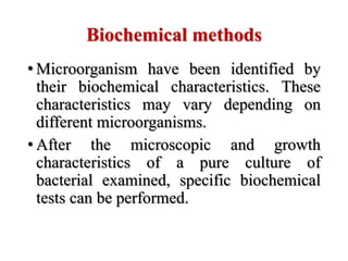
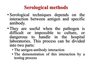
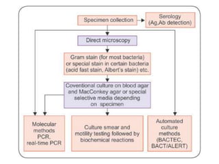
3. • Identification of unknown bacteria is one of
the major responsibilities of the
microbiologists.
• Samples of blood, tissue, food, water and
other are examined daily in laboratories
throughout the world for the presence of
microorganism.
• With some fundamental knowledge of
staining methods, isolation techniques,
bacterial nutrition, biochemical activities and
growth characterization of bacteria, it
becomes easier to identify any unknown
bacteria.
4. Collection of samples
• Body fluids, secretions and biopsy material can all
be examined to detect pathogens, antigen or
products or the immune response to them.
• Samples from the environment, e.g. water, food,
soil, etc.
• Some samples should be collected at particular
time; for example malaria parasites best sought at
the peak fever and a short time afterwards, whereas
blood for bacterial culture should be taken as fever
for begins to rise.
5. Universal safety and laboratory safety
• Specimen may contain hazardous
pathogens and must be handles with care
• General precaution is employed to reduce
the risk of transmitting blood-borne
pathogens
• Personal protective measures have to be
taken in collecting and examining
specimens
6. General rule for specimen collection
• The quantity of specimen must be adequate
• The sample should be collected from the appropriate
region of the body
• Contamination of the specimen must be avoided by using
only sterile equipment and aseptic conditions
• Always collect specimens prior to administration of
antimicrobial drugs
• Specimen maybe infectious, so take proper precautions
like wear gloves, gowns, masks and goggles, whenever
necessary.
• Specimen contains labeled with information about patient
name, code number, patient gender, patient age, specimen
source, date and time of collection, etc.
7. Collection
• Skin, nails and hair
• Mucous membrane
• Anterior nares or Throat
• Ear
• Sputum
• Urine
• Faeces
• Eye swabs
• Blood
• Cerebrospinal fluid
• Pus and wound secretions
8. Specimen handling
• Immediate after collection, the specimen
must be properly labeled and handled.
• The person collecting the specimen is
responsible for ensuring that the name,
hospital, registration number, location in
the hospital, diagnosis, current
antimicrobial therapy, name of attending
physician, admission date and types of
specimen are correctly and legibly written
or imprinted on the culture request form.
9. Specimen transportation
• Transport the specimen to the clinical
laboratory after it has been obtained from
the patients is of prime importance.
• Microbial specimens maybe transported to
laboratory by various ways.
• Special treatment requires for different
kind of microorganism.
• Transport of blood specimen to the
laboratory promptly, or place them in an
incubator at 37°C.
10. Specimen transportation
• Transport of urine specimens to the
clinical laboratory must be done as soon as
possible. Within 1 hour, specimen should
be examined, if time schedule can not be
followed, the urine sample must be
refrigerated immediately.
• CSF sample transported to the laboratory
within 15 minutes.
• Specimen for the virus are iced before
transport and can be kept at 4°C for up to
72 hours.
11. Laboratory methods
1. Direct microscopical methods
2. Cultural methods
3. Biochemical methods
4. Serological methods
5. Molecular methods
12. Microscopical methods
• The microscope has been indispensable in
the study of microorganism.
• The equipment requirement is cheap,
reagent cost is low and early result can be
obtained.
• There is no need of multiplication of
organism nor to the alive for examination.
• Microscopy is especially useful for
detecting organism that are difficult or
dangerous to grow.
13. Microscopical methods
• Magnification
• Resolution
• Parts (Eyepiece lens, Objective lens, Tube,
Arm, Base, Illuminator, Stage, Revolving
nose piece or Turret, Condenser lens and
Diaphragm or iris)
• Care of microscope
• Types of microscope
14. Microscopical methods
• Two important types of microscopy for the
diagnosis of infections are
• Stained preparation
• Unstained preparation
• Unstained preparation:
• Direct examination of unstained preparation is
suitable for rapid diagnosis in the laboratory.
• Many pathogens have a characteristic appearance,
e.g., parasites in faeces or bacteria together with
white cells in the urine.
• Unstained preparation are used to study bacterial
shape, size, arrangement and motility by using
hanging drop method.
15. Hanging drop slide method
• Objective: To observe living bacteria
motility and study the shape, size and
arrangement
• Principle: Many bacteria are show no motion
and are termed nonmotile. In an aqueous
environment, these same bacteria appear to
be moving erratically. This erratic movement
is due to Brownian movement. Brownian
movement results from the random motion of
the water molecules bombarding the bacteria
and causing them to move.
16. Hanging drop slide method
• True motility (self-propulsion) has been
recognized in other bacteria and involves several
different mechanisms.
• Bacteria that posses flagella exhibit flagellar
motion.
• Helical shaped spirochetes have axial fibrils
(modified flagella that wrap around the
bacterium) that form axial filaments. These
spirochetes move in a corkscrew and bending-
type motion.
• These type of motility or non motility can be
observed over long period in a hanging drop
slide.
17. Hanging drop slide method
• Requirements: Bacterial culture, Cavity slide, cover slip,
Vaseline or petroleum jelly, inoculating needle and Bunsen
burner
• Procedure:
• Apply Vaseline on the edge of the coverslip to provide
seal
• Use the inoculating loop to transfer a small drop of
bacterial suspension in the center of a coverslip
• Invert cavity slide and move slide onto the coverslip.
Press gently to form a seal.
• Turn the slide over and place on the stage of the
microscope
• Examination of hanging drop under low power objective
and switch to 90 – 100x objective, using immersion oil
20. Biochemical methods
• Microorganism have been identified by
their biochemical characteristics. These
characteristics may vary depending on
different microorganisms.
• After the microscopic and growth
characteristics of a pure culture of
bacterial examined, specific biochemical
tests can be performed.
21. Biochemical tests
• Carbohydrate breakdown
• Catalase production
• Citrate utilization
• Coagulase
• Decarboxylase and deamines
• Hydrogen sulfide
• Indole
• Oxidase production
• Nitrate reduction
• Proteinase production
• Urease production
22. Serological methods
• Serological techniques depends on the
interaction between antigen and specific
antibody.
• They are useful when the pathogen is
difficult or impossible to culture, or
dangerous to handle in the hospital
laboratories. This process can be divided
into two parts:
• The antigen-antibody interaction
• The demonstration of this interaction by a
testing process
23. Serological methods
• The antigen-antibody reaction depends on
the specific binding between epitopes of
the pathogens and antigen-binding sites on
the immunoglobulin molecules.
• The antigen-antibody binding was detected
by observing a natural consequences of
this interaction: precipitation,
agglutination
24. Serological methods
• Precipitation test: When a soluble antigen is mixed its
specific antibody in the presence of electrolytes at a
suitable temperature and pH, the antigen-antibody
complex forms an insoluble precipitate.
• This precipitates settle down at bottom of the tube.
• When precipitate is not sediment and remains as
floccules, the reaction is called as flocculation.
• This is done for both qualitative and quantitative
determination of both antigen and antibody.
• This is sensitive method and able to detect even little
antigen level.
• For Ring test (for anthrax), Slide test (VDRL test-
Venereal Disease Research laboratory test for Syphilis)
25. Serological methods
• Agglutination test: When an antigen or an inorganic
particle is mixed with antibody in the presence of
electrolytes at a suitable temperature and pH, the
particles are clumped or agglutinated.
• Agglutination reaction is more sensitive than
precipitation for detection of antibodies.
• Slide agglutination test (for identification of cultures of
salmonella and shigella)
• Tube agglutination test (for serological diagnosis of enteric
fever, brucellosis and typhus fevers.
• Widal test
• Immunofluorescence
• Radioimmunoassay
• Enzyme-linked Immunoassay (ELISA)
• Western blotting