The document provides details on the anatomy and physiology of the human lens:
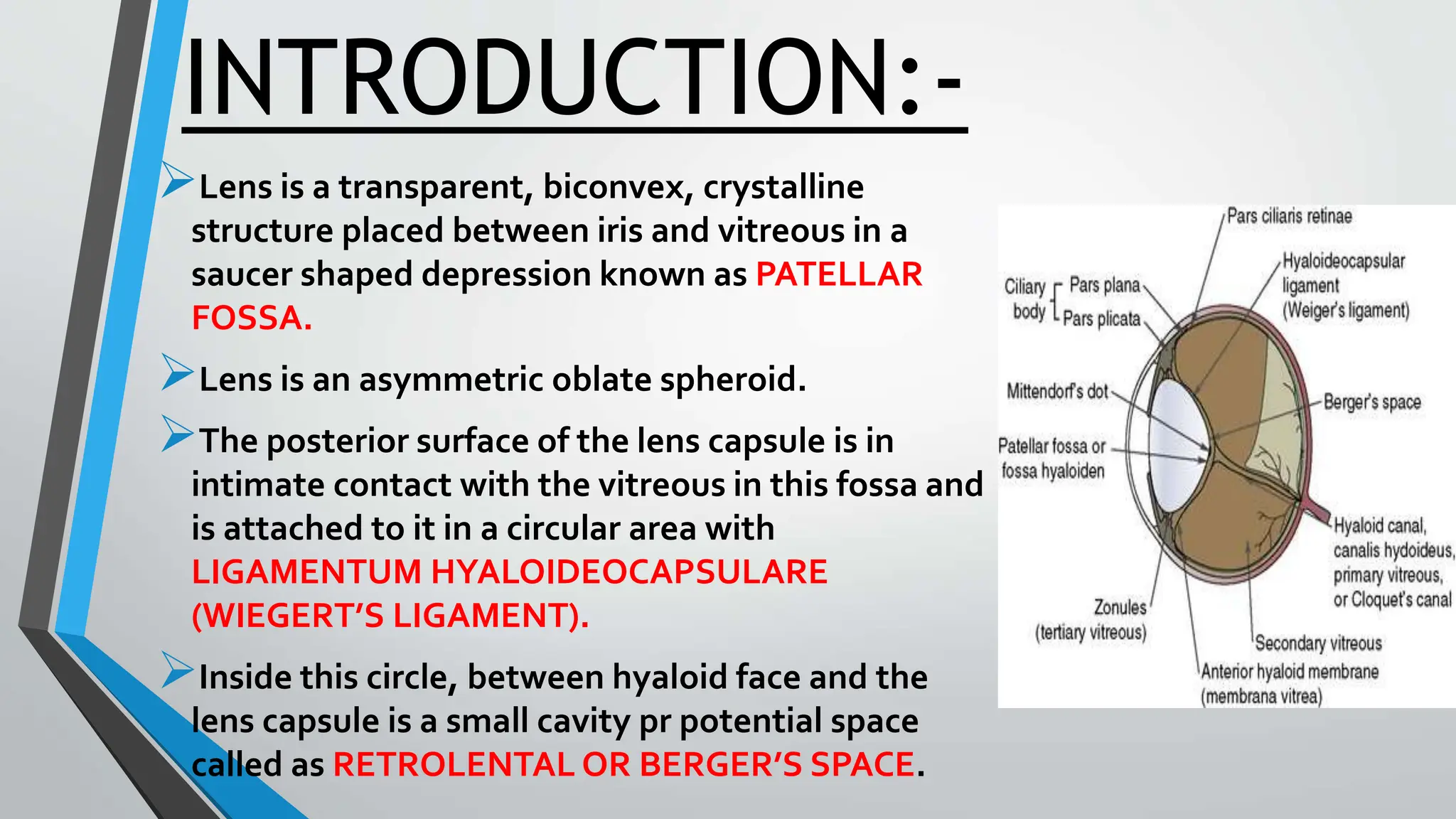
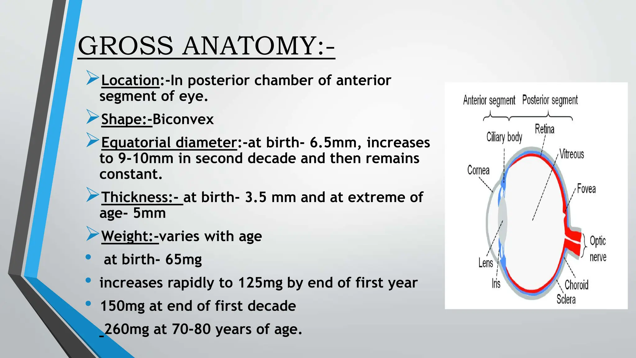
- The lens is a transparent, biconvex structure located behind the iris that focuses light onto the retina. It is encased in a collagenous capsule and becomes more yellow with age.
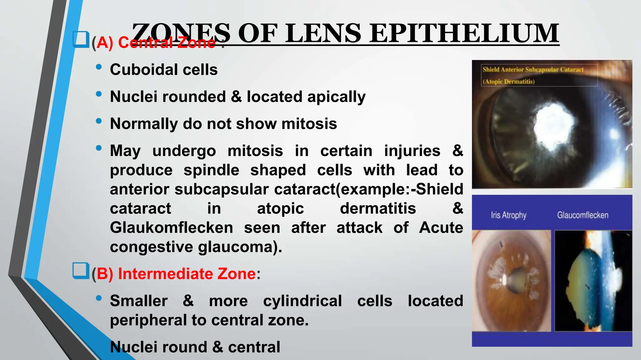
- The lens is composed of multiple layers - an outer capsule, inner epithelial cells that produce new fiber cells, and inner fiber mass. Fiber cells lose their nuclei as they mature.
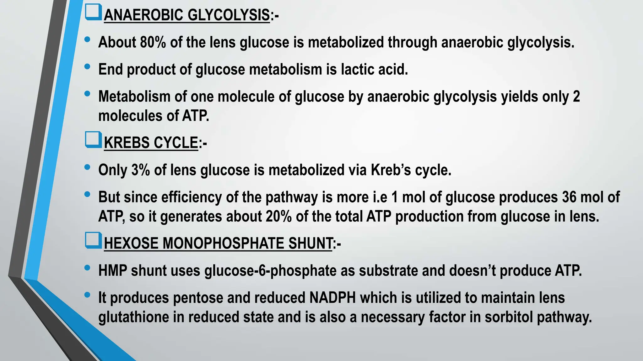
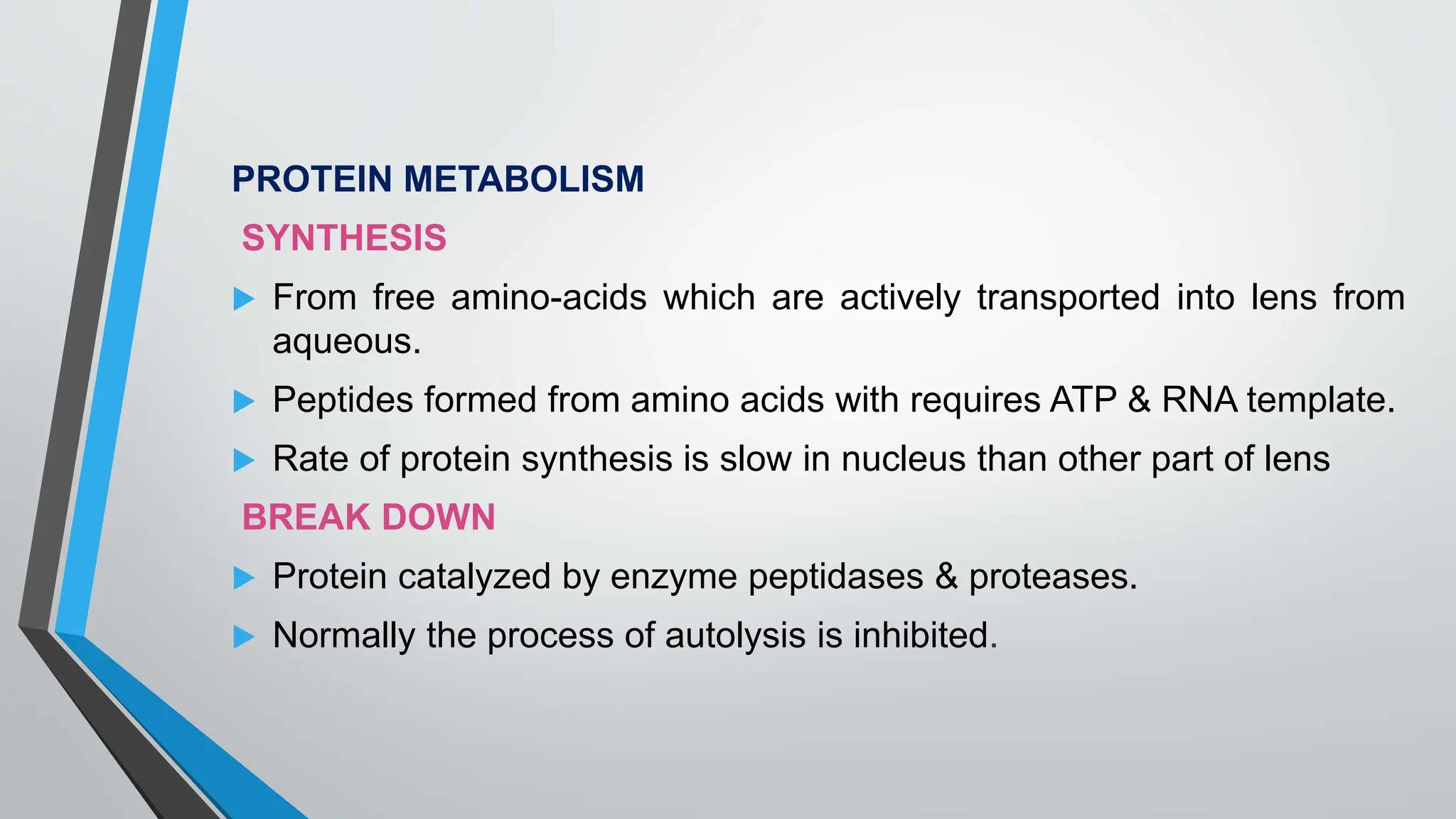
- Lens metabolism relies on glucose breakdown to produce ATP for ion transport and protein/glutathione synthesis. This occurs largely via anaerobic glycolysis within the fiber cells.
- The lens grows throughout life as new fiber cells are added at the