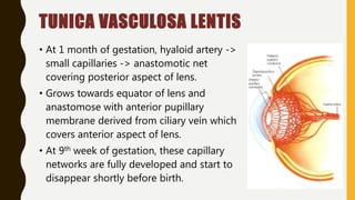
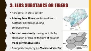
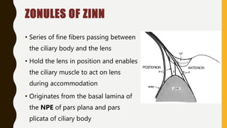
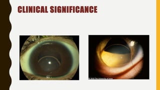
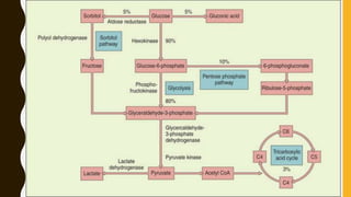
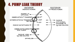
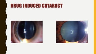
THE ANATOMY
& PHYSIOLOGY OF LENS
Dr. Ankit Bista
1st year resident
Ophthalmology
OUTLINE
• Introduction
• Embryology
• Anatomy
• Biochemical composition
• Physiology
• Clinical significance
INTRODUCTION
• Crystalline structure
• Transparent, avascular and
biconvex
• Asymmetric oblate spheroid
• Does not possess nerves, blood
vessels, or connective tissue
POSITION OF LENS
• Located between the iris and the
vitreous body
– at the pupillary area
– in the saucer shaped depression k/a
patellar fossa
ANATOMICAL RELATION
Anterior:
• AC of the eye through the pupillary
aperture, and with the posterior
surface of the iris
Posterior:
• Vitreous which is attached by
ligamentum hyaloideocapsulare
(Wiegert’s ligament)
DEVELOPMENT OF LENS
• Derived from surface ectoderm
• At 25 days of gestation, 2 lateral evaginations form from
forebrain – Optic vesicles, which closely adheres to surface
ectoderm.
LENS PLACODE
• The cells of surface ectoderm overlying the optic vesicles
become columnar at about 27th day of gestation
• This area of thickened cells is called lens plate or lens placode.
LENS PIT
• Appears at 29th day of gestation as a small
indentation or infolding of the lens placode.
• Pit deepens by cellular multiplication and
invaginates to form lens vesicle.
LENS VESICLE
• At 30 days of gestation, the lens cells separate from the surface
ectoderm.
• Becomes a single layer sphere of cuboidal cells called a lens
vesicle and is encased within a basement membrane (lens
capsule).
PRIMARY LENS FIBERS AND
EMBRYONIC NUCLEUS
• Posterior cells of lens vesicles rapidly
elongate obliterating the lumen of the cavity
• By 35th day of gestation the lumen is
completely obliterated and the cells are
called Primary lens fibers
• These fibers make up the Embryonic
nucleus at 40th day of gestation.
DEVELOPING LENS EPITHELIUM
• Cells of anterior lens vesicle still remain
cuboidal and form Lens epithelium
• Lens capsule develops as a basement
membrane.
SECONDARY LENS FIBERS
• Formed by proliferation of epithelial
cells near lens equator at around 7th
week of gestation.
• Anterior aspect of fibers – anterior
pole
• Posterior aspect – posterior pole
• Subsequently get displaced and
meet on the vertical planes, the Lens
sutures
FETAL NUCLEUS
– Secondary lens fibers
formed between 2nd to 8th
months of gestation make
up the fetal nucleus.
LENS SUTURE
• As lens fibers grow anteriorly and
posteriorly, they meet and interdigitate
at anterior and posterior pole.
• Formed only during fetal life
• As secondary fibers are added, the
sutures become more complex and
dendriform
TUNICA VASCULOSA LENTIS
• At 1 month of gestation, hyaloid artery ->
small capillaries -> anastomotic net
covering posterior aspect of lens.
• Grows towards equator of lens and
anastomose with anterior pupillary
membrane derived from ciliary vein which
covers anterior aspect of lens.
• At 9th week of gestation, these capillary
networks are fully developed and start to
disappear shortly before birth.
DEVELOPMENTAL ANOMALIES OF THE
LENS
• Lenticonus/Lentiglobu
s
• Coloboma
• Microphakia /
Microspherophakia
DEVELOPMENTAL ANOMALIES OF THE
LENS
CONGENITAL CATARACTS
• Polar cataract: Small opacities of the lens capsule & adjacent
cortex on the anterior or posterior pole of the lens.
• Capsular cataract: Small opacifications of the lens epithelium
and anterior lens capsule that spare the cortex.
• Nuclear cataract: Opacities of embryonic nucleus alone or
both embryonic or fetal nuclei.
CONGENITAL CATARACTS
• Lamellar cataract: Most common type. Round central shell
like opacity surrounding the nucleus.
• Coronary cataract: Group of club-shaped opacities in the
cortex. Arranged around the equator of the lens like a
or corona.
• Blue dot cataract: Punctate opacities in the form of rounded
bluish dots. Lies in the peripheral part of adolescent nucleus
& deeper layer of the cortex
ANATOMY OF LENS
• Anterior surface:
- radius of 10 mm (8-14 mm)
• Posterior surface:
- radius of 6 mm (4.5-7.5 mm)
• Anterior pole:
- 3 mm from back of cornea
• Posterior pole
• Equatorial diameter:
- birth (6.5mm)
- 2nd decade (9-10mm)
ANATOMY OF LENS
• Axial width/ AP diameter:
at birth – 3.5-4 mm
adult – 4.75-5mm
• Refractive index of lens: – 1.39
(nucleus - 1.42)
(cortex – 1.38)
• Refractive power: 16-17 D
• Weight of lens: at birth – 65 mg & at extreme of age –260 mg
ANATOMY OF LENS
• Accommodative power:
at birth – 14-16 D
at 25 yrs – 7-8 D
at 50 yrs – 1-2 D
• Color of lens:
at birth, infants, young adults – colorless
at about 30 yrs – yellow tinge
old age – amber color
• Consistency: Cortex is softer as compared to nucleus
STRUCTURE OF THE LENS
1. Lens capsule
2. Anterior lens
epithelium
3. Lens substance or
lens fiber
1. LENS CAPSULE
• Thin, transparent, hyaline collagenous membrane
• Elastic in nature but contain no any elastic tissue
• Anteriorly secreted by lens epithelium and posteriorly by basal
cells of elongating fibers
• Composed of type 4 collagen and 10% glycosaminoglycans
CLINICAL SIGNIFICANCE
2. ANTERIOR LENS EPITHELIUM
• Single layer of cells below the lens capsule.
• Formed of cuboidal cells but becomes columnar at equatorial
region.
• Actively dividing and elongating to form the lens fiber.
• Metabolic, synthetic and transport processes of the lens occur
here.
ZONES OF LENS EPITHELIUM
• Central Zone:
- Consists of cuboidal cells & do not mitose.
• Intermediate zone:
- Consists of cylindrical cells located peripheral to central
• Germinative zone:
- Consists of columnar cells which are most peripheral &
located just pre-equatorial and are actively dividing to form
lens fiber.
CLINICAL SIGNIFICANCE
3. LENS SUBSTANCE OR FIBERS
• Hexagonal in cross-section
• Primary lens fibers are formed from
posterior epithelium during
embryogenesis
• Formed constantly throughout life by
elongation of lens epithelium at equator
from germinative cells
• Arranged compactly as Nucleus & Cortex
NUCLEUS AND CORTEX
• Nucleus- Central part containing the oldest fibers
• Cortex- Peripheral part containing the youngest fibers
• Depending upon the period of development different zones
of nucleus are:
– Embryonic nucleus – Fetal nucleus
– Infantile nucleus – Adult nucleus
ZONULES OF ZINN
• Series of fine fibers passing between
the ciliary body and the lens
• Hold the lens in position and enables
the ciliary muscle to act on lens
during accommodation
• Originates from the basal lamina of
the NPE of pars plana and pars
plicata of ciliary body
CLINICAL SIGNIFICANCE
BIOCHEMICAL COMPOSITION
Water
• 66%
• Low amount of water helps to maintain the refractive index
• Lens dehydration maintained by active sodium pump
• Cortex more hydrated than nucleus
Proteins
• 33%
• Two Major groups:
a) Water soluble (80%): Crystallin
– alpha(32%), beta(55%) and gamma(1.5%)
b) Water insoluble(12.5%): Albuminoids
• Cortex contains more soluble proteins than nucleus which
contains more insoluble proteins.
Carbohydrates
1. Glucose
• Source is aqueous humor
• 20-120 mg%
2. Fructose
• produced from glucose
3. Glycogen
• very high in lens
• located in the nucleus
4. Inositol
5. Sorbitol
Lipids
• Cholesterol (50-60%)
• Phospholipids - sphingomyelin
• Glycolipids
• Functions-
• principal constituents of lens cell membrane
• also associated with lens epithelial cell division
Electrolytes
• Potassium is the predominant cation. 114-130 mEQ/kg
• Sodium concentration between 14-25 mEQ/kg
• Calcium lowest of all tissue calcium level with a mean value
of 0.14 mg /mg
• The main anion are chloride, bicarbonate, phosphate &
sulphates
Glutathione
• Is a tripeptide compounds
• Content varies form 3.5 - 5.5 mg/g
• Lens epithelium contains high levels of glutathione
• More than 95% of glutathione is in reduced state
PHYSIOLOGY
• Main site – lens epithelium
• Main Aims
1. Maintenance of lens transparency
2. Accommodation
3. Carbohydrate metabolism
4. Pump leak theory
5. Anti- oxidant
1. LENS TRANSPARENCY
1. Single(thin) layer of epithelial cells
2. Semi permeable lens capsule
3. Highly packed structure of lens fibers
4. Characteristic arrangement of lens protein
5. Pump mechanism of lens fibers which maintains lens
dehydration
6. Avascularity
2. ACCOMMODATION
• The mechanism by which the eye changes focus from distant
to near images
• Produced by a change in lens shape as a result of the action
of the ciliary muscle on the zonular fibers
• Lens- most malleable during childhood & the young adult
years
• In Accommodated state:
- Ciliary muscles contracts  Diameter of the muscle ring is
reduced  Relaxes the tension on the zonular fibres  Lens
become more spherical  Axial thickness of the lens increases
 Diameter of the lens decreases  Dioptric power of the eye
increases producing accommodation.
3. METABOLISM
Lens requires a continuous supply of energy (ATP) for :
• Active transport of ions & amino acids
• Maintenance of lens dehydration
• And for a continuous protein & GSH synthesis
4. PUMP LEAK THEORY
5. ANTIOXIDANTS
• Enzymatic defence mechanisms:
- High glutathione levels
- Glutathione peroxidase
- Superoxide dismutase and catalase
• Non-enzymatic defence mechanisms:
- Ascorbic acid (Vit C)
- Vit E
- Carotenoids
AGE RELATED CHANGES
Morphological Changes
• Mass & dimension of the lens increases
• Epithelial cells becomes flatter & density decreases
• Cholesterol : phospholipid ratio increases
• Increased light absorbance
Metabolic Changes
• Decreased proliferative capacity of lens epithelium
• Decreased enzymatic activity (superoxide dismutase and glucose-6-
phosphate dehydrogenase)
AGE RELATED CHANGES
Changes in Crystallin
• Increased insolubility
• loss of gamma-crystallins
• Increased disulfide bonds in gamma-crystallins
Changes in Plasma Membrane and Cytoskeletal
• loss of hexagonal cross-section
• loss of membrane proteins, lipids and cytoskeletal proteins
• Increased lens sodium and calcium with subsequent hydration
AGE RELATED CATARACT
DRUG INDUCED CATARACT
CATARACT ASSOCIATED WITH
SYSTEMIC DISEASE
Lens

Lens

  • 1.
    THE ANATOMY & PHYSIOLOGYOF LENS Dr. Ankit Bista 1st year resident Ophthalmology
  • 2.
    OUTLINE • Introduction • Embryology •Anatomy • Biochemical composition • Physiology • Clinical significance
  • 3.
    INTRODUCTION • Crystalline structure •Transparent, avascular and biconvex • Asymmetric oblate spheroid • Does not possess nerves, blood vessels, or connective tissue
  • 4.
    POSITION OF LENS •Located between the iris and the vitreous body – at the pupillary area – in the saucer shaped depression k/a patellar fossa
  • 5.
    ANATOMICAL RELATION Anterior: • ACof the eye through the pupillary aperture, and with the posterior surface of the iris Posterior: • Vitreous which is attached by ligamentum hyaloideocapsulare (Wiegert’s ligament)
  • 6.
    DEVELOPMENT OF LENS •Derived from surface ectoderm • At 25 days of gestation, 2 lateral evaginations form from forebrain – Optic vesicles, which closely adheres to surface ectoderm.
  • 7.
    LENS PLACODE • Thecells of surface ectoderm overlying the optic vesicles become columnar at about 27th day of gestation • This area of thickened cells is called lens plate or lens placode.
  • 8.
    LENS PIT • Appearsat 29th day of gestation as a small indentation or infolding of the lens placode. • Pit deepens by cellular multiplication and invaginates to form lens vesicle.
  • 9.
    LENS VESICLE • At30 days of gestation, the lens cells separate from the surface ectoderm. • Becomes a single layer sphere of cuboidal cells called a lens vesicle and is encased within a basement membrane (lens capsule).
  • 10.
    PRIMARY LENS FIBERSAND EMBRYONIC NUCLEUS • Posterior cells of lens vesicles rapidly elongate obliterating the lumen of the cavity • By 35th day of gestation the lumen is completely obliterated and the cells are called Primary lens fibers • These fibers make up the Embryonic nucleus at 40th day of gestation.
  • 11.
    DEVELOPING LENS EPITHELIUM •Cells of anterior lens vesicle still remain cuboidal and form Lens epithelium • Lens capsule develops as a basement membrane.
  • 12.
    SECONDARY LENS FIBERS •Formed by proliferation of epithelial cells near lens equator at around 7th week of gestation. • Anterior aspect of fibers – anterior pole • Posterior aspect – posterior pole • Subsequently get displaced and meet on the vertical planes, the Lens sutures
  • 13.
    FETAL NUCLEUS – Secondarylens fibers formed between 2nd to 8th months of gestation make up the fetal nucleus.
  • 14.
    LENS SUTURE • Aslens fibers grow anteriorly and posteriorly, they meet and interdigitate at anterior and posterior pole. • Formed only during fetal life • As secondary fibers are added, the sutures become more complex and dendriform
  • 15.
    TUNICA VASCULOSA LENTIS •At 1 month of gestation, hyaloid artery -> small capillaries -> anastomotic net covering posterior aspect of lens. • Grows towards equator of lens and anastomose with anterior pupillary membrane derived from ciliary vein which covers anterior aspect of lens. • At 9th week of gestation, these capillary networks are fully developed and start to disappear shortly before birth.
  • 16.
    DEVELOPMENTAL ANOMALIES OFTHE LENS • Lenticonus/Lentiglobu s • Coloboma • Microphakia / Microspherophakia
  • 17.
  • 18.
    CONGENITAL CATARACTS • Polarcataract: Small opacities of the lens capsule & adjacent cortex on the anterior or posterior pole of the lens. • Capsular cataract: Small opacifications of the lens epithelium and anterior lens capsule that spare the cortex. • Nuclear cataract: Opacities of embryonic nucleus alone or both embryonic or fetal nuclei.
  • 19.
    CONGENITAL CATARACTS • Lamellarcataract: Most common type. Round central shell like opacity surrounding the nucleus. • Coronary cataract: Group of club-shaped opacities in the cortex. Arranged around the equator of the lens like a or corona. • Blue dot cataract: Punctate opacities in the form of rounded bluish dots. Lies in the peripheral part of adolescent nucleus & deeper layer of the cortex
  • 20.
    ANATOMY OF LENS •Anterior surface: - radius of 10 mm (8-14 mm) • Posterior surface: - radius of 6 mm (4.5-7.5 mm) • Anterior pole: - 3 mm from back of cornea • Posterior pole • Equatorial diameter: - birth (6.5mm) - 2nd decade (9-10mm)
  • 21.
    ANATOMY OF LENS •Axial width/ AP diameter: at birth – 3.5-4 mm adult – 4.75-5mm • Refractive index of lens: – 1.39 (nucleus - 1.42) (cortex – 1.38) • Refractive power: 16-17 D • Weight of lens: at birth – 65 mg & at extreme of age –260 mg
  • 22.
    ANATOMY OF LENS •Accommodative power: at birth – 14-16 D at 25 yrs – 7-8 D at 50 yrs – 1-2 D • Color of lens: at birth, infants, young adults – colorless at about 30 yrs – yellow tinge old age – amber color • Consistency: Cortex is softer as compared to nucleus
  • 23.
    STRUCTURE OF THELENS 1. Lens capsule 2. Anterior lens epithelium 3. Lens substance or lens fiber
  • 24.
    1. LENS CAPSULE •Thin, transparent, hyaline collagenous membrane • Elastic in nature but contain no any elastic tissue • Anteriorly secreted by lens epithelium and posteriorly by basal cells of elongating fibers • Composed of type 4 collagen and 10% glycosaminoglycans
  • 25.
  • 26.
    2. ANTERIOR LENSEPITHELIUM • Single layer of cells below the lens capsule. • Formed of cuboidal cells but becomes columnar at equatorial region. • Actively dividing and elongating to form the lens fiber. • Metabolic, synthetic and transport processes of the lens occur here.
  • 27.
    ZONES OF LENSEPITHELIUM • Central Zone: - Consists of cuboidal cells & do not mitose. • Intermediate zone: - Consists of cylindrical cells located peripheral to central • Germinative zone: - Consists of columnar cells which are most peripheral & located just pre-equatorial and are actively dividing to form lens fiber.
  • 28.
  • 29.
    3. LENS SUBSTANCEOR FIBERS • Hexagonal in cross-section • Primary lens fibers are formed from posterior epithelium during embryogenesis • Formed constantly throughout life by elongation of lens epithelium at equator from germinative cells • Arranged compactly as Nucleus & Cortex
  • 30.
    NUCLEUS AND CORTEX •Nucleus- Central part containing the oldest fibers • Cortex- Peripheral part containing the youngest fibers • Depending upon the period of development different zones of nucleus are: – Embryonic nucleus – Fetal nucleus – Infantile nucleus – Adult nucleus
  • 31.
    ZONULES OF ZINN •Series of fine fibers passing between the ciliary body and the lens • Hold the lens in position and enables the ciliary muscle to act on lens during accommodation • Originates from the basal lamina of the NPE of pars plana and pars plicata of ciliary body
  • 32.
  • 33.
  • 34.
    Water • 66% • Lowamount of water helps to maintain the refractive index • Lens dehydration maintained by active sodium pump • Cortex more hydrated than nucleus
  • 35.
    Proteins • 33% • TwoMajor groups: a) Water soluble (80%): Crystallin – alpha(32%), beta(55%) and gamma(1.5%) b) Water insoluble(12.5%): Albuminoids • Cortex contains more soluble proteins than nucleus which contains more insoluble proteins.
  • 36.
    Carbohydrates 1. Glucose • Sourceis aqueous humor • 20-120 mg% 2. Fructose • produced from glucose 3. Glycogen • very high in lens • located in the nucleus 4. Inositol 5. Sorbitol
  • 37.
    Lipids • Cholesterol (50-60%) •Phospholipids - sphingomyelin • Glycolipids • Functions- • principal constituents of lens cell membrane • also associated with lens epithelial cell division
  • 38.
    Electrolytes • Potassium isthe predominant cation. 114-130 mEQ/kg • Sodium concentration between 14-25 mEQ/kg • Calcium lowest of all tissue calcium level with a mean value of 0.14 mg /mg • The main anion are chloride, bicarbonate, phosphate & sulphates
  • 39.
    Glutathione • Is atripeptide compounds • Content varies form 3.5 - 5.5 mg/g • Lens epithelium contains high levels of glutathione • More than 95% of glutathione is in reduced state
  • 40.
    PHYSIOLOGY • Main site– lens epithelium • Main Aims 1. Maintenance of lens transparency 2. Accommodation 3. Carbohydrate metabolism 4. Pump leak theory 5. Anti- oxidant
  • 41.
    1. LENS TRANSPARENCY 1.Single(thin) layer of epithelial cells 2. Semi permeable lens capsule 3. Highly packed structure of lens fibers 4. Characteristic arrangement of lens protein 5. Pump mechanism of lens fibers which maintains lens dehydration 6. Avascularity
  • 42.
    2. ACCOMMODATION • Themechanism by which the eye changes focus from distant to near images • Produced by a change in lens shape as a result of the action of the ciliary muscle on the zonular fibers • Lens- most malleable during childhood & the young adult years
  • 43.
    • In Accommodatedstate: - Ciliary muscles contracts  Diameter of the muscle ring is reduced  Relaxes the tension on the zonular fibres  Lens become more spherical  Axial thickness of the lens increases  Diameter of the lens decreases  Dioptric power of the eye increases producing accommodation.
  • 44.
    3. METABOLISM Lens requiresa continuous supply of energy (ATP) for : • Active transport of ions & amino acids • Maintenance of lens dehydration • And for a continuous protein & GSH synthesis
  • 46.
  • 47.
    5. ANTIOXIDANTS • Enzymaticdefence mechanisms: - High glutathione levels - Glutathione peroxidase - Superoxide dismutase and catalase • Non-enzymatic defence mechanisms: - Ascorbic acid (Vit C) - Vit E - Carotenoids
  • 48.
    AGE RELATED CHANGES MorphologicalChanges • Mass & dimension of the lens increases • Epithelial cells becomes flatter & density decreases • Cholesterol : phospholipid ratio increases • Increased light absorbance Metabolic Changes • Decreased proliferative capacity of lens epithelium • Decreased enzymatic activity (superoxide dismutase and glucose-6- phosphate dehydrogenase)
  • 49.
    AGE RELATED CHANGES Changesin Crystallin • Increased insolubility • loss of gamma-crystallins • Increased disulfide bonds in gamma-crystallins Changes in Plasma Membrane and Cytoskeletal • loss of hexagonal cross-section • loss of membrane proteins, lipids and cytoskeletal proteins • Increased lens sodium and calcium with subsequent hydration
  • 50.
  • 51.
  • 52.

Editor's Notes

  • #4 It entirely depends upon aq humor to meet its metabolic requirements and to carry off its wastes. Function: helps in refraction of light and accomodations. Refraction of light- bcz its refraction index is different from that of aq and vit. i.e 1.4 centrally and 1.36 peripherally
  • #6 Vitreous is attached to the lens posteriorly in a circular area with Wiegerts ligament..and inside this circle is a small cavity called retrolental or bergers space.
  • #7 Surface ectoderm- single layer of cuboidal cells
  • #8 Cuboidal to columnar
  • #15 Sutural Cataract Opacification of Y-suture of fetal nucleus
  • #16 Sometimes a remnant of tunica vasculosa lentis persists as small opacity or strand called Mittendorf dot.
  • #17 *Anterior lenticonus: Anterior surface of the lens become conical; a/w Alport syndrome *Posterior lenticonus: Posterior surface of the lens become conical; B/L - familial or in Lowe syndrome, Alports syndrome *Lentiglobus: Lens surface become spherical; oil droplet in retroillumination “oil droplet” red reflex is present
  • #18 *Lens coloboma- Wedge shaped defect or indentation of the lens in periphery. It mostly occurs as an isolated anomaly *Microspherophakia- Lens small in diameter and spherical in shape; Due to faulty development of secondary lens fibers *Congenital aphakia: Very rare; Complete absence of lens; Primary: lens placode fails to develop from the surface ectoderm; Secondary: More common, developing lens is spontaneously absorbed
  • #21 Ant surface is less convex than the posterior. Centre of the anterior and posterior surface is called poles. Equator is where the anterior and posterior surface meets.
  • #26 *Voissius Ring- Imprinted iris pigments in the anterior surface of anterior lens capsule due to blunt trauma to eye *Radiation Induced Cataract- Punctate opacities within posterior lens capsule; Feathery anterior sub capsular opacities radiating towards the equator
  • #27 Posterior lens Epithelium absent because they are used up in filling the central cavity of lens vesicle during development period
  • #29 *Anterior Subcapsular Cataract- Metaplasia of central zone cells into spindle shaped myofibroblast like cell; eg: Shield cataract in atopic dermatitis; Glaukomflecken after an attack of acute angle closure glaucoma *Posterior Subcapsular Cataract- Dysplasia of germinative zone cells; eg radiation cataract
  • #31 *Embryonic nucleus – It is the innermost part of nucleus ( 3 months of gestation); It consists of primary lens fibers *Fetal nucleus – lie around the embryonic nucleus & corresponds to the lens from 3 months of gestation till birth; Its fibers meet around sutures which are anteriorly Y shaped & posteriorly inverted Y shape *Infantile nucleus : lens from birth to puberty *Adult nucleus : lens fibers formed after puberty to rest of the life
  • #32 as the Anterior fibers as the Equatorial fibers and as Posterior fibers
  • #33 Developmental : Deficient development of zonules causes ectopia lentis Partial dislocation of lens: Subluxation Complete dislocation of lens: Dislocated
  • #34 Lens has High K, Lactic acid, Glutathione, AA and proteins than that of Aq humor and Less Na, water, Cl, Glucose
  • #35 Lens is relatively dehydrated organ Water content of the lens is 66%
  • #36 *Protein content is higher in lens than in any organ. *The physical state of proteins is imp for the transparency of lens. *Overtime the lens protein aggregates to form very large particles that become water insoluble and thus increasing the opacity of the lens.
  • #39 K maybe high because of the large intracellular space in the lens.
  • #42 *Transmits 80% of light *During early stage of embryogenesis, lens is opaque due to tunica vasculosa. But later vascularity is lost thus becomes transparent. *High conc of reduced glutathione maintains the lens protein in reduced state and ensures the integrity of cell membrane pump.
  • #44 Hardening of the lens with age reduces this accommodation and causes presbyopia.
  • #46 *Anaerobic Glycolysis 80%; 2 molecules of net ATP per glucose molecule *TCA Cycle 3%; requires o2 so only limited to lens epithelium where there is adequate o2; 36 molecules of net ATP per glucose molecule; 25 % of the lens ATP *Pentose Phosphate Pathway (HMP Shunt) 5-10%; provides NADPH for fatty acid biosynthesis, glutathione reductase & aldose reductase; provides ribose for nucleotide biosynthesis *Sorbitol Pathway <5%; increased accumulation of sorbitol creates a hyperosmotic effect that results in an infusion of fluid; intracellular accumulation of polyols leads to a collapse and liquefaction of lens fibers, which ultimately results in the formation of lens opacities
  • #47 K & AA are actively transported into the anterior lens via the epithelium anteriorly. They then diffuse out with the concentration gradient through the back of the lens, where there are no active transport mechanisms. Conversely, sodium flows in through the back of the lens with the concentration gradient and then is actively exchanged for potassium by the epithelium. In support of this theory, an anteroposterior gradient was found for both ions: potassium was concentrated in the anterior lens; sodium, in the posterior lens. Most of the Na+,K+-ATPase activity is found in the lens epithelium and the superficial cortical fiber cells. The active transport mechanisms are lost if the capsule and attached epithelium are removed from the lens but not if the capsule alone is removed by enzymatic degradation with collagenase. These findings support the hypothesis that the epithelium is the primary site for active transport in the lens.
  • #48 Prevents oxidation of the lens
  • #52 Systemic or topical steroids cause posterior subcapsular cataract Chlorpromazine cause central, anterior capsular granules
  • #53 *Diabetic snowflake cataract *Stellate posterior subcapsular cataract in Myotonic dystrophy *Shield-like anterior subcapsular cataract in Atopic dermatitis