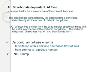
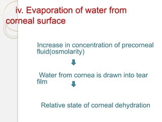
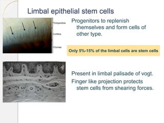
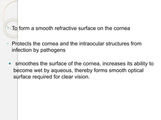
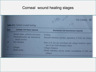
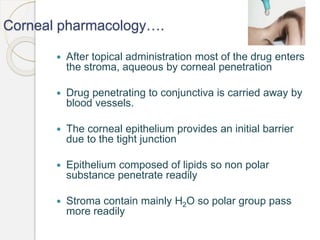
PHYSIOLOGY OF
CORNEA
Amrit Acharya
1st Year
Resident, NEH
2016, Jan. 24
History
The word cornea has come
from kerato.
Kerato in greek mean horn or
shield like
Ancient greek used to believe
that cornea is derived from
thinly sliced horn of animal
Functions of cornea
 It is the most important refractive media in the eye .
 The transparency of the cornea results from the uniform
spacing of the collagen fibrils in the substantia propria.
 Any increase in tissue fluid between the fibrils cause
cloudiness of the cornea.
 The endothelium and epithelium play a major role in
limiting fluid uptake by the corneal stroma.
Layers of cornea
Composition of cornea
Water 78%
Collagen 15%
Type I 50-55%
type III <1%
Type IV 8-10%
TypeVI 25-30%
other protein 5%
Keratan sulphate 0.7%
Chondroitin/dermatan sulphate 0.3%
Hyaluronic acid and salts 1%
Biochemical composition of epithelium
 10% of the total weight of cornea.
 Water - 70%of wet weight.
 protein synthesis in epithelium is highest.
 Lipids:phospholipid and cholesterol
 Contain enzymes necessary for krebs cycle, glycolysis
 Acetylcholine and ATP, glycogen ,glutathione
 Electrolyte:K, Na ,Cl .
Biochemical composition of stroma
 Contains 80% water and 20 % solids
 Collagen(type I,V,XII,XIV)
 Soluble proteins- albumin,immunoglobulins and glycoprotein.
 Proteoglycans(GAG fractions- keratan sulfate 50%,chondroitin
sulfate 25% and chondroitin 25%)
 Glycolytic and Krebs cycle enzymes
 Matrix metalloproteinases :MMP-1(collagenase-I),MMP-
2(Gelatinase A,MMP-3(Stromelysin I)
 Electrolytes and salts
Biochemical composition of descemet´s membrane
 Composed of collagen(73%)and glycoproteins
 Collagen of descemet’s membrane is insoluble and
extremely resistant to chemical and enzymatic action.
 Descemet’s membrane doesn’t contain GAG
 Single cell layered structure
 Contains enzymes for glycolysis and Krebs cycle.
Biochemical composition of endothelium
Metabolism in cornea
 The most active layers for metabolism are epithelium and
endothelium.Energy in the form of ATP is generated by
breakdown of glucose.
 Sources of nutrients for the corneal metabolism and
metabolic pathway are –
= Oxygen
= Glucose
= Amino acids
Oxygen
 Epithelium: derives O2 from tear film and
limbal capillaries
 Oxygen required by epithelium is 1/10th of that
available from atmosphere when the eyes are open
and about ¼ of that available from palprebral
conjunctiva when eyes are closed
 Endothelium: derives from aqueous humour
 Total corneal oxygen consumption:9.5ml 02 /cm2
/hr
Glucose
• Primary metabolic substrate for epithelial
cells,stromal keratocytes and endothelium
 In absence of an exogenous supply of glucose
,glycogen stored in corneal epithelium is broken
down
 Rate of glucose consumption of cornea:
100mg/cm2/hr 90% being consumed by epithelium
 Glucose is metabolized in the cornea by 3
metabolic pathway
= Anaerobic glycolysis
 Glucose
Lactic
acid 2 ATP
= Tricarboxylic acid( Krebs's cycle)
 36 ATP produced from a molecule of glucose
 Only 12% of glucose metabolise through TCA cycle
Accumulation of lactate even in aerobic condition
Lacate is eliminated from cornea by diffusion through
epithelium
Glucose 36 ATP CO2 & H2O
= Hexose monophosphate(HMP)
shunt.
NADPH produced is utilized in lipids
synthesis by cornea
Ribose produced is used to build
DNA,RNA & nucleic Acid
….continued
 In epithelium and endothelium, the
HMP pathway breaks down 35%-65%
of the glucose but stroma metabolize
very little via this pathway
Pyruvic acid is end product of glucose
Amino acids
Supplied from aqueous humour by
passive diffusion
• Requirement: For synthesis 10 mg/hr
of protein , for constant shedding and
replacement of epithelial cells of
cornea.
 Maurice theory
 Theory of goldman
Arrangement of stromal lamellae
Maurice theory(1957)
The stromal collagen fibrills are of equal diameter
(275-350 Å) and are equally distant from each other,
arranged as a lattice with the inner fibrillar spacing less
than a wave length of light (4000-7000 Å)
He explains , because of their small diameter and regularity of
separation ,back scattered light would be almost completely supressed
by destructive interference.
Goldman and Benedek’s theory (1967) :
He suggest that , a perfect crystalline lattice
periodicity
Is not always necessary for the sufficient
destructive interference .
He explains, If fibril separation and diameter is less
than a third of the wavelength of the incident light,
almost perfect transparency will issue.
The stromal fibrils are small in relation to the light
and do not interfere with the light transmission ,
unless they are larger than one half a wave length
of light.
Corneal transparency
Physiological factors :
-stromal swelling pressure
-metabolic pump
-barrier function
-evaporation from the corneal
surface
-intraocular pressure.
Corneal Hydration
State of relative dehydration that is necessary for
corneal transparency. Normal water content of cornea
( 80%) is kept constant by balance of factors that draw
water in cornea(swelling pressure and IOP), factor which
prevent flow of water in cornea(epithelial barrier) and
that draw water out of cornea(Endothelial pump)
Factors affecting corneal hydration
i. Stromal swelling pressure (SP)
- 50mmHG exerted by GAGs and
collagen of corneal stroma.
- Imbibition pressure (IP) is equal
to SP in vitro but not in vivo .
ii. Barrier function of epithelium and endothelium
- semi permeable in nature.
- calcium dependent
iii. Active pump mechanisms-
Mechanism by which endothelium
Removes fluid from stroma referred as
endothelial pump.
a) Active process :
Na-K-ATPase pump
 - Essential component of endothelial pump
function.
- Integral membrane protein located in the
basolateral aspect of endothelium
– its action is vital in the maintenance of corneal hydration.
-Central to all the system as maintains the sodium gradient
required for the Na -H exchange thereby promoting
bicarbonate production.
 Bicarbonate dependant ATPase:
- Is essential for the maintenance of the corneal thickness
-The bicarbonate transported by the endothelium is generated
intracellularly via the action of carbonic anhydrase.
- CO2 diffuses into the cell from the extra cellular space combines with
the water in presence of the carbonic anhydrase . The carbonic
anhydrase dissociates into H + and bicarbonate ions.
 Carbonic anhydrase enzyme
Inhibitation of this enzyme decreases flow of fluid
from stroma to aqueous humour
 Na-H pump.
b) Passive process :
 K+, Cl-,Hco3- ions diffuse into the aqueous
 Na+,Cl-,Hco3- diffuse from aqueous into cornea
iv. Evaporation of water from
corneal surface
Increase in concentration of precorneal
fluid(osmolarity)
Water from cornea is drawn into tear
film
Relative state of corneal dehydration
v. Intra ocular pressure
IOP > Swelling pressure = corneal oedema occur
- The relation of swelling pressure of stroma to IOP is
- IP(imbibition pressure of corneal stroma) =IOP- SP
- As stromal pressure decreases precipitiously by increase in corneal
thickness, mild corneal edema combined with increase in IOP leads
to high imbibitions pressure and subsequent microbullous formation
and epithelial edema.
Cellular factor affecting transparency
- keratocytes maintain transparency by producing
collagens and proteoglycans
- keratocytes contain enzymes involved in assembly
of stromal matrix
Specific enzyme defects are associated with corneal
opacification eg- mucopolysaccharidoses
Limbal epithelial stem cells
Progenitors to replenish
themselves and form cells of
other type.
Present in limbal palisade of vogt.
Finger like projection protects
stem cells from shearing forces.
Only 5%-15% of the limbal cells are stem cells
…...…stem cells
Highly vascularized and potential source
of nutrient and growth factors for stem
cells.
Are slow cycling cells and retain DNA
labels for longer time.But in events of
injury they become highly proliferative.
Prevents corneal neovascu-
larization and conjunctival ingrowth
Causes of
limbal stem
cells
deficiency
Congenital
-Aniridia
Aquired
-chemical burn
-thermal burn
-Multiple corneal
surface surgery
-prolonged contact
lens use
Corneal Epithelium
 Functions:
 To form a Barrier between the environment and the
stroma of the cornea.
Barrier formed as the cells move superficially to the
surface of the cornea ,differentiating until the
superficial cells form two layers of the flattened cells
encircled by the tight junction, which serves as a
high resistance, semi permeable membrane.
Barrier functions to prevent the movement of the
fluid from tears to the stroma .
 To form a smooth refractive surface on the cornea
 Protects the cornea and the intraocular structures from
infection by pathogens
 smoothes the surface of the cornea, increases its ability to
become wet by aqueous, thereby forms smooth optical
surface required for clear vision.
Maintenance of the Epithelium:
 Maintained by a balance among:
(X,Y,Z Hypothesis)
Process of cell migration:
. originate from stem cells in limbal epithelium
. Migration of new basal cells into cornea from
limbus.
.Migrate centripetally at about 120 μm/wk
Prolifera-
tion of
basal
cells (x)
Centripetal
migration
of cells
(Y)
Epithelial loss
from corneal
surface
(Z)
Mitosis:
 Occurs only in basal cell layer
Daughter cells move upward from basal
layer, differenciating into wing cells and
finally into superficial cells
Shedding of superficial cells:
Corneal epithelium: Stratified Squamous
epithelium from which terminally
differentiated, superficial cells
continuously shed
Epithelium turn over completely every
week
Epithelial wound repair
Mitosis resume and epithelial thickness is re-
established
Re-establishment of adhesion between basal epithelial
cells and bowman´s layer
Cells separate from basal lamina and travel in amoeboid
manner unless halted by contact inhibition
Injury inhibits mitosis of epithelial cells
Centripetal migration of marginal cells by
rearangement of actin fibrils in filopodial extension of
cells
Stromal wound healing
Deposition of fibrin within the stromal wound
Rapid epithelization of wound
Activation of keratocytes to divide and
synthesize collagen and GAGs
Initial lay out of irregular fibroblast
Production of normal corneal matrix to restore
clarity in small wound
Stromal wound healing continued…….
 Keratocyte at injured site undergo apoptosis
peaking 4 hrs. after initial insult. Modulate wound
healing by activating adjacent keratocytes
 In 1-2 wks remodeling starts. Which sometime
continue over prolong period and eventually
restore corneal transparency.
Corneal wound healing stages
Corneal injury
Corneal opacities
Nebular scar
Macular scar
Leucomatous
Endothelial injury and repair
 When endothelial cells are lost defect is covered by
spreading of cells from adjacent areas of wound
 If single cell lost the cells surrounding the defect
spread to fill in the area left by missing cell
• If large defect-cell migration toward the center of
wound followed by remodeling into hexagonal
shape.
• The pump of endothelium are reestablished when
monolayer is restored allowing cornea to return to
normal thickness
Corneal Vascularization-
 Chemical theory
- Destruction of vasoinhibitory factor(VIF)
- Release of vasostimulatory factor(VSF)
 Mechanical theory:( Cogan) blood vessels cannot invade stroma
due to its compactness
 Combined theory:( Maurice et al) role of both VIF & compactness of
the cornea.
 Role of leucocytes – stimulate corneal vascular growth.
Pathogenesis of corneal vascularization
New capillaries arise from the perilimbal capillaries and parent
venules by focal degradation of the venular basement
membrane.
Migration of endothelial cells toward angiogenic stimulus
Endothelial cells elongate and form solid sprout which later
develop lumen
The outer surface is lined by pericytes and blood flow begin
Clinical correlation:
• Cornea is immunologically privileged for keratoplasty
due to avascularity, absence of lymphatics and few
antigen presenting cells.
• Degree and depth of corneal vascularization are prognostic in
keratoplasty.
 Deep vascularization of more than 2 quadrants is considered
as high risk of graft rejection following keratoplasty
Types of corneal vascularisation
Superficial
Deep
Superficial Deep
Present below
epithelial layer
Lie in corneal stroma
Can be traced with
conjunctival vessels
Continuity can´t be
traced beyond
limbus
Pattern- commonly
arborising
Terminal loops ,
brush, umbel
Causes- Trachoma ,
Phlyctenular kerato -
conjunctivitis, superficial
corneal ulcer
Interestial keratitis ,
disciform keratitis,
chemical burn, deep
corneal ulcer
Corneal pharmacology
 The volume of normal adult tear film 7-9μL
and the maximum amount cul de sac can
maintain : 25 to 30μL
 Volume of a drop is approx. 40 μL .
 Most of the medication is immediately lost to eye lashes
and runs out of the eye and the remainder is diluted by
tear film to approx. 25%.
 Both volume and concentration of drop are reduced.
 Ointment is retained in cul de sac, gradually melts, releasing
the drug into the tears, purpose is to increase the time the
drug is present in tears
Corneal pharmacology….
 After topical administration most of the drug enters
the stroma, aqueous by corneal penetration
 Drug penetrating to conjunctiva is carried away by
blood vessels.
 The corneal epithelium provides an initial barrier
due to the tight junction
 Epithelium composed of lipids so non polar
substance penetrate readily
 Stroma contain mainly H2O so polar group pass
more readily
 Drugs must pass through both barriers, those soluble in
both lipids and water exhibit best penetration.
 Some preservatives present in drug e.g. benzalkonium
chloride impair the integrity of epithelial barrier and
increase penetration.
Drug permeability across cornea
 Depends on:
 Solubility- epithelium and endothelium easily
penetrated by lipid soluble substances. Stroma is
hydrophilic so allow water soluble substance.
Hence to go through drug should be amphipathic.
 Ioniation- Drug must have the capacity to exist
both in ionized and non-ionized form for better
penetration,non ionised drugs can penetrate
through epithelium and ionized through stroma.
 pH: affect on electric charges and stability of
solution. Solution outside the range of 4-10
increases the permeability.
 Molecular size: not important for lipid soluble
substance but for water soluble size should be
less than 4 A.
 Molecular weight: less than 500 can pass through
 Wetting agents: They increases permeability by
reducing surface tension.
Drug deposits in cornea
 Vortex keratopathy : whorl like corneal epithelial
deposits.
 Antimalarials
 Amiodarone
 Chlorpromazine:yellowish brown deposits in
endothelium and deep stroma
 Argyrosis
 due to silver deposit
 greyish brown deposits in
descemets
membrane
 Chrysiasis:dust like
deposits in corneal stroma
Aging changes in cornea
 By advancing age cornea becomes less translucent & dust like
opacities due to condensation in stroma.
 Bowman's & Descemet’s membranes also increase in
thickness .
 Arcus senilis is present due to infiltration of extra cellular lipid
and in almost every person over 60yrs.
 The small protrusion at the periphery of Descemet’s
membrane occur and known as Hassal Henle bodies and do
not interfere with vision.
References
Adler’s Physiology of eye. 7th ed.
Internet resources
Fundamentals and External disease & cornea- American academy
of Ophthalmology. 2011-2012
A.K.Khurana Anatomy and Physiology of Eye
Clinical anatomy of eye Richard S. snell, Michael A.Lemp 2nd
edition
 cornea physiology

cornea physiology

  • 1.
    PHYSIOLOGY OF CORNEA Amrit Acharya 1stYear Resident, NEH 2016, Jan. 24
  • 2.
    History The word corneahas come from kerato. Kerato in greek mean horn or shield like Ancient greek used to believe that cornea is derived from thinly sliced horn of animal
  • 3.
    Functions of cornea It is the most important refractive media in the eye .  The transparency of the cornea results from the uniform spacing of the collagen fibrils in the substantia propria.  Any increase in tissue fluid between the fibrils cause cloudiness of the cornea.  The endothelium and epithelium play a major role in limiting fluid uptake by the corneal stroma.
  • 4.
  • 6.
    Composition of cornea Water78% Collagen 15% Type I 50-55% type III <1% Type IV 8-10% TypeVI 25-30% other protein 5% Keratan sulphate 0.7% Chondroitin/dermatan sulphate 0.3% Hyaluronic acid and salts 1%
  • 7.
    Biochemical composition ofepithelium  10% of the total weight of cornea.  Water - 70%of wet weight.  protein synthesis in epithelium is highest.  Lipids:phospholipid and cholesterol  Contain enzymes necessary for krebs cycle, glycolysis  Acetylcholine and ATP, glycogen ,glutathione  Electrolyte:K, Na ,Cl .
  • 8.
    Biochemical composition ofstroma  Contains 80% water and 20 % solids  Collagen(type I,V,XII,XIV)  Soluble proteins- albumin,immunoglobulins and glycoprotein.  Proteoglycans(GAG fractions- keratan sulfate 50%,chondroitin sulfate 25% and chondroitin 25%)  Glycolytic and Krebs cycle enzymes  Matrix metalloproteinases :MMP-1(collagenase-I),MMP- 2(Gelatinase A,MMP-3(Stromelysin I)  Electrolytes and salts
  • 9.
    Biochemical composition ofdescemet´s membrane  Composed of collagen(73%)and glycoproteins  Collagen of descemet’s membrane is insoluble and extremely resistant to chemical and enzymatic action.  Descemet’s membrane doesn’t contain GAG  Single cell layered structure  Contains enzymes for glycolysis and Krebs cycle. Biochemical composition of endothelium
  • 10.
    Metabolism in cornea The most active layers for metabolism are epithelium and endothelium.Energy in the form of ATP is generated by breakdown of glucose.  Sources of nutrients for the corneal metabolism and metabolic pathway are – = Oxygen = Glucose = Amino acids
  • 11.
    Oxygen  Epithelium: derivesO2 from tear film and limbal capillaries  Oxygen required by epithelium is 1/10th of that available from atmosphere when the eyes are open and about ¼ of that available from palprebral conjunctiva when eyes are closed  Endothelium: derives from aqueous humour  Total corneal oxygen consumption:9.5ml 02 /cm2 /hr
  • 12.
    Glucose • Primary metabolicsubstrate for epithelial cells,stromal keratocytes and endothelium  In absence of an exogenous supply of glucose ,glycogen stored in corneal epithelium is broken down  Rate of glucose consumption of cornea: 100mg/cm2/hr 90% being consumed by epithelium  Glucose is metabolized in the cornea by 3 metabolic pathway
  • 13.
    = Anaerobic glycolysis Glucose Lactic acid 2 ATP
  • 14.
    = Tricarboxylic acid(Krebs's cycle)  36 ATP produced from a molecule of glucose  Only 12% of glucose metabolise through TCA cycle Accumulation of lactate even in aerobic condition Lacate is eliminated from cornea by diffusion through epithelium Glucose 36 ATP CO2 & H2O
  • 15.
    = Hexose monophosphate(HMP) shunt. NADPHproduced is utilized in lipids synthesis by cornea Ribose produced is used to build DNA,RNA & nucleic Acid
  • 16.
    ….continued  In epitheliumand endothelium, the HMP pathway breaks down 35%-65% of the glucose but stroma metabolize very little via this pathway Pyruvic acid is end product of glucose
  • 17.
    Amino acids Supplied fromaqueous humour by passive diffusion • Requirement: For synthesis 10 mg/hr of protein , for constant shedding and replacement of epithelial cells of cornea.
  • 18.
     Maurice theory Theory of goldman Arrangement of stromal lamellae
  • 19.
    Maurice theory(1957) The stromalcollagen fibrills are of equal diameter (275-350 Å) and are equally distant from each other, arranged as a lattice with the inner fibrillar spacing less than a wave length of light (4000-7000 Å) He explains , because of their small diameter and regularity of separation ,back scattered light would be almost completely supressed by destructive interference.
  • 20.
    Goldman and Benedek’stheory (1967) : He suggest that , a perfect crystalline lattice periodicity Is not always necessary for the sufficient destructive interference . He explains, If fibril separation and diameter is less than a third of the wavelength of the incident light, almost perfect transparency will issue. The stromal fibrils are small in relation to the light and do not interfere with the light transmission , unless they are larger than one half a wave length of light.
  • 21.
    Corneal transparency Physiological factors: -stromal swelling pressure -metabolic pump -barrier function -evaporation from the corneal surface -intraocular pressure.
  • 22.
    Corneal Hydration State ofrelative dehydration that is necessary for corneal transparency. Normal water content of cornea ( 80%) is kept constant by balance of factors that draw water in cornea(swelling pressure and IOP), factor which prevent flow of water in cornea(epithelial barrier) and that draw water out of cornea(Endothelial pump)
  • 23.
    Factors affecting cornealhydration i. Stromal swelling pressure (SP) - 50mmHG exerted by GAGs and collagen of corneal stroma. - Imbibition pressure (IP) is equal to SP in vitro but not in vivo .
  • 24.
    ii. Barrier functionof epithelium and endothelium - semi permeable in nature. - calcium dependent
  • 25.
    iii. Active pumpmechanisms- Mechanism by which endothelium Removes fluid from stroma referred as endothelial pump. a) Active process : Na-K-ATPase pump  - Essential component of endothelial pump function. - Integral membrane protein located in the basolateral aspect of endothelium – its action is vital in the maintenance of corneal hydration. -Central to all the system as maintains the sodium gradient required for the Na -H exchange thereby promoting bicarbonate production.
  • 26.
     Bicarbonate dependantATPase: - Is essential for the maintenance of the corneal thickness -The bicarbonate transported by the endothelium is generated intracellularly via the action of carbonic anhydrase. - CO2 diffuses into the cell from the extra cellular space combines with the water in presence of the carbonic anhydrase . The carbonic anhydrase dissociates into H + and bicarbonate ions.  Carbonic anhydrase enzyme Inhibitation of this enzyme decreases flow of fluid from stroma to aqueous humour  Na-H pump.
  • 27.
    b) Passive process:  K+, Cl-,Hco3- ions diffuse into the aqueous  Na+,Cl-,Hco3- diffuse from aqueous into cornea
  • 28.
    iv. Evaporation ofwater from corneal surface Increase in concentration of precorneal fluid(osmolarity) Water from cornea is drawn into tear film Relative state of corneal dehydration
  • 29.
    v. Intra ocularpressure IOP > Swelling pressure = corneal oedema occur - The relation of swelling pressure of stroma to IOP is - IP(imbibition pressure of corneal stroma) =IOP- SP - As stromal pressure decreases precipitiously by increase in corneal thickness, mild corneal edema combined with increase in IOP leads to high imbibitions pressure and subsequent microbullous formation and epithelial edema. Cellular factor affecting transparency - keratocytes maintain transparency by producing collagens and proteoglycans - keratocytes contain enzymes involved in assembly of stromal matrix Specific enzyme defects are associated with corneal opacification eg- mucopolysaccharidoses
  • 30.
    Limbal epithelial stemcells Progenitors to replenish themselves and form cells of other type. Present in limbal palisade of vogt. Finger like projection protects stem cells from shearing forces. Only 5%-15% of the limbal cells are stem cells
  • 31.
    …...…stem cells Highly vascularizedand potential source of nutrient and growth factors for stem cells. Are slow cycling cells and retain DNA labels for longer time.But in events of injury they become highly proliferative. Prevents corneal neovascu- larization and conjunctival ingrowth
  • 32.
    Causes of limbal stem cells deficiency Congenital -Aniridia Aquired -chemicalburn -thermal burn -Multiple corneal surface surgery -prolonged contact lens use
  • 33.
    Corneal Epithelium  Functions: To form a Barrier between the environment and the stroma of the cornea. Barrier formed as the cells move superficially to the surface of the cornea ,differentiating until the superficial cells form two layers of the flattened cells encircled by the tight junction, which serves as a high resistance, semi permeable membrane. Barrier functions to prevent the movement of the fluid from tears to the stroma .
  • 34.
     To forma smooth refractive surface on the cornea  Protects the cornea and the intraocular structures from infection by pathogens  smoothes the surface of the cornea, increases its ability to become wet by aqueous, thereby forms smooth optical surface required for clear vision.
  • 35.
    Maintenance of theEpithelium:  Maintained by a balance among: (X,Y,Z Hypothesis) Process of cell migration: . originate from stem cells in limbal epithelium . Migration of new basal cells into cornea from limbus. .Migrate centripetally at about 120 μm/wk Prolifera- tion of basal cells (x) Centripetal migration of cells (Y) Epithelial loss from corneal surface (Z)
  • 36.
    Mitosis:  Occurs onlyin basal cell layer Daughter cells move upward from basal layer, differenciating into wing cells and finally into superficial cells
  • 37.
    Shedding of superficialcells: Corneal epithelium: Stratified Squamous epithelium from which terminally differentiated, superficial cells continuously shed Epithelium turn over completely every week
  • 38.
    Epithelial wound repair Mitosisresume and epithelial thickness is re- established Re-establishment of adhesion between basal epithelial cells and bowman´s layer Cells separate from basal lamina and travel in amoeboid manner unless halted by contact inhibition Injury inhibits mitosis of epithelial cells Centripetal migration of marginal cells by rearangement of actin fibrils in filopodial extension of cells
  • 39.
    Stromal wound healing Depositionof fibrin within the stromal wound Rapid epithelization of wound Activation of keratocytes to divide and synthesize collagen and GAGs Initial lay out of irregular fibroblast Production of normal corneal matrix to restore clarity in small wound
  • 40.
    Stromal wound healingcontinued…….  Keratocyte at injured site undergo apoptosis peaking 4 hrs. after initial insult. Modulate wound healing by activating adjacent keratocytes  In 1-2 wks remodeling starts. Which sometime continue over prolong period and eventually restore corneal transparency.
  • 41.
  • 42.
  • 43.
  • 44.
    Endothelial injury andrepair  When endothelial cells are lost defect is covered by spreading of cells from adjacent areas of wound  If single cell lost the cells surrounding the defect spread to fill in the area left by missing cell • If large defect-cell migration toward the center of wound followed by remodeling into hexagonal shape. • The pump of endothelium are reestablished when monolayer is restored allowing cornea to return to normal thickness
  • 45.
    Corneal Vascularization-  Chemicaltheory - Destruction of vasoinhibitory factor(VIF) - Release of vasostimulatory factor(VSF)  Mechanical theory:( Cogan) blood vessels cannot invade stroma due to its compactness  Combined theory:( Maurice et al) role of both VIF & compactness of the cornea.  Role of leucocytes – stimulate corneal vascular growth.
  • 46.
    Pathogenesis of cornealvascularization New capillaries arise from the perilimbal capillaries and parent venules by focal degradation of the venular basement membrane. Migration of endothelial cells toward angiogenic stimulus Endothelial cells elongate and form solid sprout which later develop lumen The outer surface is lined by pericytes and blood flow begin
  • 47.
    Clinical correlation: • Corneais immunologically privileged for keratoplasty due to avascularity, absence of lymphatics and few antigen presenting cells. • Degree and depth of corneal vascularization are prognostic in keratoplasty.  Deep vascularization of more than 2 quadrants is considered as high risk of graft rejection following keratoplasty
  • 48.
    Types of cornealvascularisation Superficial Deep Superficial Deep Present below epithelial layer Lie in corneal stroma Can be traced with conjunctival vessels Continuity can´t be traced beyond limbus Pattern- commonly arborising Terminal loops , brush, umbel Causes- Trachoma , Phlyctenular kerato - conjunctivitis, superficial corneal ulcer Interestial keratitis , disciform keratitis, chemical burn, deep corneal ulcer
  • 49.
    Corneal pharmacology  Thevolume of normal adult tear film 7-9μL and the maximum amount cul de sac can maintain : 25 to 30μL  Volume of a drop is approx. 40 μL .  Most of the medication is immediately lost to eye lashes and runs out of the eye and the remainder is diluted by tear film to approx. 25%.  Both volume and concentration of drop are reduced.  Ointment is retained in cul de sac, gradually melts, releasing the drug into the tears, purpose is to increase the time the drug is present in tears
  • 50.
    Corneal pharmacology….  Aftertopical administration most of the drug enters the stroma, aqueous by corneal penetration  Drug penetrating to conjunctiva is carried away by blood vessels.  The corneal epithelium provides an initial barrier due to the tight junction  Epithelium composed of lipids so non polar substance penetrate readily  Stroma contain mainly H2O so polar group pass more readily
  • 51.
     Drugs mustpass through both barriers, those soluble in both lipids and water exhibit best penetration.  Some preservatives present in drug e.g. benzalkonium chloride impair the integrity of epithelial barrier and increase penetration.
  • 52.
    Drug permeability acrosscornea  Depends on:  Solubility- epithelium and endothelium easily penetrated by lipid soluble substances. Stroma is hydrophilic so allow water soluble substance. Hence to go through drug should be amphipathic.  Ioniation- Drug must have the capacity to exist both in ionized and non-ionized form for better penetration,non ionised drugs can penetrate through epithelium and ionized through stroma.
  • 53.
     pH: affecton electric charges and stability of solution. Solution outside the range of 4-10 increases the permeability.  Molecular size: not important for lipid soluble substance but for water soluble size should be less than 4 A.  Molecular weight: less than 500 can pass through  Wetting agents: They increases permeability by reducing surface tension.
  • 54.
    Drug deposits incornea  Vortex keratopathy : whorl like corneal epithelial deposits.  Antimalarials  Amiodarone  Chlorpromazine:yellowish brown deposits in endothelium and deep stroma
  • 55.
     Argyrosis  dueto silver deposit  greyish brown deposits in descemets membrane  Chrysiasis:dust like deposits in corneal stroma
  • 56.
    Aging changes incornea  By advancing age cornea becomes less translucent & dust like opacities due to condensation in stroma.  Bowman's & Descemet’s membranes also increase in thickness .  Arcus senilis is present due to infiltration of extra cellular lipid and in almost every person over 60yrs.  The small protrusion at the periphery of Descemet’s membrane occur and known as Hassal Henle bodies and do not interfere with vision.
  • 57.
    References Adler’s Physiology ofeye. 7th ed. Internet resources Fundamentals and External disease & cornea- American academy of Ophthalmology. 2011-2012 A.K.Khurana Anatomy and Physiology of Eye Clinical anatomy of eye Richard S. snell, Michael A.Lemp 2nd edition