The document summarizes the anatomy of the crystalline lens. It discusses the lens's structure, composition, dimensions, and surgical anatomy. Key points include:
- The lens is a transparent biconvex structure composed mainly of specialized cells and proteins. It helps focus light onto the retina to allow vision.
- Structurally, it has an outer lens capsule enclosing lens epithelium cells and elongated lens fibers in concentric layers. The fibers are arranged in a nucleus and surrounding cortex.
- Dimensions vary with age but the lens is roughly 10mm in diameter and weighs around 258mg in adults. It provides around 16-17 diopters of refractive power and accommodates vision changes.
- Surgically
The tear film constitutes Three layers :- An outermost lipid (oily) layer An aqueous (watery) layer that makes up 90% of the tear film volume; and A mucin layer that coats the corneal surface.
3. To form smooth optical surface on cornea. To keep the surface of cornea & conjunctiva moist It serve as lubricant It transfer oxygen Provide antibacterial action Wash debris out It provides a pathway for WBC in case of injury
4. Functions of lipid layer Retards evaporation of tear film Prevents the overflow of tears
5. Function of Aqueous Layer Flushes, buffers and lubricates the corneal surface Delivers oxygen and other nutrients to the corneal surface Wash out debris Delivers antibacterial enzymes and antibodies such as lysozyme.
6. Functions of Mucin Layer Spreads tears over corneal surface. Protects the cornea against foreign substances . Makes corneal surface smooth by filling in surface irregularities
1. Introduction Gross anatomy Layers Blood supply, drainage and nerve supply
2. INTRODUCTION • Sclera forms posterior 5/6th of external tunic , connective tissue coat of eyeball. • it continues with duramater and cornea • Its whole surface covered by tenon’s capsule • Anteriorly covered by- bulbar conjunctiva • Inner surface lies in contact with choroid • With a potential suprachoroidal space in between
3. Equa THICKNESS OF SCLERA
4. • Thickness varies with individual, with age • Thinner- children, elder, F> M • Thickest posteriorly • Gradually becomes thinner when traced anteriorly • Thin at insertion of extraocular muscle
Lens is a transparent, biconvex, crystalline structure placed between iris and the vitreous in a saucer-shaped depression, the patellar fossa. The lens is a crystalline structure that is avascular and is devoid of nerves and connective tissue
It consists of three distinct part:
Lens capsule
Anterior lens epithelium, and
Lens substance or lens fibres
This lecture includes anatomy and Physiology of Cornea, if u like it kindly share it with colleagues and like it. I will share more lectures related to eye anatomy and optometry.
Thank You.
The Atlas of the eye is a B.sc. degree research
It contains three parts:
- Anatomy & Physiology of the eye
- Pathology & errors in the eye
- Photography of the eye
enjoy it!
Lung Cancer: Artificial Intelligence, Synergetics, Complex System Analysis, S...Oleg Kshivets
RESULTS: Overall life span (LS) was 2252.1±1742.5 days and cumulative 5-year survival (5YS) reached 73.2%, 10 years – 64.8%, 20 years – 42.5%. 513 LCP lived more than 5 years (LS=3124.6±1525.6 days), 148 LCP – more than 10 years (LS=5054.4±1504.1 days).199 LCP died because of LC (LS=562.7±374.5 days). 5YS of LCP after bi/lobectomies was significantly superior in comparison with LCP after pneumonectomies (78.1% vs.63.7%, P=0.00001 by log-rank test). AT significantly improved 5YS (66.3% vs. 34.8%) (P=0.00000 by log-rank test) only for LCP with N1-2. Cox modeling displayed that 5YS of LCP significantly depended on: phase transition (PT) early-invasive LC in terms of synergetics, PT N0—N12, cell ratio factors (ratio between cancer cells- CC and blood cells subpopulations), G1-3, histology, glucose, AT, blood cell circuit, prothrombin index, heparin tolerance, recalcification time (P=0.000-0.038). Neural networks, genetic algorithm selection and bootstrap simulation revealed relationships between 5YS and PT early-invasive LC (rank=1), PT N0—N12 (rank=2), thrombocytes/CC (3), erythrocytes/CC (4), eosinophils/CC (5), healthy cells/CC (6), lymphocytes/CC (7), segmented neutrophils/CC (8), stick neutrophils/CC (9), monocytes/CC (10); leucocytes/CC (11). Correct prediction of 5YS was 100% by neural networks computing (area under ROC curve=1.0; error=0.0).
CONCLUSIONS: 5YS of LCP after radical procedures significantly depended on: 1) PT early-invasive cancer; 2) PT N0--N12; 3) cell ratio factors; 4) blood cell circuit; 5) biochemical factors; 6) hemostasis system; 7) AT; 8) LC characteristics; 9) LC cell dynamics; 10) surgery type: lobectomy/pneumonectomy; 11) anthropometric data. Optimal diagnosis and treatment strategies for LC are: 1) screening and early detection of LC; 2) availability of experienced thoracic surgeons because of complexity of radical procedures; 3) aggressive en block surgery and adequate lymph node dissection for completeness; 4) precise prediction; 5) adjuvant chemoimmunoradiotherapy for LCP with unfavorable prognosis.
Pulmonary Thromboembolism - etilogy, types, medical- Surgical and nursing man...VarunMahajani
Disruption of blood supply to lung alveoli due to blockage of one or more pulmonary blood vessels is called as Pulmonary thromboembolism. In this presentation we will discuss its causes, types and its management in depth.
New Directions in Targeted Therapeutic Approaches for Older Adults With Mantl...i3 Health
i3 Health is pleased to make the speaker slides from this activity available for use as a non-accredited self-study or teaching resource.
This slide deck presented by Dr. Kami Maddocks, Professor-Clinical in the Division of Hematology and
Associate Division Director for Ambulatory Operations
The Ohio State University Comprehensive Cancer Center, will provide insight into new directions in targeted therapeutic approaches for older adults with mantle cell lymphoma.
STATEMENT OF NEED
Mantle cell lymphoma (MCL) is a rare, aggressive B-cell non-Hodgkin lymphoma (NHL) accounting for 5% to 7% of all lymphomas. Its prognosis ranges from indolent disease that does not require treatment for years to very aggressive disease, which is associated with poor survival (Silkenstedt et al, 2021). Typically, MCL is diagnosed at advanced stage and in older patients who cannot tolerate intensive therapy (NCCN, 2022). Although recent advances have slightly increased remission rates, recurrence and relapse remain very common, leading to a median overall survival between 3 and 6 years (LLS, 2021). Though there are several effective options, progress is still needed towards establishing an accepted frontline approach for MCL (Castellino et al, 2022). Treatment selection and management of MCL are complicated by the heterogeneity of prognosis, advanced age and comorbidities of patients, and lack of an established standard approach for treatment, making it vital that clinicians be familiar with the latest research and advances in this area. In this activity chaired by Michael Wang, MD, Professor in the Department of Lymphoma & Myeloma at MD Anderson Cancer Center, expert faculty will discuss prognostic factors informing treatment, the promising results of recent trials in new therapeutic approaches, and the implications of treatment resistance in therapeutic selection for MCL.
Target Audience
Hematology/oncology fellows, attending faculty, and other health care professionals involved in the treatment of patients with mantle cell lymphoma (MCL).
Learning Objectives
1.) Identify clinical and biological prognostic factors that can guide treatment decision making for older adults with MCL
2.) Evaluate emerging data on targeted therapeutic approaches for treatment-naive and relapsed/refractory MCL and their applicability to older adults
3.) Assess mechanisms of resistance to targeted therapies for MCL and their implications for treatment selection
These lecture slides, by Dr Sidra Arshad, offer a quick overview of physiological basis of a normal electrocardiogram.
Learning objectives:
1. Define an electrocardiogram (ECG) and electrocardiography
2. Describe how dipoles generated by the heart produce the waveforms of the ECG
3. Describe the components of a normal electrocardiogram of a typical bipolar leads (limb II)
4. Differentiate between intervals and segments
5. Enlist some common indications for obtaining an ECG
Study Resources:
1. Chapter 11, Guyton and Hall Textbook of Medical Physiology, 14th edition
2. Chapter 9, Human Physiology - From Cells to Systems, Lauralee Sherwood, 9th edition
3. Chapter 29, Ganong’s Review of Medical Physiology, 26th edition
4. Electrocardiogram, StatPearls - https://www.ncbi.nlm.nih.gov/books/NBK549803/
5. ECG in Medical Practice by ABM Abdullah, 4th edition
6. ECG Basics, http://www.nataliescasebook.com/tag/e-c-g-basics
Report Back from SGO 2024: What’s the Latest in Cervical Cancer?bkling
Are you curious about what’s new in cervical cancer research or unsure what the findings mean? Join Dr. Emily Ko, a gynecologic oncologist at Penn Medicine, to learn about the latest updates from the Society of Gynecologic Oncology (SGO) 2024 Annual Meeting on Women’s Cancer. Dr. Ko will discuss what the research presented at the conference means for you and answer your questions about the new developments.
Tom Selleck Health: A Comprehensive Look at the Iconic Actor’s Wellness Journeygreendigital
Tom Selleck, an enduring figure in Hollywood. has captivated audiences for decades with his rugged charm, iconic moustache. and memorable roles in television and film. From his breakout role as Thomas Magnum in Magnum P.I. to his current portrayal of Frank Reagan in Blue Bloods. Selleck's career has spanned over 50 years. But beyond his professional achievements. fans have often been curious about Tom Selleck Health. especially as he has aged in the public eye.
Follow us on: Pinterest
Introduction
Many have been interested in Tom Selleck health. not only because of his enduring presence on screen but also because of the challenges. and lifestyle choices he has faced and made over the years. This article delves into the various aspects of Tom Selleck health. exploring his fitness regimen, diet, mental health. and the challenges he has encountered as he ages. We'll look at how he maintains his well-being. the health issues he has faced, and his approach to ageing .
Early Life and Career
Childhood and Athletic Beginnings
Tom Selleck was born on January 29, 1945, in Detroit, Michigan, and grew up in Sherman Oaks, California. From an early age, he was involved in sports, particularly basketball. which played a significant role in his physical development. His athletic pursuits continued into college. where he attended the University of Southern California (USC) on a basketball scholarship. This early involvement in sports laid a strong foundation for his physical health and disciplined lifestyle.
Transition to Acting
Selleck's transition from an athlete to an actor came with its physical demands. His first significant role in "Magnum P.I." required him to perform various stunts and maintain a fit appearance. This role, which he played from 1980 to 1988. necessitated a rigorous fitness routine to meet the show's demands. setting the stage for his long-term commitment to health and wellness.
Fitness Regimen
Workout Routine
Tom Selleck health and fitness regimen has evolved. adapting to his changing roles and age. During his "Magnum, P.I." days. Selleck's workouts were intense and focused on building and maintaining muscle mass. His routine included weightlifting, cardiovascular exercises. and specific training for the stunts he performed on the show.
Selleck adjusted his fitness routine as he aged to suit his body's needs. Today, his workouts focus on maintaining flexibility, strength, and cardiovascular health. He incorporates low-impact exercises such as swimming, walking, and light weightlifting. This balanced approach helps him stay fit without putting undue strain on his joints and muscles.
Importance of Flexibility and Mobility
In recent years, Selleck has emphasized the importance of flexibility and mobility in his fitness regimen. Understanding the natural decline in muscle mass and joint flexibility with age. he includes stretching and yoga in his routine. These practices help prevent injuries, improve posture, and maintain mobilit
TEST BANK for Operations Management, 14th Edition by William J. Stevenson, Ve...kevinkariuki227
TEST BANK for Operations Management, 14th Edition by William J. Stevenson, Verified Chapters 1 - 19, Complete Newest Version.pdf
The prostate is an exocrine gland of the male mammalian reproductive system
It is a walnut-sized gland that forms part of the male reproductive system and is located in front of the rectum and just below the urinary bladder
Function is to store and secrete a clear, slightly alkaline fluid that constitutes 10-30% of the volume of the seminal fluid that along with the spermatozoa, constitutes semen
A healthy human prostate measures (4cm-vertical, by 3cm-horizontal, 2cm ant-post ).
It surrounds the urethra just below the urinary bladder. It has anterior, median, posterior and two lateral lobes
It’s work is regulated by androgens which are responsible for male sex characteristics
Generalised disease of the prostate due to hormonal derangement which leads to non malignant enlargement of the gland (increase in the number of epithelial cells and stromal tissue)to cause compression of the urethra leading to symptoms (LUTS
Ozempic: Preoperative Management of Patients on GLP-1 Receptor Agonists Saeid Safari
Preoperative Management of Patients on GLP-1 Receptor Agonists like Ozempic and Semiglutide
ASA GUIDELINE
NYSORA Guideline
2 Case Reports of Gastric Ultrasound
ARTIFICIAL INTELLIGENCE IN HEALTHCARE.pdfAnujkumaranit
Artificial intelligence (AI) refers to the simulation of human intelligence processes by machines, especially computer systems. It encompasses tasks such as learning, reasoning, problem-solving, perception, and language understanding. AI technologies are revolutionizing various fields, from healthcare to finance, by enabling machines to perform tasks that typically require human intelligence.
Title: Sense of Taste
Presenter: Dr. Faiza, Assistant Professor of Physiology
Qualifications:
MBBS (Best Graduate, AIMC Lahore)
FCPS Physiology
ICMT, CHPE, DHPE (STMU)
MPH (GC University, Faisalabad)
MBA (Virtual University of Pakistan)
Learning Objectives:
Describe the structure and function of taste buds.
Describe the relationship between the taste threshold and taste index of common substances.
Explain the chemical basis and signal transduction of taste perception for each type of primary taste sensation.
Recognize different abnormalities of taste perception and their causes.
Key Topics:
Significance of Taste Sensation:
Differentiation between pleasant and harmful food
Influence on behavior
Selection of food based on metabolic needs
Receptors of Taste:
Taste buds on the tongue
Influence of sense of smell, texture of food, and pain stimulation (e.g., by pepper)
Primary and Secondary Taste Sensations:
Primary taste sensations: Sweet, Sour, Salty, Bitter, Umami
Chemical basis and signal transduction mechanisms for each taste
Taste Threshold and Index:
Taste threshold values for Sweet (sucrose), Salty (NaCl), Sour (HCl), and Bitter (Quinine)
Taste index relationship: Inversely proportional to taste threshold
Taste Blindness:
Inability to taste certain substances, particularly thiourea compounds
Example: Phenylthiocarbamide
Structure and Function of Taste Buds:
Composition: Epithelial cells, Sustentacular/Supporting cells, Taste cells, Basal cells
Features: Taste pores, Taste hairs/microvilli, and Taste nerve fibers
Location of Taste Buds:
Found in papillae of the tongue (Fungiform, Circumvallate, Foliate)
Also present on the palate, tonsillar pillars, epiglottis, and proximal esophagus
Mechanism of Taste Stimulation:
Interaction of taste substances with receptors on microvilli
Signal transduction pathways for Umami, Sweet, Bitter, Sour, and Salty tastes
Taste Sensitivity and Adaptation:
Decrease in sensitivity with age
Rapid adaptation of taste sensation
Role of Saliva in Taste:
Dissolution of tastants to reach receptors
Washing away the stimulus
Taste Preferences and Aversions:
Mechanisms behind taste preference and aversion
Influence of receptors and neural pathways
Impact of Sensory Nerve Damage:
Degeneration of taste buds if the sensory nerve fiber is cut
Abnormalities of Taste Detection:
Conditions: Ageusia, Hypogeusia, Dysgeusia (parageusia)
Causes: Nerve damage, neurological disorders, infections, poor oral hygiene, adverse drug effects, deficiencies, aging, tobacco use, altered neurotransmitter levels
Neurotransmitters and Taste Threshold:
Effects of serotonin (5-HT) and norepinephrine (NE) on taste sensitivity
Supertasters:
25% of the population with heightened sensitivity to taste, especially bitterness
Increased number of fungiform papillae
2. It is a highly organised system of specialsed cells.
It constitutes an important component of the optical
system of the eye.
Introduction
1. It allows the passage of incident light to the
retina as it is transparent.
2. It enables the eye to focus the images of the
objects on the retina lying at distances from near
to infinity ( accomodation)
3. Lens helps in refraction of light and it constitutes
one fourth of total diopteric power of the eye.
3. Anatomy
It is a transparent, avascular, biconvex,semisolid,intraocular,
crystalline structure placed between the iris and vitreous in a
saucer shaped depression the patellar fossa.
It is attached posteriorly to the vitreous in a circular manner
with Ligamentum Hyaloideocapsulare also called Wiegerts`s
ligament.
Between the hyaloid face and lens capsule there there is a
small potential space called retrolental or Bergers space.
It is supported in its position by zonules of zinn or suspensory
ligaments.
4.
5. Like any other lens the crystalline lens has 2 surfaces , anterior and
posterior
Anterior surface is less convex as compared to the posterior surface
The centre of anterior and posterior surfaces is called as the
anterior and posterior pole respectively.
The anterior pole is about 3mm from the back of cornea
6. The lens is unique among organs in that is contains cells solely
of a single type , in various stages of cytodifferentiation and
retains within it all the cells formed during life time .
As cells become older and more embedded they undergo several
changes, losing organelles and to some extent their structural
integrity and becoming progressively more inert metabolically.
As no cells are shed the lens demonstrates cells at varying states
of senescence.
7. Dimensions
1. Radius of anterior surface - 10 mm (8-14mm)
2. Radius of posterior surface - 6 mm (4.5- 7.5mm)
3. Anterior pole- 3 mm from the back of cornea.
4. Equatorial diameter
6.5mm at birth
10mm in adults
8. 6. Axial width ( AP width)
At birth- 3.5-4 mm
In adults- 4.75- 5 mm
7.Refractive index of lens
As a whole – 1.39
Nucleus – 1.42
Cortex- 1.38
8. Refractive power – 16-17 D
9. Weight of lens
at birth- 65 mg
at around 10 years 135 mg
at extremes of age- 258 mg
9. 10. Accomodative power
at birth - 14-16 D
at 25 yrs - 7-8 D
at 50 yrs - 1-2 D
11. Colour of lens
Infants – Transparent
Young adults - Colourless
After about 30 yrs of age - yellowish tinge
Old age- amber colour
12. Consistency of lens- Semisolid cortex is softer as compared to the
nucleus
10. It is composed of 64% water,35% proteins and 1 %
lipids, carbohydrates and other trace elements.
Structure of lens:
1. Lens capsule
2. Lens epithelium
3. Lens Fibres
11. Lens capsule
It is a thin , transparent, hyaline collagenous membrane which
surrounds the lens completely.
It is secreted by the basal cells of anterior lens epithelium anteriorly and by
the basal area of elongating lens fibers posteriorly.
It is the thickest basement membrane of body.
It is an inverted basement membrane facing inside of closed cavity.
It is more thick anteriorly than posteriorly and at equator than poles.
It is thinnest at the posterior pole.
It is mainly made up of of type-IV collagen and Glycomaminoglycans.
12.
13. Capsule thickness increases anteriorly with age but there is little change
at the posterior pole.
On microscopy the capsule appears as a homogenous, transparent
structure showing a Lamellar appearance with fibres arranged parallel to
its surface
14. There are upto 40 lamellae , each of which is about 40 nm thick .
The lamellar structure becomes modified with age since it
disappears from the posterior pole during the first decade and
from the anterior aspect four to five decades later.
Basement membrane origins of the capsule are displayed by a
positive PAS reagent which stains the glycoprotein matrix
The capsule is permeable to water, ions and other small molecules
and offers a barrier to large protein molecules.
15. Anterior lens
epithelium
It is a single layer of cuboidal nucleated epithelial cells.
Contains all the organelles found in a typical epithelial cell
Cells have a high metabolic rate ( content of ATP and enzymes
highest in this area of lens
Almost all metabolic, synthetic and transport process of lens occurs
here.
In the equatorial region these cells become columnar and are
actively dividing and elongating to form new lens fibres through out
life
There is no corresponding posterior epithelium
16. There are about 500000 epithelial cells in a mature
lens .
These cells have a prominent well characterised
cytoskeleton network consisting of Actin , myosin.
Alpha actinin , vimentin , spectrin & microtubules .
This well defined cytoskeleton compartmentalizes the
cell interior .
The cytoskeletal network is in the form of polygonal
array of geodomes located subjacent and attached to
their apical membrane .
17.
18. Central zone :-
It consists of cuboidal cells.
These cells are stable and their number reduces with the age.
Normally, these cells do not mitose.
But can do so in response to various insults like uveitis, atopic dermatitis.
Intermidiate zone :-
It contains small but more cylindrical cells.
They can mitose occasionally.
Germinative zone :-
It consists of coloumnar cells.
These are actively dividing and elongating to form new lens fibres.
Very susceptible to irradiation.
21. Lens fibres
Anterior lens epithelium elongates to form the lens fibres.
They form the main bulk of the lens
They are long thin transparent cells firmly packed with diameter of
4-7 micron and length upto 12mm
New cell fibres are laid on the older deeper fibres
Superficial new fibres are nucleated with elongation of the cell
The nuclei assume a more anterior position
Anterior shifted nucleus of the newly formed cells form the line
convex forward at equator, called as nuclear or lens bow.
22.
23. On cross section the lens fibres are hexagonal in shape and are bound
together by ground substance .
The cytoplasm of lens fibres are devoid of nuclei with very few organelles
24. There are interlocking processes between cells as ball-and-socket and
tongue-and-groove interdigitations.
Interdigitations are less complicated in superficial zone to permit
moulding of the lens in accomodation.
Lens fibers exhibit numerous gap junctions
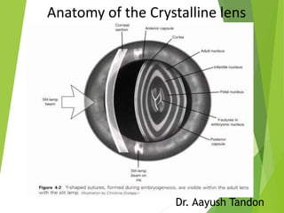
25. Initial fibres forming the foetal nucleus terminate with two Y-shaped
sutures, anteriorly upright Y and posteriorly inverted Y.
26. In adults continuously forming fibres are arranged compactly as nucleus
and cortex.
With further growh there is symmetrical branching of sutures forming
the 9 point star of the mature cortex
27. Nucleus :
Central part containing the oldest fibres
Parts of nucleus-
1. Embryonic nucleus (1-3 months of
Gestation)
1. Fetal nucleus (3 months – birth)
2. Infantile nucleus (Birth- puberty)
3. Adult nucleus.
28. Cortex
Peripheral part
It has the youngest fibres.
Histologically ,section through the equator shows the hexagonal structure
of lens fibres and bound together by ground substance.
30. Grading of nucleus hardness
Grade 1 – Green yellow
Grade 2 – Yellow
Grade 3 – Amber
Grade 4 –Brown
Grade 5 – Black
31. Ciliary Zonules
The ciliary zonules ( Zonules of Zinn or Suspensory ligaments )
consist of a series of stiff, non elasitic fibres which run from ciliary
body and fuse into the outer later of the lens capsule
The ciliary epithelial cells of the eye probably synthesize portions of
the zonules
The zonules are primarily made of fibrillin, a connective tissue
protein
Each zonular fibre has a diameter of about 0.35 – 1 microns
It is composed of microfibrils, glycoproteins and
mucopolysaccharides
They hold the lens in postion and enable the ciliary muscles to act on
it
32. Gross appearance
Grossly, the ciliary zonules from a complete ring of fibres, which extend
from ciliary body to the lens equator circumferentially.
On cut section the ciliary zonules appear to be arranged in a triangular
form.
33. The base of the triangle is towards the equator of the lens and apex towards
the ciliary body .
As the zonular fibres insert on the lens capsule a narrow space is created
around the equator which is called the ‘The canal of Hanover
34.
35. Structurally , 3 different types of fibres-
1) First type fibres- thick,wavy and lie near
vitreous.
2) second type fibres-thin and flat.
3) third type fibres- very fine & run circular
course.
39. Arrangement of zonular fibres (New
concept)
> Majority of zonules arise from the posterior end of the pars plana upto 1.5 mm from the ora serrata
> They run a continues course from ora serrata to the edge of lens
40. Applied Anatomy
1. Capsule
2. Superficial cortex :
a) C1α- subcapsular clear zone
b) C1β- first zone of disjunction seen as bright,
narrow, scattering zone of discontinuity
c) C2 – second cortical clear zone.
3. Deep cortex :
a) C3 – bright light scattering zone
b) C4- clear zone of cortex.
4. Nucleus.
41. In a can opener capsulotomy, improper joining of the radial cuts can lead
to a pull on the still attached capsule, as it is removed causing a posterior
extension of the tear
Extension of the tear to the periphery may occur if the attempted rhexis
size is larger than 6mm.
If the CCC created is larger than the proposed optic size ,the odds of
posterior capsular opacification are higher
Creation of small rhexis (less than 4mm) carries a risk of fluid entrapment
while performing hydrodissection leading to ruptureof the posterior
capsule .
Nucleus rotation in case of inadequate hydroprocedures can lead to
zonular dialysis
Editor's Notes
This causes any new lens fibres to be added on the inside of the lens
This shows that the seceretory source of the basement membrane ( Anterior Epithelium) is itself situated anteriorly and is involved in remodelling of the lens capsule which occurs with lens growth .
Albumin Mr 70kDA
HB Mr 66,7 kDA
There is no posterior epithelium as the cells are used up in filling the central cavity of the lens vesicle
Central density of about 5009/mm2 in men and 5781/mm2 in women .
Central Zone – Metplasia of these central zone epithelial cells can lead to Anterior subcapsular cataract eg. Shield cataract in Atopic dermatitis & glaukomflekon after acute attack of Acute angle closure glaucoma
Dysplasia of these cells of the germinative zone can lead to PSC as seen in radiation cataract, myotonic dystrophy and neurofibromatosis type 2
At first the lens fibres are forms from posterior epithelium but later on lens fibres are drived from the cells of the equatorial region of anterior epithelium
Size of embryonic and fetal nucleus remain constant whie adult nucleus is always increasing
This stratification is due to the optical difference between the older more sclerotic regions of central lens and newer transparent peripheral areas
Important for setting the parameters of the machine for effective phacoemulsification
Equatorial fibres are present in large numbers in young eyes but tend to become less numerous as age advances
Orbiculo-posterior capsular fibres
- most posterior and innermost fibres
- origin- ora serrata
- insertion- on posterior capsule of lens alonwith Weigerts ligament.
- second type fibres
Orbiculo-anterior capsular fibres
-thickest and strongest
- arise from pars plana
- inserts anterior to the equator of lens
- first type fibres
Cilio-posterior capsular fibres-
-most numerous zonular fibres
- arise mainly from the valleys
- inserts on posterior capsule
Cilio-equatorial capsular fibres-
-- arise from valleys of ciliary process
- directly inserts on the equator of lens
- third type of fibres
- abundent in youthful eyes andreduces in number with advancing age
Pars orbicularis –
Arise from post end of pars plana ,pass forward over the plans plana
Zonular Plexus –
Reaching the post margin of of pars plicata the zonular fibres segment into zonular plexus which pass throught valleys between the ciliary process . Known as tension fibres
Zonular fork –
After reaching anterior margin of pars plicata fibres consolidate into budles which bend at right angle
Zonular limbs
Ant – analogous to orbicularis anterior fibres
Equatorial – analogous to cilio equatorial fibres
Posterior – Analogous to orbiculo posterior fibres
Zonular dialysis likely to happen in presence tof capsule cortical adhesions