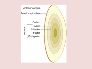
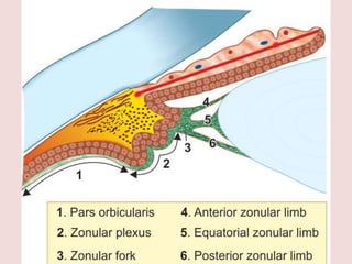
Lens
By Priyanka ( MBBS Student )
• The lens is a transparent, biconvex, crystalline
structure placed between iris and the vitreous in a
saucer shaped depression, the patellar fossa.
• Diameter is 9–10 mm.
• Thickness varies with age from 3.5 mm (at birth) to 5 mm (at extreme of
age).
• Weight varies from 135 mg (0–9 years) to 255 mg (40–80 years of age).
• Surfaces. Anterior surface is less convex (radius of curvature 10 mm) than the
posterior (radius of curvature 6 mm). These two surfaces meet at the
equator.
• Refractive index is 1.39.
• Total dioptric power is about 18 D (range 16–20D).
• Accommodative power of lens varies with age, being 14–16 D (at birth); 7–8
D (at 25 years of age) and 1–2 D (at 50 years of age).
1. Lens capsule. It is a thin, transparent, hyaline
membrane surrounding the lens which is thicker
over the anterior than the posterior surface. The lens
capsule is thickest at pre-equator regions (14 mu) and
thinnest at the posterior pole (3 mu).
2. Anterior epithelium. It is a single layer of cuboidal
cells which lies deep to the anterior capsule. In the
equatorial region these cells become columnar, are actively dividing
and elongating to form new
lens fibres throughout the life.
There is no posterior
epithelium, as these cells are used up in filling the
central cavity of lens vesicle during development of
the lens
3. Lens fibres. The epithelial cells elongate to form
lens fibres which have a complicated structural form.
Mature lens fibres are cells which have lost their
nuclei. As the lens fibres are formed throughout the
life, these are arranged compactly as nucleus and
cortex of the lens
i. Nucleus. It is the central part containing the oldest
fibres. It consists of different zones, which are laid
down successively as the development proceeds.
In the beam of slit-lamp these are seen as zones
of discontinuity.
• Embryonic nucleus. It is the innermost part of
nucleus which corresponds to the lens upto first
3 months of gestation. It consists of primary lens
fibres which are formed by elongation of the cells
of posterior wall of lens vesicle,
Fetal nucleus. It lies around the embryonic
nucleus and corresponds to the lens from 3
months of gestation till birth. Its fibres meet
around sutures which are anteriorly Y-shaped
and posteriorly inverted Y-shaped
• Infantile nucleus corresponds to the lens from birth
to puberty,
• Adult nucleus corresponds to the lens fibres
formed after puberty to rest of the life.
Cortex. It is the peripheral part which comprises
the youngest lens fibres.
Suspensory ligaments of lens
Suspensory ligaments of lens (Zonules of Zinn),
also called as ciliary zonules, consist essentially of a
series of fibres passing from ciliary body to the lens.
These hold the lens in position and enable the ciliary
muscle to act on it.
1. Pars orbicularis refers to the feltwork of zonular
fibres which line pars plana part of ciliary body.
2. Zonular plexuses refers to the intervening network
of zonular fibres which are attached between ciliary
processes in the region of pars plicata part of ciliary
body.
3. Zonular fork refers to the consolidated bundle of
zonular fibres which bend at right angle from the
anterior margin of pars plicata towards the lens after
dividing into three zonular limbs:
• Anterior zonular limb fibres are denser and insert
on the anterior lens capsule about 1.5 mm from
the equator.
• Equatorial zonular limb fibres are sparse and fan
out in a brush-like manner to get inserted into the
lens capsule of equatorial region.
• Posterior zonular limb fibres are inserted on the
posterior lens capsule about 1.25 mm from the
equator.
Lens transparency
Factors that play significant role in maintaining outstanding clarity and
transparency of the crystalline lens are:
• Avascularity of the lens.
• Characteristic of lens fibre:
–– Tightly-packed nature of lens cells,
–– Narrow lens fibre membranes,
–– Loss of organelles
• Role of lens proteins. Major intrinsic proteins of the
lens belong to superfamily of aquaporines and is
classified as aquaporine MIP26 or aquaporin 0.
• Lens capsule. Semipermeable character.
• Pump mechanism of lens fibre membranes that
regulate the electrolyte and water balance in the
lens, maintaining relative dehydration
• Auto-oxidation and high concentration of reduced
glutathione in the lens maintains the lens proteins
in a reduced state and ensures the integrity of the
cell membrane pump.
Lens requires a continuous supply of energy (ATP) for
active transport of ions and aminoacids,
maintenance of lens dehydration, and for a
continuous protein and GSH synthesis.
Source of nutrient supply.
The crystalline lens, being an avascular structure is
dependent for its metabolism on chemical exchanges
with the aqueous humour.
Some nutrition is also derived from the vitreous
humour, but it is much less.
• Glycolytic pathway. In the lens, 80% glucose is
metabolised anaerobically by this pathway.
• Pentose, Hexose, Monophosphate (HMP) shunt is
responsible for metabolism of 15% of glucose.
• Kreb’s citric acid cycle metabolises only a small
proportion by the oxidative process.
Preventive antioxidant mechanisms of lens are:
• Enzymatic defence mechanism, which includes
reduced glutathione (most important), glutathione
peroxide, superoxide dismutase and catalase.
• Non-enzymatic defence mechanism includes
vitamin C, vitamin E and possibly carotenoids.
Thank You!
Acknowledgement – A K Khurana
Comprehensive Ophthalomology

Lens ( Ophthalmology- Quick Revision )

  • 1.
    Lens By Priyanka (MBBS Student )
  • 2.
    • The lensis a transparent, biconvex, crystalline structure placed between iris and the vitreous in a saucer shaped depression, the patellar fossa.
  • 3.
    • Diameter is9–10 mm. • Thickness varies with age from 3.5 mm (at birth) to 5 mm (at extreme of age). • Weight varies from 135 mg (0–9 years) to 255 mg (40–80 years of age). • Surfaces. Anterior surface is less convex (radius of curvature 10 mm) than the posterior (radius of curvature 6 mm). These two surfaces meet at the equator. • Refractive index is 1.39. • Total dioptric power is about 18 D (range 16–20D). • Accommodative power of lens varies with age, being 14–16 D (at birth); 7–8 D (at 25 years of age) and 1–2 D (at 50 years of age).
  • 5.
    1. Lens capsule.It is a thin, transparent, hyaline membrane surrounding the lens which is thicker over the anterior than the posterior surface. The lens capsule is thickest at pre-equator regions (14 mu) and thinnest at the posterior pole (3 mu).
  • 6.
    2. Anterior epithelium.It is a single layer of cuboidal cells which lies deep to the anterior capsule. In the equatorial region these cells become columnar, are actively dividing and elongating to form new lens fibres throughout the life. There is no posterior epithelium, as these cells are used up in filling the central cavity of lens vesicle during development of the lens
  • 7.
    3. Lens fibres.The epithelial cells elongate to form lens fibres which have a complicated structural form. Mature lens fibres are cells which have lost their nuclei. As the lens fibres are formed throughout the life, these are arranged compactly as nucleus and cortex of the lens
  • 8.
    i. Nucleus. Itis the central part containing the oldest fibres. It consists of different zones, which are laid down successively as the development proceeds. In the beam of slit-lamp these are seen as zones of discontinuity.
  • 9.
    • Embryonic nucleus.It is the innermost part of nucleus which corresponds to the lens upto first 3 months of gestation. It consists of primary lens fibres which are formed by elongation of the cells of posterior wall of lens vesicle,
  • 10.
    Fetal nucleus. Itlies around the embryonic nucleus and corresponds to the lens from 3 months of gestation till birth. Its fibres meet around sutures which are anteriorly Y-shaped and posteriorly inverted Y-shaped
  • 12.
    • Infantile nucleuscorresponds to the lens from birth to puberty,
  • 13.
    • Adult nucleuscorresponds to the lens fibres formed after puberty to rest of the life.
  • 14.
    Cortex. It isthe peripheral part which comprises the youngest lens fibres.
  • 15.
    Suspensory ligaments oflens Suspensory ligaments of lens (Zonules of Zinn), also called as ciliary zonules, consist essentially of a series of fibres passing from ciliary body to the lens. These hold the lens in position and enable the ciliary muscle to act on it.
  • 16.
    1. Pars orbicularisrefers to the feltwork of zonular fibres which line pars plana part of ciliary body. 2. Zonular plexuses refers to the intervening network of zonular fibres which are attached between ciliary processes in the region of pars plicata part of ciliary body.
  • 17.
    3. Zonular forkrefers to the consolidated bundle of zonular fibres which bend at right angle from the anterior margin of pars plicata towards the lens after dividing into three zonular limbs: • Anterior zonular limb fibres are denser and insert on the anterior lens capsule about 1.5 mm from the equator. • Equatorial zonular limb fibres are sparse and fan out in a brush-like manner to get inserted into the lens capsule of equatorial region. • Posterior zonular limb fibres are inserted on the posterior lens capsule about 1.25 mm from the equator.
  • 19.
    Lens transparency Factors thatplay significant role in maintaining outstanding clarity and transparency of the crystalline lens are: • Avascularity of the lens. • Characteristic of lens fibre: –– Tightly-packed nature of lens cells, –– Narrow lens fibre membranes, –– Loss of organelles • Role of lens proteins. Major intrinsic proteins of the lens belong to superfamily of aquaporines and is classified as aquaporine MIP26 or aquaporin 0. • Lens capsule. Semipermeable character.
  • 20.
    • Pump mechanismof lens fibre membranes that regulate the electrolyte and water balance in the lens, maintaining relative dehydration • Auto-oxidation and high concentration of reduced glutathione in the lens maintains the lens proteins in a reduced state and ensures the integrity of the cell membrane pump.
  • 21.
    Lens requires acontinuous supply of energy (ATP) for active transport of ions and aminoacids, maintenance of lens dehydration, and for a continuous protein and GSH synthesis.
  • 22.
    Source of nutrientsupply. The crystalline lens, being an avascular structure is dependent for its metabolism on chemical exchanges with the aqueous humour. Some nutrition is also derived from the vitreous humour, but it is much less.
  • 24.
    • Glycolytic pathway.In the lens, 80% glucose is metabolised anaerobically by this pathway. • Pentose, Hexose, Monophosphate (HMP) shunt is responsible for metabolism of 15% of glucose. • Kreb’s citric acid cycle metabolises only a small proportion by the oxidative process.
  • 25.
    Preventive antioxidant mechanismsof lens are: • Enzymatic defence mechanism, which includes reduced glutathione (most important), glutathione peroxide, superoxide dismutase and catalase. • Non-enzymatic defence mechanism includes vitamin C, vitamin E and possibly carotenoids.
  • 26.
    Thank You! Acknowledgement –A K Khurana Comprehensive Ophthalomology