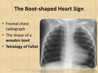
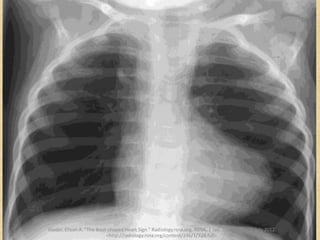
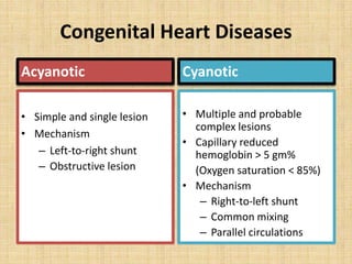
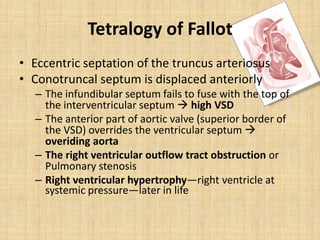
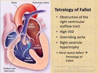
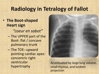
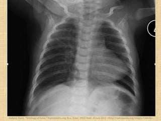
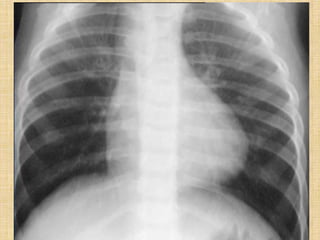
SlideShare a Scribd company logo
The Boot-shaped Heart Sign
Thorsang Chayovan
Outline
• Overview of the
boot-shaped heart
sign
• Tetralogy of Fallot
– Overview
– Radiology
– Prognosis and
Treatment
Haider, Ehsan A. "The Boot-shaped Heart Sign." Radiology.rsna.org. RSNA, 1 Jan. 2008. Web. 23 July 2012.
<http://radiology.rsna.org/content/246/1/328.full>.
The Boot-shaped Heart Sign
• Frontal chest
radiograph
• The shape of a
wooden boot
• Tetralogy of Fallot
Haider, Ehsan A. "The Boot-shaped Heart Sign." Radiology.rsna.org. RSNA, 1 Jan. 2008. Web. 23 July 2012.
<http://radiology.rsna.org/content/246/1/328.full>.
Haider, Ehsan A. "The Boot-shaped Heart Sign." Radiology.rsna.org. RSNA, 1 Jan. 2008. Web. 23 July 2012.
<http://radiology.rsna.org/content/246/1/328.full>.
Haider, Ehsan A. "The Boot-shaped Heart Sign." Radiology.rsna.org. RSNA, 1 Jan. 2008. Web. 23 July 2012.
<http://radiology.rsna.org/content/246/1/328.full>.
Tetralogy of Fallot
• One of the most common cyanotic congenital
heart conditions (75%)
• Accounts for 10% of all congenital heart
diseases
• Has estimated prevalence of 1 in 2,000 birth
Congenital Heart Diseases
Congenital
Heart Diseases
Acyanotic Cyanotic
Congenital Heart Diseases
Acyanotic
• Simple and single lesion
• Mechanism
– Left-to-right shunt
– Obstructive lesion
Cyanotic
• Multiple and probable
complex lesions
• Capillary reduced
hemoglobin > 5 gm%
(Oxygen saturation < 85%)
• Mechanism
– Right-to-left shunt
– Common mixing
– Parallel circulations
Tetralogy of Fallot
• Eccentric septation of the truncus arteriosus
• Conotruncal septum is displaced anteriorly
– The infundibular septum fails to fuse with the top of
the interventricular septum  high VSD
– The anterior part of aortic valve (superior border of
the VSD) overrides the ventricular septum 
overiding aorta
– The right ventricular outflow tract obstruction or
Pulmonary stenosis
– Right ventricular hypertrophy—right ventricle at
systemic pressure—later in life
Embryogenesis of the heart
Witmer, Lawrence M. "Developmental Anatomy of the Heart and the Embryological Basis for Cardiac Defects." Ohio University, Athens.23 July 2012. Lecture.
Tetralogy of Fallot
• Obstruction of the
right ventricular
outflow tract
• High VSD
• Overriding aorta
• Right ventricle
hypertrophy
+ Atrial septal defect 
Pentalogy of
Fallot
Cukukoglu, Sardar M. "Cyanotic Diseases." Escardio.org. N.p., 2010.
Web. 23 July 2012.
<http://www.escardio.org/congresses/euroecho2010/Documents/teach
ing-course-slides/euroecho2010-cyaotic-disease-Kucukoglu-591.pdf>.
Tetralogy of Fallot
• Cyanosis
– Severe cases ↔ at birth ↔ severe PS
– Mild cases ↔ much later ↔ mild PS
• Squatting
• Dyspnea
• Failure to thrive
Tetralogy of Fallot
• Degree of pulmonary stenosis determines the
clinical picture
– If mild, the clinical picture of VSD is shown instead
– However the muscular element of pulmonary
stenosis is nearly always progressive
– Eventually reversing the left to right shunt to be
permanently right to left before the first birthday
Radiology in Tetralogy of Fallot
• The Boot-shaped
Heart sign
“coeur en sabot”
– The UPPER part of the
Boot: flat / concave
pulmonary trunk
– The TOE--upward
pointing cardiac apex:
concentric right
ventricular
hypertrophy
Accentuated by large lung volume,
small thymus, and lordotic
projection
Radiology in Tetralogy of Fallot
• Heart size
– The heart is usually not enlarged at birth
Unless there is a large left-to-right shunt
– Could be enlarged in later life from biventricular
hypoxic failure
• Pulmonary vasculature—decreased
• Aortic arch
– Always enlarged
– 25% has right-sided aortic arch
• might be associated with absent left pulmonary artery
• resulting in less pulmonary vasculature in the left lung
Upturned cardiac apex sign
• Concentric right ventricular hypertrophy
• However not seen in severe pulmonic stenosis
with intact interventricular septum
(RV systolic pressure >>> systemic systolic pressure)
• Maybe related to right ventricular hypertrophy
with altered development of the left ventricle
and abnormal horizontal ventricular septum
• Only encountered in congenital heart lesions and
not in acquired right ventricular hypertrophy
Gallard, Frank. "Tetralogy of Fallot." Radiopaedia.org. N.p., 5 Jan. 2010. Web. 23 July 2012. <http://radiopaedia.org/images/156636>.
"Cyanotic Heart Diseases." Learningradiology.com. N.p., n.d. Web. 23 July 2012. <www.learningradiology.com/.../cyanoticheartdz.pdf>.
MRI
• Exquisite anatomical and functional information
• Detailed assessment of the pulmonary artery is
particularly important
– Repair of the cardiac defects without addressing
pulmonary artery hypoplasia / stenosis has a poor
outcome
• The main pulmonary artery or right pulmonary
artery/ascending aorta < 0.3
– Primary repair would be unsuccessful
– Bridging shunt operation may be of benefit
• Assessment of coronary artery origin
– essential to surgical planning
Prognosis
• Depends on timing
• Best outcome seen in patients repaired before
the age of 5
• 90% of untreated tetralogy patients succumb
by the age of 10 years
• Residual right ventricular dysfunction is
common
– Up to 10% of patients require re-operation within
20 years
Treatment
• Primary repair
– Preferred
–usually performed at the time of diagnosis
• Palliative shunt
– typically in pulmonary arterial hypoplasia
• Post-surgical complications
– conduction dysfunction
– valvular dysfunction
Reference
• Haider, Ehsan A. "The Boot-shaped Heart Sign." Radiology.rsna.org. RSNA,
1 Jan. 2008. Web. 23 July 2012.
<http://radiology.rsna.org/content/246/1/328.full>.
• Grainger, Ronald G. Grainger & Alison's Diagnostic Radiology. 4th ed. Vol.
1. N.p.: Churchill Livingstone, 2003. Print.
• Cukukoglu, Sardar M. "Cyanotic Diseases." Escardio.org. N.p., 2010. Web.
23 July 2012.
<http://www.escardio.org/congresses/euroecho2010/Documents/teachin
g-course-slides/euroecho2010-cyaotic-disease-Kucukoglu-591.pdf>.
• Gallard, Frank. "Tetralogy of Fallot." Radiopaedia.org. N.p., 5 Jan. 2010.
Web. 23 July 2012. <http://radiopaedia.org/images/156636>.
• "Cyanotic Heart Diseases." Learningradiology.com. N.p., n.d. Web. 23 July
2012. <www.learningradiology.com/.../cyanoticheartdz.pdf>.
• Witmer, Lawrence M. "Developmental Anatomy of the Heart and the
Embryological Basis for Cardiac Defects." Ohio University, Athens. 23 July
2012. Lecture.

More Related Content

What's hot

Patent Ductus Arteriosus (PDA)
Patent Ductus Arteriosus (PDA)Patent Ductus Arteriosus (PDA)
Patent Ductus Arteriosus (PDA)
Dr.Sayeedur Rumi
 
cyanotic and acyanotic Congenital heart disease for undergraduated student uo...
cyanotic and acyanotic Congenital heart disease for undergraduated student uo...cyanotic and acyanotic Congenital heart disease for undergraduated student uo...
cyanotic and acyanotic Congenital heart disease for undergraduated student uo...
Azad Haleem
 
EBSTEIN ANOMALY
EBSTEIN ANOMALYEBSTEIN ANOMALY
Diaphragmatic hernia
Diaphragmatic herniaDiaphragmatic hernia
Diaphragmatic hernia
sudarshan731
 
Chest X-rays for Undergraduates
Chest X-rays for UndergraduatesChest X-rays for Undergraduates
Chest X-rays for Undergraduates
Abdullah Ansari
 
Congenital heart disease and vascular abnormality(x-ray findings)
Congenital heart disease and vascular abnormality(x-ray findings)Congenital heart disease and vascular abnormality(x-ray findings)
Congenital heart disease and vascular abnormality(x-ray findings)
z2jeetendra
 
Ductus dependent circulation
Ductus dependent circulationDuctus dependent circulation
Ductus dependent circulation
Raghu Kishore Galla
 
Omphalocele vs gastroschisis
Omphalocele vs gastroschisisOmphalocele vs gastroschisis
Omphalocele vs gastroschisis
Rusila Divere
 
Acute Decompensated Heart Failure
Acute Decompensated Heart FailureAcute Decompensated Heart Failure
Acute Decompensated Heart Failure
SCGH ED CME
 
basics of chest X- ray interpretation
basics of chest X- ray interpretationbasics of chest X- ray interpretation
basics of chest X- ray interpretation
Maha Yousif
 
Collapse and consolidation Lung Radiology
Collapse and consolidation Lung RadiologyCollapse and consolidation Lung Radiology
Collapse and consolidation Lung RadiologyNeelam Ashar
 
Surgical management of tetralogy of fallot
Surgical management of tetralogy of fallotSurgical management of tetralogy of fallot
Surgical management of tetralogy of fallot
rahul arora
 
Approach to a patient with Haemoptysis
Approach to a patient with HaemoptysisApproach to a patient with Haemoptysis
Approach to a patient with Haemoptysis
Khairul Jessy
 
Abnormal Chest xray
Abnormal Chest xray Abnormal Chest xray
Abnormal Chest xray
Roshan Valentine
 
Chest xray
Chest xray  Chest xray
Chest xray
Abino David
 
Newborn chest reid
Newborn chest reidNewborn chest reid
Newborn chest reid
CHOP
 
Abdominal wall defects
Abdominal wall defectsAbdominal wall defects
Abdominal wall defects
Tarek Kotb
 
Eisenmenger syndrome
Eisenmenger syndromeEisenmenger syndrome
Eisenmenger syndrome
Feba
 

What's hot (20)

Patent Ductus Arteriosus (PDA)
Patent Ductus Arteriosus (PDA)Patent Ductus Arteriosus (PDA)
Patent Ductus Arteriosus (PDA)
 
cyanotic and acyanotic Congenital heart disease for undergraduated student uo...
cyanotic and acyanotic Congenital heart disease for undergraduated student uo...cyanotic and acyanotic Congenital heart disease for undergraduated student uo...
cyanotic and acyanotic Congenital heart disease for undergraduated student uo...
 
EBSTEIN ANOMALY
EBSTEIN ANOMALYEBSTEIN ANOMALY
EBSTEIN ANOMALY
 
Atrial Fibrillation
Atrial FibrillationAtrial Fibrillation
Atrial Fibrillation
 
Cyanotic spell.
Cyanotic spell.Cyanotic spell.
Cyanotic spell.
 
Diaphragmatic hernia
Diaphragmatic herniaDiaphragmatic hernia
Diaphragmatic hernia
 
Chest X-rays for Undergraduates
Chest X-rays for UndergraduatesChest X-rays for Undergraduates
Chest X-rays for Undergraduates
 
Congenital heart disease and vascular abnormality(x-ray findings)
Congenital heart disease and vascular abnormality(x-ray findings)Congenital heart disease and vascular abnormality(x-ray findings)
Congenital heart disease and vascular abnormality(x-ray findings)
 
Ductus dependent circulation
Ductus dependent circulationDuctus dependent circulation
Ductus dependent circulation
 
Omphalocele vs gastroschisis
Omphalocele vs gastroschisisOmphalocele vs gastroschisis
Omphalocele vs gastroschisis
 
Acute Decompensated Heart Failure
Acute Decompensated Heart FailureAcute Decompensated Heart Failure
Acute Decompensated Heart Failure
 
basics of chest X- ray interpretation
basics of chest X- ray interpretationbasics of chest X- ray interpretation
basics of chest X- ray interpretation
 
Collapse and consolidation Lung Radiology
Collapse and consolidation Lung RadiologyCollapse and consolidation Lung Radiology
Collapse and consolidation Lung Radiology
 
Surgical management of tetralogy of fallot
Surgical management of tetralogy of fallotSurgical management of tetralogy of fallot
Surgical management of tetralogy of fallot
 
Approach to a patient with Haemoptysis
Approach to a patient with HaemoptysisApproach to a patient with Haemoptysis
Approach to a patient with Haemoptysis
 
Abnormal Chest xray
Abnormal Chest xray Abnormal Chest xray
Abnormal Chest xray
 
Chest xray
Chest xray  Chest xray
Chest xray
 
Newborn chest reid
Newborn chest reidNewborn chest reid
Newborn chest reid
 
Abdominal wall defects
Abdominal wall defectsAbdominal wall defects
Abdominal wall defects
 
Eisenmenger syndrome
Eisenmenger syndromeEisenmenger syndrome
Eisenmenger syndrome
 

Similar to The boot shaped heart sign

Stroke & Balint's syndrome
Stroke & Balint's syndromeStroke & Balint's syndrome
Stroke & Balint's syndromemeducationdotnet
 
Congenital heart disease for undergraduates student uod 2015
Congenital heart disease for undergraduates student uod 2015Congenital heart disease for undergraduates student uod 2015
Congenital heart disease for undergraduates student uod 2015
Azad Haleem
 
Management of congenital heart disease in infants
Management of congenital heart disease in infantsManagement of congenital heart disease in infants
Management of congenital heart disease in infants
SMSRAZA
 
Approach to patient with congenital heart disease
Approach to patient with congenital heart diseaseApproach to patient with congenital heart disease
Approach to patient with congenital heart disease
Annamaneni Vamshi
 
4 Congenital Heart Disease
4 Congenital Heart Disease4 Congenital Heart Disease
4 Congenital Heart Diseaseghalan
 
4 Congenital Heart Disease
4 Congenital Heart Disease4 Congenital Heart Disease
4 Congenital Heart Diseaseghalan
 
Acyanotic heart defects
Acyanotic heart defectsAcyanotic heart defects
Acyanotic heart defects
SatthyNAGAPrathyusha
 
Transposition of the great arteries
Transposition of the great arteriesTransposition of the great arteries
Transposition of the great arteries
jagan _jaggi
 
Cardiac Murmers -2014 - Jacob
Cardiac Murmers -2014 - JacobCardiac Murmers -2014 - Jacob
Cardiac Murmers -2014 - Jacob
LejayJacob
 
An approach to a patient with Atrial septal defect (ASD)
An approach to a patient with Atrial  septal defect (ASD)An approach to a patient with Atrial  septal defect (ASD)
An approach to a patient with Atrial septal defect (ASD)
PROFESSOR DR. MD. TOUFIQUR RAHMAN
 
Congenital heart disease
Congenital heart disease Congenital heart disease
Congenital heart disease
manaswi debbarma
 
CAROTID STENOSIS .pptx
CAROTID STENOSIS                   .pptxCAROTID STENOSIS                   .pptx
CAROTID STENOSIS .pptx
Juma675663
 
Pediatric_Cardiac_Disorders.ppt
Pediatric_Cardiac_Disorders.pptPediatric_Cardiac_Disorders.ppt
Pediatric_Cardiac_Disorders.ppt
Salam467227
 
X-RAY OF THE HEART.pptx
X-RAY OF THE HEART.pptxX-RAY OF THE HEART.pptx
X-RAY OF THE HEART.pptx
NovitaDewi28
 
heart newborn.pptx
heart newborn.pptxheart newborn.pptx
heart newborn.pptx
ShambelNegese
 
Acynotic congenital heart disease 2021
Acynotic congenital heart disease 2021Acynotic congenital heart disease 2021
Acynotic congenital heart disease 2021
Imran Iqbal
 
Congenital heart diseases
Congenital heart diseasesCongenital heart diseases
Congenital heart diseases
MWIZERWA JEAN-LUC
 
04 2 25 Cardiac新2009
04 2 25 Cardiac新200904 2 25 Cardiac新2009
04 2 25 Cardiac新2009Deep Deep
 
seminar PDA
seminar PDAseminar PDA
seminar PDA
Dr. Habibur Rahim
 
Congenital heart disease
Congenital heart diseaseCongenital heart disease
Congenital heart disease
zabron charles saguda
 

Similar to The boot shaped heart sign (20)

Stroke & Balint's syndrome
Stroke & Balint's syndromeStroke & Balint's syndrome
Stroke & Balint's syndrome
 
Congenital heart disease for undergraduates student uod 2015
Congenital heart disease for undergraduates student uod 2015Congenital heart disease for undergraduates student uod 2015
Congenital heart disease for undergraduates student uod 2015
 
Management of congenital heart disease in infants
Management of congenital heart disease in infantsManagement of congenital heart disease in infants
Management of congenital heart disease in infants
 
Approach to patient with congenital heart disease
Approach to patient with congenital heart diseaseApproach to patient with congenital heart disease
Approach to patient with congenital heart disease
 
4 Congenital Heart Disease
4 Congenital Heart Disease4 Congenital Heart Disease
4 Congenital Heart Disease
 
4 Congenital Heart Disease
4 Congenital Heart Disease4 Congenital Heart Disease
4 Congenital Heart Disease
 
Acyanotic heart defects
Acyanotic heart defectsAcyanotic heart defects
Acyanotic heart defects
 
Transposition of the great arteries
Transposition of the great arteriesTransposition of the great arteries
Transposition of the great arteries
 
Cardiac Murmers -2014 - Jacob
Cardiac Murmers -2014 - JacobCardiac Murmers -2014 - Jacob
Cardiac Murmers -2014 - Jacob
 
An approach to a patient with Atrial septal defect (ASD)
An approach to a patient with Atrial  septal defect (ASD)An approach to a patient with Atrial  septal defect (ASD)
An approach to a patient with Atrial septal defect (ASD)
 
Congenital heart disease
Congenital heart disease Congenital heart disease
Congenital heart disease
 
CAROTID STENOSIS .pptx
CAROTID STENOSIS                   .pptxCAROTID STENOSIS                   .pptx
CAROTID STENOSIS .pptx
 
Pediatric_Cardiac_Disorders.ppt
Pediatric_Cardiac_Disorders.pptPediatric_Cardiac_Disorders.ppt
Pediatric_Cardiac_Disorders.ppt
 
X-RAY OF THE HEART.pptx
X-RAY OF THE HEART.pptxX-RAY OF THE HEART.pptx
X-RAY OF THE HEART.pptx
 
heart newborn.pptx
heart newborn.pptxheart newborn.pptx
heart newborn.pptx
 
Acynotic congenital heart disease 2021
Acynotic congenital heart disease 2021Acynotic congenital heart disease 2021
Acynotic congenital heart disease 2021
 
Congenital heart diseases
Congenital heart diseasesCongenital heart diseases
Congenital heart diseases
 
04 2 25 Cardiac新2009
04 2 25 Cardiac新200904 2 25 Cardiac新2009
04 2 25 Cardiac新2009
 
seminar PDA
seminar PDAseminar PDA
seminar PDA
 
Congenital heart disease
Congenital heart diseaseCongenital heart disease
Congenital heart disease
 

More from Thorsang Chayovan

COVID-19 Findings on Chest CT
COVID-19 Findings on Chest CTCOVID-19 Findings on Chest CT
COVID-19 Findings on Chest CT
Thorsang Chayovan
 
Dyspepsia endoscopy guideline
Dyspepsia endoscopy guidelineDyspepsia endoscopy guideline
Dyspepsia endoscopy guideline
Thorsang Chayovan
 
Common respiratory problems
Common respiratory problemsCommon respiratory problems
Common respiratory problems
Thorsang Chayovan
 
Avascular necrosis
Avascular necrosisAvascular necrosis
Avascular necrosis
Thorsang Chayovan
 
Shoulder injury
Shoulder injuryShoulder injury
Shoulder injury
Thorsang Chayovan
 
Pediatric pneumonia Thai
Pediatric pneumonia ThaiPediatric pneumonia Thai
Pediatric pneumonia Thai
Thorsang Chayovan
 
Pediatric upper urinary tract infection in Thai
Pediatric upper urinary tract infection in ThaiPediatric upper urinary tract infection in Thai
Pediatric upper urinary tract infection in Thai
Thorsang Chayovan
 
Tokyo guidelines for cholangitis and cholecystitis
Tokyo guidelines for cholangitis and cholecystitis Tokyo guidelines for cholangitis and cholecystitis
Tokyo guidelines for cholangitis and cholecystitis
Thorsang Chayovan
 
The role of ercp in diseases of the biliary tract and pancreas
The role of ercp in diseases of the biliary tract and pancreasThe role of ercp in diseases of the biliary tract and pancreas
The role of ercp in diseases of the biliary tract and pancreas
Thorsang Chayovan
 
Role of endoscopy in dyspepsia
Role of endoscopy in dyspepsiaRole of endoscopy in dyspepsia
Role of endoscopy in dyspepsia
Thorsang Chayovan
 
Role of endoscopy in choledocholithiasis
Role of endoscopy in choledocholithiasis Role of endoscopy in choledocholithiasis
Role of endoscopy in choledocholithiasis
Thorsang Chayovan
 
Pneumonia in children
Pneumonia in childrenPneumonia in children
Pneumonia in children
Thorsang Chayovan
 
คู่มือเวชปฏิบัติหัตถการ
คู่มือเวชปฏิบัติหัตถการคู่มือเวชปฏิบัติหัตถการ
คู่มือเวชปฏิบัติหัตถการ
Thorsang Chayovan
 
Febrile neutropenia in children
Febrile neutropenia in childrenFebrile neutropenia in children
Febrile neutropenia in children
Thorsang Chayovan
 
Exanthematous fever in children
Exanthematous fever in childrenExanthematous fever in children
Exanthematous fever in children
Thorsang Chayovan
 
Diseases of the rectum and anal canal in Thai
Diseases of the rectum and anal canal in ThaiDiseases of the rectum and anal canal in Thai
Diseases of the rectum and anal canal in Thai
Thorsang Chayovan
 
Thai PALS manual 2009
Thai PALS manual 2009Thai PALS manual 2009
Thai PALS manual 2009
Thorsang Chayovan
 
NEJM Cholecystitis
NEJM CholecystitisNEJM Cholecystitis
NEJM Cholecystitis
Thorsang Chayovan
 
Thai hemorrhagic stroke guideline 2008
Thai hemorrhagic stroke guideline 2008Thai hemorrhagic stroke guideline 2008
Thai hemorrhagic stroke guideline 2008
Thorsang Chayovan
 

More from Thorsang Chayovan (20)

COVID-19 Findings on Chest CT
COVID-19 Findings on Chest CTCOVID-19 Findings on Chest CT
COVID-19 Findings on Chest CT
 
Dyspepsia endoscopy guideline
Dyspepsia endoscopy guidelineDyspepsia endoscopy guideline
Dyspepsia endoscopy guideline
 
Common respiratory problems
Common respiratory problemsCommon respiratory problems
Common respiratory problems
 
Avascular necrosis
Avascular necrosisAvascular necrosis
Avascular necrosis
 
Shoulder injury
Shoulder injuryShoulder injury
Shoulder injury
 
Pediatric pneumonia Thai
Pediatric pneumonia ThaiPediatric pneumonia Thai
Pediatric pneumonia Thai
 
Pediatric upper urinary tract infection in Thai
Pediatric upper urinary tract infection in ThaiPediatric upper urinary tract infection in Thai
Pediatric upper urinary tract infection in Thai
 
Tokyo guidelines for cholangitis and cholecystitis
Tokyo guidelines for cholangitis and cholecystitis Tokyo guidelines for cholangitis and cholecystitis
Tokyo guidelines for cholangitis and cholecystitis
 
The role of ercp in diseases of the biliary tract and pancreas
The role of ercp in diseases of the biliary tract and pancreasThe role of ercp in diseases of the biliary tract and pancreas
The role of ercp in diseases of the biliary tract and pancreas
 
Role of endoscopy in dyspepsia
Role of endoscopy in dyspepsiaRole of endoscopy in dyspepsia
Role of endoscopy in dyspepsia
 
Role of endoscopy in choledocholithiasis
Role of endoscopy in choledocholithiasis Role of endoscopy in choledocholithiasis
Role of endoscopy in choledocholithiasis
 
Pneumonia in children
Pneumonia in childrenPneumonia in children
Pneumonia in children
 
คู่มือเวชปฏิบัติหัตถการ
คู่มือเวชปฏิบัติหัตถการคู่มือเวชปฏิบัติหัตถการ
คู่มือเวชปฏิบัติหัตถการ
 
Febrile neutropenia in children
Febrile neutropenia in childrenFebrile neutropenia in children
Febrile neutropenia in children
 
Exanthematous fever in children
Exanthematous fever in childrenExanthematous fever in children
Exanthematous fever in children
 
Diseases of the rectum and anal canal in Thai
Diseases of the rectum and anal canal in ThaiDiseases of the rectum and anal canal in Thai
Diseases of the rectum and anal canal in Thai
 
Thai PALS manual 2009
Thai PALS manual 2009Thai PALS manual 2009
Thai PALS manual 2009
 
NEJM Cholecystitis
NEJM CholecystitisNEJM Cholecystitis
NEJM Cholecystitis
 
Thai hemorrhagic stroke guideline 2008
Thai hemorrhagic stroke guideline 2008Thai hemorrhagic stroke guideline 2008
Thai hemorrhagic stroke guideline 2008
 
ACLS Thai
ACLS ThaiACLS Thai
ACLS Thai
 

The boot shaped heart sign

  • 1. The Boot-shaped Heart Sign Thorsang Chayovan
  • 2. Outline • Overview of the boot-shaped heart sign • Tetralogy of Fallot – Overview – Radiology – Prognosis and Treatment Haider, Ehsan A. "The Boot-shaped Heart Sign." Radiology.rsna.org. RSNA, 1 Jan. 2008. Web. 23 July 2012. <http://radiology.rsna.org/content/246/1/328.full>.
  • 3. The Boot-shaped Heart Sign • Frontal chest radiograph • The shape of a wooden boot • Tetralogy of Fallot Haider, Ehsan A. "The Boot-shaped Heart Sign." Radiology.rsna.org. RSNA, 1 Jan. 2008. Web. 23 July 2012. <http://radiology.rsna.org/content/246/1/328.full>.
  • 4. Haider, Ehsan A. "The Boot-shaped Heart Sign." Radiology.rsna.org. RSNA, 1 Jan. 2008. Web. 23 July 2012. <http://radiology.rsna.org/content/246/1/328.full>. Haider, Ehsan A. "The Boot-shaped Heart Sign." Radiology.rsna.org. RSNA, 1 Jan. 2008. Web. 23 July 2012. <http://radiology.rsna.org/content/246/1/328.full>.
  • 5. Tetralogy of Fallot • One of the most common cyanotic congenital heart conditions (75%) • Accounts for 10% of all congenital heart diseases • Has estimated prevalence of 1 in 2,000 birth
  • 6. Congenital Heart Diseases Congenital Heart Diseases Acyanotic Cyanotic
  • 7. Congenital Heart Diseases Acyanotic • Simple and single lesion • Mechanism – Left-to-right shunt – Obstructive lesion Cyanotic • Multiple and probable complex lesions • Capillary reduced hemoglobin > 5 gm% (Oxygen saturation < 85%) • Mechanism – Right-to-left shunt – Common mixing – Parallel circulations
  • 8. Tetralogy of Fallot • Eccentric septation of the truncus arteriosus • Conotruncal septum is displaced anteriorly – The infundibular septum fails to fuse with the top of the interventricular septum  high VSD – The anterior part of aortic valve (superior border of the VSD) overrides the ventricular septum  overiding aorta – The right ventricular outflow tract obstruction or Pulmonary stenosis – Right ventricular hypertrophy—right ventricle at systemic pressure—later in life
  • 9. Embryogenesis of the heart Witmer, Lawrence M. "Developmental Anatomy of the Heart and the Embryological Basis for Cardiac Defects." Ohio University, Athens.23 July 2012. Lecture.
  • 10. Tetralogy of Fallot • Obstruction of the right ventricular outflow tract • High VSD • Overriding aorta • Right ventricle hypertrophy + Atrial septal defect  Pentalogy of Fallot Cukukoglu, Sardar M. "Cyanotic Diseases." Escardio.org. N.p., 2010. Web. 23 July 2012. <http://www.escardio.org/congresses/euroecho2010/Documents/teach ing-course-slides/euroecho2010-cyaotic-disease-Kucukoglu-591.pdf>.
  • 11. Tetralogy of Fallot • Cyanosis – Severe cases ↔ at birth ↔ severe PS – Mild cases ↔ much later ↔ mild PS • Squatting • Dyspnea • Failure to thrive
  • 12. Tetralogy of Fallot • Degree of pulmonary stenosis determines the clinical picture – If mild, the clinical picture of VSD is shown instead – However the muscular element of pulmonary stenosis is nearly always progressive – Eventually reversing the left to right shunt to be permanently right to left before the first birthday
  • 13. Radiology in Tetralogy of Fallot • The Boot-shaped Heart sign “coeur en sabot” – The UPPER part of the Boot: flat / concave pulmonary trunk – The TOE--upward pointing cardiac apex: concentric right ventricular hypertrophy Accentuated by large lung volume, small thymus, and lordotic projection
  • 14. Radiology in Tetralogy of Fallot • Heart size – The heart is usually not enlarged at birth Unless there is a large left-to-right shunt – Could be enlarged in later life from biventricular hypoxic failure • Pulmonary vasculature—decreased • Aortic arch – Always enlarged – 25% has right-sided aortic arch • might be associated with absent left pulmonary artery • resulting in less pulmonary vasculature in the left lung
  • 15. Upturned cardiac apex sign • Concentric right ventricular hypertrophy • However not seen in severe pulmonic stenosis with intact interventricular septum (RV systolic pressure >>> systemic systolic pressure) • Maybe related to right ventricular hypertrophy with altered development of the left ventricle and abnormal horizontal ventricular septum • Only encountered in congenital heart lesions and not in acquired right ventricular hypertrophy
  • 16. Gallard, Frank. "Tetralogy of Fallot." Radiopaedia.org. N.p., 5 Jan. 2010. Web. 23 July 2012. <http://radiopaedia.org/images/156636>.
  • 17. "Cyanotic Heart Diseases." Learningradiology.com. N.p., n.d. Web. 23 July 2012. <www.learningradiology.com/.../cyanoticheartdz.pdf>.
  • 18. MRI • Exquisite anatomical and functional information • Detailed assessment of the pulmonary artery is particularly important – Repair of the cardiac defects without addressing pulmonary artery hypoplasia / stenosis has a poor outcome • The main pulmonary artery or right pulmonary artery/ascending aorta < 0.3 – Primary repair would be unsuccessful – Bridging shunt operation may be of benefit • Assessment of coronary artery origin – essential to surgical planning
  • 19. Prognosis • Depends on timing • Best outcome seen in patients repaired before the age of 5 • 90% of untreated tetralogy patients succumb by the age of 10 years • Residual right ventricular dysfunction is common – Up to 10% of patients require re-operation within 20 years
  • 20. Treatment • Primary repair – Preferred –usually performed at the time of diagnosis • Palliative shunt – typically in pulmonary arterial hypoplasia • Post-surgical complications – conduction dysfunction – valvular dysfunction
  • 21. Reference • Haider, Ehsan A. "The Boot-shaped Heart Sign." Radiology.rsna.org. RSNA, 1 Jan. 2008. Web. 23 July 2012. <http://radiology.rsna.org/content/246/1/328.full>. • Grainger, Ronald G. Grainger & Alison's Diagnostic Radiology. 4th ed. Vol. 1. N.p.: Churchill Livingstone, 2003. Print. • Cukukoglu, Sardar M. "Cyanotic Diseases." Escardio.org. N.p., 2010. Web. 23 July 2012. <http://www.escardio.org/congresses/euroecho2010/Documents/teachin g-course-slides/euroecho2010-cyaotic-disease-Kucukoglu-591.pdf>. • Gallard, Frank. "Tetralogy of Fallot." Radiopaedia.org. N.p., 5 Jan. 2010. Web. 23 July 2012. <http://radiopaedia.org/images/156636>. • "Cyanotic Heart Diseases." Learningradiology.com. N.p., n.d. Web. 23 July 2012. <www.learningradiology.com/.../cyanoticheartdz.pdf>. • Witmer, Lawrence M. "Developmental Anatomy of the Heart and the Embryological Basis for Cardiac Defects." Ohio University, Athens. 23 July 2012. Lecture.