PRESENTATION TOPIC: CYANOTIC
CONGENITALHEART DISEASE
• PREPARED BY:
• FATIMA ZAINAB KAMARA ID 22044
• ZAINAB KADIATU KAMARA ID 22045
• MODULE: PEDIATRICS
• LECTURER: DR AMADU JALLOH
2.
INTRODUCTION
• Cyanotic congenitalheart disease {CCHD}
encompasses a group of congenital heart defects that
lead to a low level of oxygen in the blood, resulting
in cyanosis{ a bluish tint in the skin, lips, and nails}.
This happens because these defects allow
deoxygenated {blue} blood to bypass the lugs,
mixing with oxygenated blood, or because blood has
difficulty reaching the lugs for oxygenation.
3.
TYPES OF CYANOTIC
CONGENITALHEART DISEASE
Tetralogy of Fallot {TOF}
Transposition of the Great Arteries {TGA}
Tricuspid Atresia
Total Anomalus Pulmonary Venous Connection
{TAPCV}
Truncus Arteriosus
4.
Tetralogy of Fallot
Tetralogyof fallot {TOF} is the most common type of
cyanotic congenital heart disease. This condition is
characterized by the combination of four defects:
Ventricular Septal defect
Pulmonary Stenosis
Dextroposition of the aorta so that it overrides the
ventricular septum
Right ventricular hypertrophy
5.
PATHOPHYSIOLOGY
• The initialdefect in TOF is a narrowing of the right ventricular
outflow tract into the pulmonary artery {Pulmonary Stenosis}.
• Prevents deoxygenated blood from entering the pulmonary circuit.
• In response to this outflow obstruction RV pressure is high as the
LV, a right to left shunt appear{VSD}.
• VSD allows shunting of the blood between ventricles.
6.
CONT
• The aortaoverrides the VSD
• Mixed blood immediately exist the heart
• Severity and clinical consequences of TOF is the
degree of right ventricular outflow obstruction.
7.
Clinical features
• Cyanosis,tet spell
• Dyspnea
• Clubbing of the fingers and toes
• Slow weight gain {Poor growth}
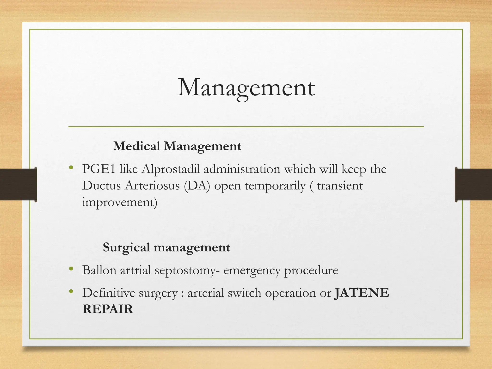
Management
• Treatment ofTOF depends on the severity of the
RVOT obstruction. Infants with severe tetralogy
require urgent medical treatment and surgical
intervention in the neonatal period. Therapy is aimed
at providing an immediate increase in pulmonary
blood flow to prevent the sequelae of severe hypoxia.
• Pott’s procedure: the upper descending aorta is
anastomosed with the left pulmonary artery.
11.
CONT
• Brock’s procedure:Pulmonary valvotomy done to
correct PS
The IV administration of prostaglandin E1 (PGE1 ;
0.01-0.20μg/kg/min)
TRICUSPID ATRESIA
Congenital absenceof the tricuspid valve is called tricuspid
atresia.
There is an absence of right atrioventricular connection.
The right ventricle is hypoplastic {undersized or absent}.
This condition is associated with PS and PD.
There is complete mixing of unoxygenated and oxygenated
blood in the left Atrium and so have a mandatory ASD.
A PDA or VSD is necessary for PBF and survival.
14.
• Pulmonary bloodflow (and thus the degree of cyanosis) depends on the size
of the VSD and the presence and severity of any associated pulmonic
stenosis.
• In patients with tricuspid atresia and transposition of the great
arteries (TGA) , LV blood flows directly into the pulmonary
artery, whereas systemic blood must traverse the VSD and
right ventricle to reach the aorta.
• In these patients, pulmonary blood flow is usually massively
increased and heart failure develops early. If the VSD is
restrictive, aortic blood flow may be compromised.
• Coarctation of the aorta is often noted in this setting.
Transposition of theGreat
Arteries
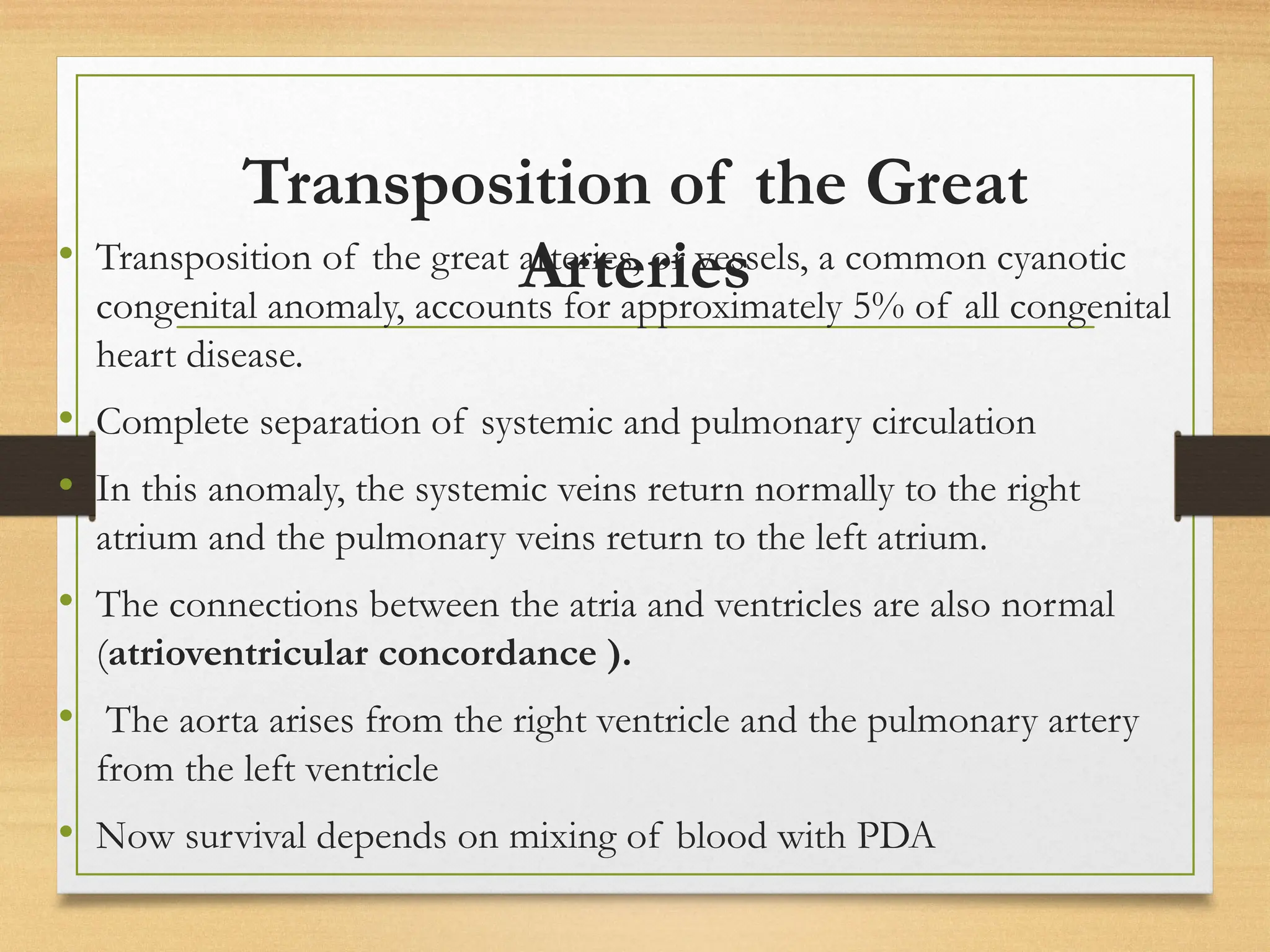
• Transposition of the great arteries, or vessels, a common cyanotic
congenital anomaly, accounts for approximately 5% of all congenital
heart disease.
• Complete separation of systemic and pulmonary circulation
• In this anomaly, the systemic veins return normally to the right
atrium and the pulmonary veins return to the left atrium.
• The connections between the atria and ventricles are also normal
(atrioventricular concordance ).
• The aorta arises from the right ventricle and the pulmonary artery
from the left ventricle
• Now survival depends on mixing of blood with PDA
20.
Clinical Manifestation
• Timeof presentation depends on weather ventricular septum is
intact or VSD is present.
• Patient with TGA with intact septum are cyanotic at birth with
CHF occuring in first week of life.
• Patients with TGA with VSD presents with mild cyanosis and
CHF occur in 6-8weeks ( or 4-10 weeks).
• Cyanosis
• Tachypnoea
• hypoxemia
Total anomalous pulmonary
venousconnection (TAPVC)
• Pulmonary veins instead of draining into left atrium,
drain into either
• Superior Vena Cava
• Right Atrium
• Inferior Vena Cava/ Portal vein/ Hepatic veins
26.
TYPES
• Supracardiac TAPVC
•Cardiac TAPVC
• Infracardiac TAPVC
• Supracardiac TAPVC is the most type
• Whereas Infracardiac is the most obstructed type.
Diagnosis
• History taking
•Clinical Examination
Cyanosis is always present even with supplemental
oxygen.
Examination may reveal signs of right heart overload,
such as a prominent right ventricular impulse and
possibly a widely spilt and fixed S2{second heart
sound}.
Complications
• Onset ofCCF occurs at 4-10 weeks of life
• Respiratory distress syndrome
• Pneumonia
• Hypoplastic left heart syndrome
32.
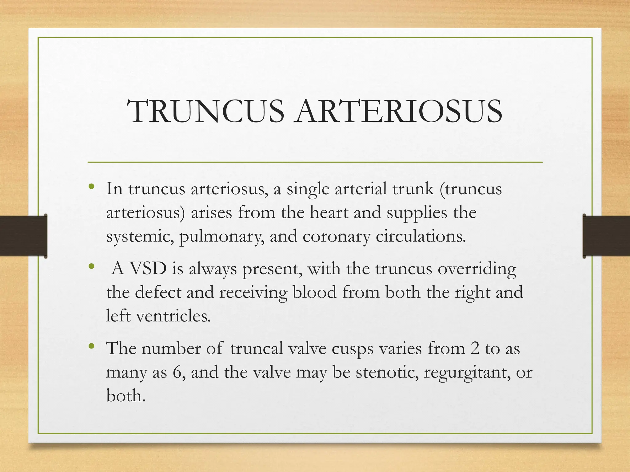
TRUNCUS ARTERIOSUS
• Intruncus arteriosus, a single arterial trunk (truncus
arteriosus) arises from the heart and supplies the
systemic, pulmonary, and coronary circulations.
• A VSD is always present, with the truncus overriding
the defect and receiving blood from both the right and
left ventricles.
• The number of truncal valve cusps varies from 2 to as
many as 6, and the valve may be stenotic, regurgitant, or
both.
33.
• The pulmonaryarteries can arise together from the posterior left
side of the persistent truncus arteriosus and then divide into left
and right pulmonary arteries (type I ).
• In types II and III truncus arteriosus, no main pulmonary
artery is present, and the right and left pulmonary arteries arise
from separate orifices on the posterior (type II ) or lateral (type
III ) aspects of the truncus arteriosus.
• Type IV truncus is a term no longer used because, in this case,
there is no identifiable connection between the heart and
pulmonary arteries, and pulmonary blood flow is derived from
major aortopulmonary collateral arteries arising from the transverse
or descending aorta; this is essentially a form of pulmonary atresia.
Treatment
• In thefirst few weeks of life, many of these infants can
be managed with anticongestive medications, as PVR
falls, heart failure symptoms worsen and surgery is
indicated, usually within the 1st few mo.
• Delay of surgery much beyond this time period may
increase the likelihood of pulmonary vascular disease;
many centers now perform routine neonatal repair at the
time of diagnosis.
• Transcatheter stent-valve.


![cyanotic congenital heart disease[1].pptx](https://cdn.slidesharecdn.com/ss_thumbnails/cyanoticcongenitalheartdisease1-250421184527-f500caa9-thumbnail.jpg?width=640&height=640&fit=bounds)


![ACUTE_AND_CHRONIC_RENAL_FAILxxxxx1].pptx](https://cdn.slidesharecdn.com/ss_thumbnails/acuteandchronicrenalfail1-250803044151-1ee4884c-thumbnail.jpg?width=640&height=640&fit=bounds)


