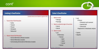
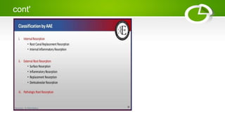
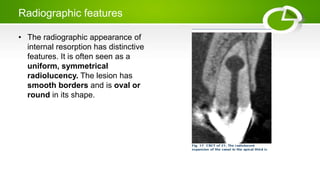
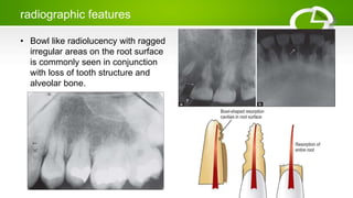
TOOTH RESORPTION
Makerere University
BDS IV
Outline
• Definition
• Classification of tooth resorption
• mechanism of resorption
• Internal resorption
• External resorption
• Differences between external and internal tooth resorption
• management of internal and external tooth resorption
Definition
• Resorption of human oral hard tissue is described as the physiologic or
pathologic dissolution of mineralized tissues in bone, dentin or cementum by
osteoclastic-type. (According to the American Association of Endodontics).
• This process may occur in a tooth internally or externally, so it is important for
a dentist to clearly differiate between the two so as to offer appropriate
treatment.
• The occurrence of resorption cannot be predicted, it can be identified
radiographically.
Classification of tooth resorption
The literature talks of many classifications but the major ones include the
following:
• Cohen's classification
• Classification by american association of endodontists
• Andreasen classification of tooth resorption
• Lindskog's classification
cont'
cont'
Mechanism of tooth resorption
Resorption of hard tissues takes place in two events:
• Degradation of inorganic crystal structures (hydroxyapatite)
• Degradation of organic matrix
Degradation of inorganic crystal structures
It is initiated by the creation of an acidic pH of 3 to 4.5 at the site of resorption.
This is created by the polarized proton pump which is produced within the
border of the clast cells. Below the pH of 5,the dissolution of hydroxyapatite
occurs.Enzymes carbonic anhydrase II which catalyzes the conversion of CO2
and H2 CO3 intracellularly also maintains an acidic environment at the site of
resorption which is a readily available source of H+ ions.
cont'
• Degradation of organic matrix
Three main enzymes involved in this process are collagenase,matrix metallo
proteinases (MMP) and cysteine proteinases. Collagenase and MMP act at a
neutral or just below neutral pH—7.4.MMP is involved more in the odontoblastic
action.Cysteine proteinases work more in an acidic pH.
Methods used to detect tooth resorption
• convetional radiograph
• digital radiographs
• cone beam computed tomography(CBCT)
Internal resorption
• Internal resorption is caused by damage to the predentin and odontoblastic
layer, which can lead to exposure of the mineralized dentin.Loss of predentin
protecting the internal dentin wall is important for initiating the internal
resorption.
• This is pulpally related problem that triggers resorption of the dentin from the
pulp outward. The tooth often has history of trauma or pulp capping.
Etiology
• longstanding chronic inflammation of the pulp
• caries related injuries
• traumatic injuries ( luxation injuries)
• Iatrogenic causes
deep restorative procedures
cont'
Application of heat over the pulp
preparation of a tooth for a crown
• Idiopathic
• Once exposed, the mineralized dentin wall can be resorbed by
multinucleated cells termed dentinoclasts.The ongoing inflammatory process
within the pulp stimulates the dentinoclasts to continue their resorptive
process as long as they receive blood supply from vital apical pulpal tissue.
Clinical features
• Internal root resorption is usually asymptomatic until root has been perforated
and become necrotic and is detected coincidentally through routine
radiographs.
• Patient may complain of pain when the lesion perforates and tissue is
exposed to oral fluids.
• Internal resorption can be found in all areas of the root canal but is most
commonly found in cervical region and most commonly seen in maxillary
central incisors
• The granulation tissue can clinically manifest itself as a ‘pink spot’ in cases in
which crown dentin destruction is severe and this appearance is called pink
tooth
Radiographic features
• The radiographic appearance of
internal resorption has distinctive
features. It is often seen as a
uniform, symmetrical
radiolucency. The lesion has
smooth borders and is oval or
round in its shape.
Types of internal resorption
Root Canal Replacement Resorption (Metaplastic Resorption)
• Resorption of dentin and subsequent deposition of hard tissues are found
that resembles bone or cementum or osteodentin, but not dentin. They
represent areas of destruction and repair. This occurs mainly due to low
grade irritation of pulpal tissue.
Radiographic Features
• Radiographically the tooth shows enlargement of the canal space. This
space latter gets engorged with a material of radiopaque appearance giving
the expression of hard tissue.
Internal Inflammatory Resorption
• This is that form of internal resorption in which progressive loss of dentin is
present without the deposition of any form of hard tissue in the resorption
cavity.
Clinical features
• If it occurs in or near the crown, a pinkish or reddish color is seen through the
crown, and appears gray/dark gray if the pulp becomes necrotic.
• In advance cases of resorption, perforation of the root is usually followed by
the development of a sinus tract.
• Internal resorption is active only in teeth where a part of the pulp remains
vital.
Radiographic features
• It presents round or ovoid radiolucent area in the central portion of the tooth
with smooth well defined margins
Management of internal resorption
Treatment options of teeth with internal resorption:
• Without perforation: Endodontic therapy
• With perforation
• – Nonsurgical: Ca(OH)2 therapy—obturation
• – Surgical:
i. Surgical flap
ii. Root resection
External tooth resorption
• External root resorption is a process that leads to an irreversible loss of cementum and
dentin.It has been observed in both vital and nonvital teeth.
• External resorption is initiated with the mechanical or chemical injury to the precementum
covering the external surface of the root.
Injuries can result from:
• dental trauma
• surgical procedures
• excessive pressure of an impacted tooth or tumor
• chemical irritation following whitening procedures
cont'
Types of external tooth resorption
surface resorption
• External surface resorption is a transient phenomenon in
which the root surface undergoes spontaneous destruction
and repair . It is the least destructive form of external root résorption and is a
selflimiting process; hence, it requires no treatment.
radiographic evaluation
External surface resorption is usually not visible on radiographs because of its
small size. Later it appears as small excavations on the root surface with
normal lamina dura and periodontal space. These excavations can be found on
the lateral surface of the root or at the apex, resulting in the appearance of
shorter roots
cont'
• no treatment is required.
External Inflammatory Root Resorption
• It is the most common and most destructive type of resorption and is thought
to be caused by presence of infected or necrotic pulp tissue in the root canal.
It is best described as a bowlshaped resorptive defect that penetrates dentin.
clinical features
• Patient gives history of trauma—recent or past
• Necrotic pulp/irreversible pulpitis are frequently seen
• Tooth is usually mobile in most of the cases
• Inflammation of the periodontal tissue is commonly seen
• Percussion sensitivity is present
• Pocket formation may or may not be there.
radiographic features
• Bowl like radiolucency with ragged
irregular areas on the root surface
is commonly seen in conjunction
with loss of tooth structure and
alveolar bone.
Treatment
Treatment of external inflammatory root resorption is dependent on the etiology.
• Resorption as a result of orthodontic treatment, removal of the pressure of
orthodontic movement will arrest the resorption.
• Cervically located resorption in which the pulp is nonvital. Nonsurgical root
canal therapy is performed.
• In case of infected gingival tissues, appropriate periodontal care consisting of
removal of plaque and calculus followed by periodontal maintenance is
indicated.
• If the sustaining infection is pulpal, root canal therapy has been shown to be
a very successful means of treatment of inflammatory resorption. It has been
recommended to include a calcium hydroxide paste.
Replacement Resorption/Dentoalveolar ankylosis
• This form of external root resorption occurs secondary to
traumatic injury to the external root surface.This may
either be a localized injury where the healing occurs with
cementum repair or a diffuse injury where healing occurs
by osseous replacement of the resorbed root.
• Healing occurs without an intermediate attachment
apparatus and the bone comes into contact with the root
surface.
cont'
• In more severe cases, the condition is progressive and
will result in eventual tooth loss.
• During this process, cells of the alveolar bone replace the
periodontal attachment and resorb the root.
• These ankylosed teeth do not have physiological mobility
and this is recognized clinically by a metallic sound on
percussion.
• Radiographically, no radiolucent areas are present and
lamina dura and periodontal ligament space are absent.
differences btn external and internal tooth resorption
internal resorption
radiographic features
• There is enlargement ofroot canal
which is well demarcated, enlarged
‘Ballooning area’ of resorption.
• Does not involve bone, so
radiolucency is confined to root.
Bone resorption is seen only if
lesion perforates
external resorption
• There is ragged area,i.e. ‘scooped
out’ area on the side of the root.
• It is almost always accompanied by
resorption of bone, so radiolucency
appears in both root and adjacent
bone
cont'
pulp testing
• Commonly occurs in teeth with vital
pulp so gives positive response to
pulp tests but negative response is
seen when pulp gets involved
• canal outline is interrupted
• Involves commonly infected pulp
space, so negative response to
pulp tests.
• canal outline remains visible and
intact

Tooth resorption

  • 1.
  • 2.
    Outline • Definition • Classificationof tooth resorption • mechanism of resorption • Internal resorption • External resorption • Differences between external and internal tooth resorption • management of internal and external tooth resorption
  • 3.
    Definition • Resorption ofhuman oral hard tissue is described as the physiologic or pathologic dissolution of mineralized tissues in bone, dentin or cementum by osteoclastic-type. (According to the American Association of Endodontics). • This process may occur in a tooth internally or externally, so it is important for a dentist to clearly differiate between the two so as to offer appropriate treatment. • The occurrence of resorption cannot be predicted, it can be identified radiographically.
  • 4.
    Classification of toothresorption The literature talks of many classifications but the major ones include the following: • Cohen's classification • Classification by american association of endodontists • Andreasen classification of tooth resorption • Lindskog's classification
  • 5.
  • 6.
  • 7.
    Mechanism of toothresorption Resorption of hard tissues takes place in two events: • Degradation of inorganic crystal structures (hydroxyapatite) • Degradation of organic matrix Degradation of inorganic crystal structures It is initiated by the creation of an acidic pH of 3 to 4.5 at the site of resorption. This is created by the polarized proton pump which is produced within the border of the clast cells. Below the pH of 5,the dissolution of hydroxyapatite occurs.Enzymes carbonic anhydrase II which catalyzes the conversion of CO2 and H2 CO3 intracellularly also maintains an acidic environment at the site of resorption which is a readily available source of H+ ions.
  • 8.
    cont' • Degradation oforganic matrix Three main enzymes involved in this process are collagenase,matrix metallo proteinases (MMP) and cysteine proteinases. Collagenase and MMP act at a neutral or just below neutral pH—7.4.MMP is involved more in the odontoblastic action.Cysteine proteinases work more in an acidic pH. Methods used to detect tooth resorption • convetional radiograph • digital radiographs • cone beam computed tomography(CBCT)
  • 9.
    Internal resorption • Internalresorption is caused by damage to the predentin and odontoblastic layer, which can lead to exposure of the mineralized dentin.Loss of predentin protecting the internal dentin wall is important for initiating the internal resorption. • This is pulpally related problem that triggers resorption of the dentin from the pulp outward. The tooth often has history of trauma or pulp capping. Etiology • longstanding chronic inflammation of the pulp • caries related injuries • traumatic injuries ( luxation injuries) • Iatrogenic causes deep restorative procedures
  • 10.
    cont' Application of heatover the pulp preparation of a tooth for a crown • Idiopathic • Once exposed, the mineralized dentin wall can be resorbed by multinucleated cells termed dentinoclasts.The ongoing inflammatory process within the pulp stimulates the dentinoclasts to continue their resorptive process as long as they receive blood supply from vital apical pulpal tissue.
  • 11.
    Clinical features • Internalroot resorption is usually asymptomatic until root has been perforated and become necrotic and is detected coincidentally through routine radiographs. • Patient may complain of pain when the lesion perforates and tissue is exposed to oral fluids. • Internal resorption can be found in all areas of the root canal but is most commonly found in cervical region and most commonly seen in maxillary central incisors • The granulation tissue can clinically manifest itself as a ‘pink spot’ in cases in which crown dentin destruction is severe and this appearance is called pink tooth
  • 12.
    Radiographic features • Theradiographic appearance of internal resorption has distinctive features. It is often seen as a uniform, symmetrical radiolucency. The lesion has smooth borders and is oval or round in its shape.
  • 13.
    Types of internalresorption Root Canal Replacement Resorption (Metaplastic Resorption) • Resorption of dentin and subsequent deposition of hard tissues are found that resembles bone or cementum or osteodentin, but not dentin. They represent areas of destruction and repair. This occurs mainly due to low grade irritation of pulpal tissue. Radiographic Features • Radiographically the tooth shows enlargement of the canal space. This space latter gets engorged with a material of radiopaque appearance giving the expression of hard tissue.
  • 14.
    Internal Inflammatory Resorption •This is that form of internal resorption in which progressive loss of dentin is present without the deposition of any form of hard tissue in the resorption cavity. Clinical features • If it occurs in or near the crown, a pinkish or reddish color is seen through the crown, and appears gray/dark gray if the pulp becomes necrotic. • In advance cases of resorption, perforation of the root is usually followed by the development of a sinus tract. • Internal resorption is active only in teeth where a part of the pulp remains vital. Radiographic features • It presents round or ovoid radiolucent area in the central portion of the tooth with smooth well defined margins
  • 15.
    Management of internalresorption Treatment options of teeth with internal resorption: • Without perforation: Endodontic therapy • With perforation • – Nonsurgical: Ca(OH)2 therapy—obturation • – Surgical: i. Surgical flap ii. Root resection
  • 16.
    External tooth resorption •External root resorption is a process that leads to an irreversible loss of cementum and dentin.It has been observed in both vital and nonvital teeth. • External resorption is initiated with the mechanical or chemical injury to the precementum covering the external surface of the root. Injuries can result from: • dental trauma • surgical procedures • excessive pressure of an impacted tooth or tumor • chemical irritation following whitening procedures
  • 17.
    cont' Types of externaltooth resorption surface resorption • External surface resorption is a transient phenomenon in which the root surface undergoes spontaneous destruction and repair . It is the least destructive form of external root résorption and is a selflimiting process; hence, it requires no treatment. radiographic evaluation External surface resorption is usually not visible on radiographs because of its small size. Later it appears as small excavations on the root surface with normal lamina dura and periodontal space. These excavations can be found on the lateral surface of the root or at the apex, resulting in the appearance of shorter roots
  • 18.
    cont' • no treatmentis required. External Inflammatory Root Resorption • It is the most common and most destructive type of resorption and is thought to be caused by presence of infected or necrotic pulp tissue in the root canal. It is best described as a bowlshaped resorptive defect that penetrates dentin. clinical features • Patient gives history of trauma—recent or past • Necrotic pulp/irreversible pulpitis are frequently seen • Tooth is usually mobile in most of the cases • Inflammation of the periodontal tissue is commonly seen • Percussion sensitivity is present • Pocket formation may or may not be there.
  • 19.
    radiographic features • Bowllike radiolucency with ragged irregular areas on the root surface is commonly seen in conjunction with loss of tooth structure and alveolar bone.
  • 20.
    Treatment Treatment of externalinflammatory root resorption is dependent on the etiology. • Resorption as a result of orthodontic treatment, removal of the pressure of orthodontic movement will arrest the resorption. • Cervically located resorption in which the pulp is nonvital. Nonsurgical root canal therapy is performed. • In case of infected gingival tissues, appropriate periodontal care consisting of removal of plaque and calculus followed by periodontal maintenance is indicated. • If the sustaining infection is pulpal, root canal therapy has been shown to be a very successful means of treatment of inflammatory resorption. It has been recommended to include a calcium hydroxide paste.
  • 21.
    Replacement Resorption/Dentoalveolar ankylosis •This form of external root resorption occurs secondary to traumatic injury to the external root surface.This may either be a localized injury where the healing occurs with cementum repair or a diffuse injury where healing occurs by osseous replacement of the resorbed root. • Healing occurs without an intermediate attachment apparatus and the bone comes into contact with the root surface.
  • 22.
    cont' • In moresevere cases, the condition is progressive and will result in eventual tooth loss. • During this process, cells of the alveolar bone replace the periodontal attachment and resorb the root. • These ankylosed teeth do not have physiological mobility and this is recognized clinically by a metallic sound on percussion. • Radiographically, no radiolucent areas are present and lamina dura and periodontal ligament space are absent.
  • 23.
    differences btn externaland internal tooth resorption internal resorption radiographic features • There is enlargement ofroot canal which is well demarcated, enlarged ‘Ballooning area’ of resorption. • Does not involve bone, so radiolucency is confined to root. Bone resorption is seen only if lesion perforates external resorption • There is ragged area,i.e. ‘scooped out’ area on the side of the root. • It is almost always accompanied by resorption of bone, so radiolucency appears in both root and adjacent bone
  • 24.
    cont' pulp testing • Commonlyoccurs in teeth with vital pulp so gives positive response to pulp tests but negative response is seen when pulp gets involved • canal outline is interrupted • Involves commonly infected pulp space, so negative response to pulp tests. • canal outline remains visible and intact