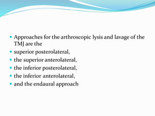
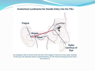
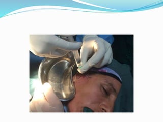
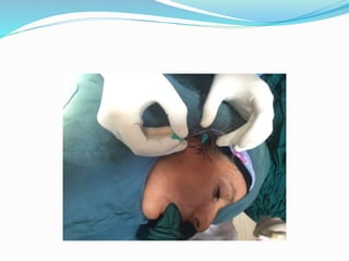
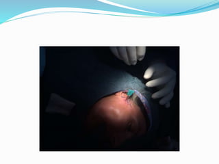
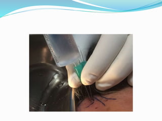
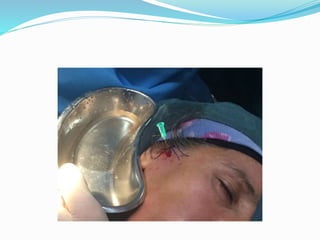
1. Arthroscopic lysis and lavage is a technique used to treat painful clicking or popping in the temporomandibular joint (TMJ) by releasing adhesions and repositioning displaced discs.
2. There are several methods for performing arthroscopic lysis and lavage of the TMJ, involving the use of one or two needles or cannulas to irrigate and lavage the joint.
3. Common solutions used in lysis and lavage include saline, local anesthetics, sodium hyaluronate, and glucocorticoids which help reduce inflammation.
10. Arthroscopic lysis and lavage can be used to treat those
patients with
painful clicking or
popping,
to release intra articular adhesions and anteriorly
displaced non-reducing discs, and to confirm other
diagnostic findings that couldwar- rant surgical
intervention.
11. There were 8 different methods for the lysis and lavage of
1. arthroscopic lysis and lavage,
2. two-needle arthrocentesis using an irrigation pump,
3. modified two-needle arthrocentesis,
4. the double-needle cannula method,
5. single-needle arthrocentesis, use of a single Shepard
cannula with two ports and two lumens,
6. two-needle arthrocentesis,
7. single-puncture arthrocentesis,
8. two-needle arthrocentesis with new anatomical
landmarks1
12. Approaches for the arthroscopic lysis and lavage of the
TMJ are the
superior posterolateral,
the superior anterolateral,
the inferior posterolateral,
the inferior anterolateral,
and the endaural approach
30. Treatment of TMJ static disk with arthroscopic lysis and lavage: a
comparison between MRI arthroscopic findings and clinical
results.
1999 -Casares G1,
Arthroscopic surgery using the lysis and lavage technique was performed on 30
temporomandibular joints (TMJs) of 26 patients with the clinical and
radiological (pseudodynamic Magnetic Resonance Imaging) diagnosis of static
disk (SD).
Parameters of condylar movement, position of the disk in relation to the condyle
in open and closed mouth positions and mobility of the articular disk were
evaluated both clinically and radiologically using pseudodynamic Magnetic
Resonance Imaging (MRI) before and after surgery.
Subjective parameters of pain and functional limitation were also evaluated
clinically.
The results showed that all the clinical and radiological parameters analyzed
improved significantly in all the patients included in the study.
In view of the results, the authors concluded that SD should be added to the
existing categories of TMJ internal derangements.
Pseudodynamic MRI is the diagnostic technique of choice in these cases and
arthroscopic surgery with lysis and lavage followed by active physical therapy is
an effective treatment modality in this type of pathology.
31. A review of techniques of lysis and lavage of the TMJ
Sinan Tozoglu a,∗, Fouad A. Al-Belasy b, M. Franklin Dolwic ca
Department of Oral and Maxillofacial Surgery, Ataturk University,
Dentistry Faculty, Ataturk University Campus, Erzurum, Turkey
b Faculty of Dentistry, Mansoura University, Egyptc
College of Dentistry, University of Florida, Gainesville, FL 32610,
United States
Accepted 25 March 2010