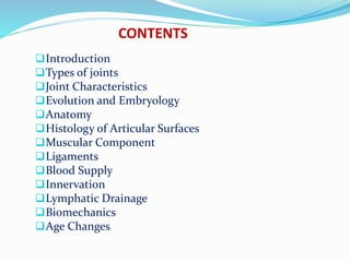
This document provides an overview of the temporomandibular joint (TMJ), including its:
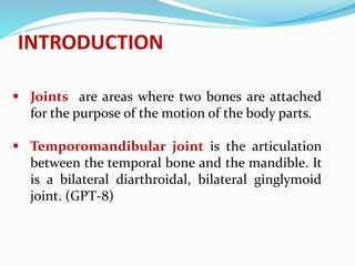
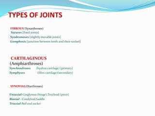
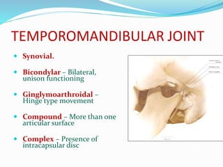
- Types (synovial, bicondylar, ginglymoarthroidal)
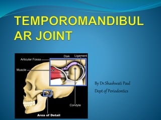
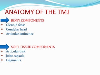
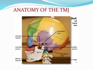
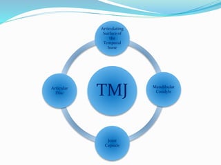
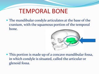
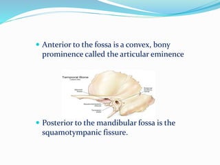
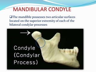
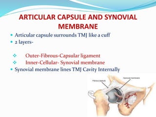
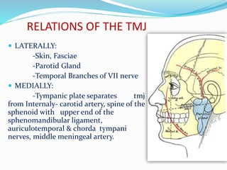
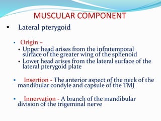
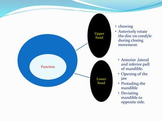
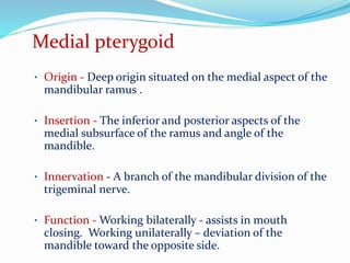
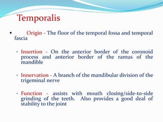
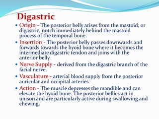
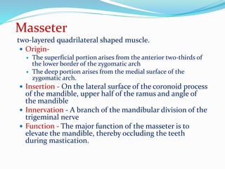
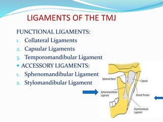
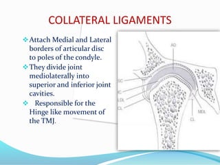
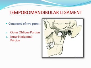
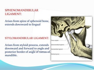
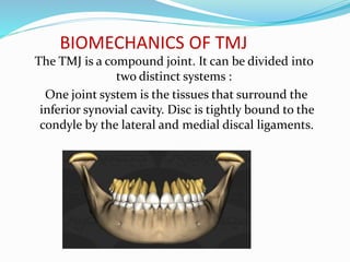
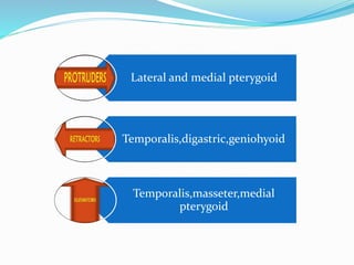
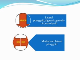
- Anatomy (bones, articular disc, ligaments, muscles)
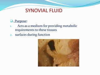
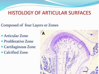
- Histology of the articular surfaces
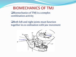
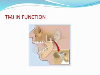
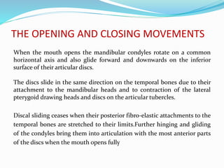
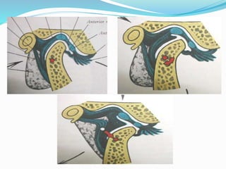
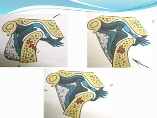
- Biomechanics and functions like opening and closing the mouth
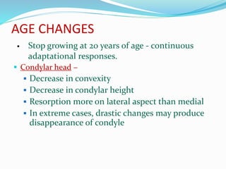
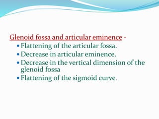
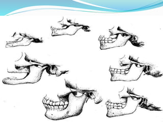
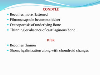
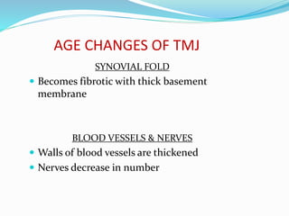
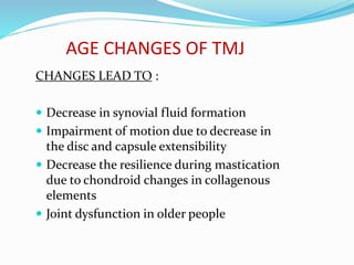
- Age-related changes like flattening of bones and thinning of tissues


![Temporomandibular joint [Autosaved].pptx](https://cdn.slidesharecdn.com/ss_thumbnails/temporomandibularjointautosaved-230521065437-1cbd4148-thumbnail.jpg?width=640&height=640&fit=bounds)


