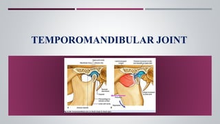
TEMPOROMANDIBULAR JOINT
1. INTRODUCTION
2. DEVELOPMENT OF TMJ
3. JOINTS
4. ANATOMY OF TMJ
5. MOVEMENTS AND MUSCLES INVOLVED
6. NEUROVASCULATURE OF TMJ
7. AGE CHANGES
8. DIAGNOSTIC AIDS
9. TMJ DISORDERS
10.EXAMINATION OF TMJ
CONTENTS
11. HYPO & HYPERPLASIA
12.LUXATION/SUB LUXATION
13.ANKYLOSIS
14. OSTEO & RHEUMATOID ARTHRITIS
15. MPDS
16. NEOPLASTIC TUMORS
17.SURGICAL APPROACH
18.CONCLUSION
19.REFERNCES
INTRODUCTION
 Connects the jaw bone to the skull.
 TMJ is a compound , synovial , bicondylar,
ginglymo diarthroidal joint.
 It’s the articulation between the squamous portion of
the temporal bone and the condyle of the mandible
 Only movable joint in the entire craniofacial complex.
 One of the most complex joints in the human body.
DEVELOPMENT OF TMJ
 In contrast to other diarthroidal joints TMJ is last joint to start its development, in
about 7th week in utero.
 Meckel’s cartilage provides skeletal support.
 Development of TMJ , begins with condensation of the developing mesenchymal
matrix around the Meckel’s cartilage – 6th to 7th week of IU.
WHEN???
There are 3 stages that define the normal embryologic development of the TMJ:
1.Blastematic stage
( 7-8 weeks of IU)
2. Cavitation stage
(9-11 weeks of IU)
3.Maturation stage
( 12th week of IU)
 Temporal blastema forms articular surface of temporal bone.
 Condylar blastema forms condylar cartilage, aponeurosis of lateral pterygoid muscle,
Articular disc & TMJ capsule.
 Inferior joint space is formed by cavitations that develops between the condylar
blastema & the mesenchymal connecting band of Meckel’s cartilage.
 Superior joint space is formed by cavitation between fibrous band & articular fossa.
 Condylar process of mandible develops by endochondral ossification.
 Glenoid fossa & articular eminence form by intra membranous ossification.
JOINTS
JOINTS
Fibrous joint
Cartilaginous joint
Synovial joint
SYNARTHROSIS
AMPHIARTHROSIS
DIARTHROSIS
STRUCTURAL
CLASSIFICATION
FUNCTIONAL
CLASSIFICATION
 Joint is an articulation between two bones.
ANATOMY OF TMJ
TMJ is made up of
Bony components
-Condylar head
-Glenoid fossa
-Articular eminence
Soft tissue components
-Joint capsule
-Articular disc
-Ligaments
Muscles associated with TMJ
CONDYLAR HEAD
 Portion of mandible that articulates with the cranium.
 15 to 20 mm & rounded - mediolaterally
 8 to 10 mm & convex - anteroposteriorly.
 Articular part of the mandible – ovoid condylar
process.
 Majority of human condyles are convex superiorly.
 Anterior view – it has medial and lateral projections
called poles.
 Medial pole is usually prominent than lateral.
GLENOID FOSSA
 The squamous portion of the temporal bone is made up
of the concave mandibular fossa, in which the condyle
is situated also called the articular or Glenoid fossa.
 Surface – smooth, oval and deeply hallowed out
 Bone is very thin at the depth of the fossa.
 Roof – creates partition between middle cranial fossa
and the joint.
 Lined by dense avascular fibrocartilage.
 The articular surfaces of the joint has temporal
component and condylar component.
 The upper articular surface is formed by the
 (a) articularfossa,
 (b) articular eminence of the temporal bone. This surface
is concavo-convex from behind forwards.
 The lower articular surface is formed by the head
(condyle) of the mandible. This surface is elliptical in
shape.
 The articular surfaces are covered by fibrocartilage, hence
temporomandibular joint is an atypical synovial joint.
ARTICULAR SURFACES
ARTICULAR EMINENCE
 Bony prominence at the base of zygomatic process of
the temporal bone anterior to the glenoid fossa.
 Convex – anteroposteriorly
 Concave - mediolaterall
JOINT CAPSULE
 Completely encloses the articular surface of the temporal bone and the condyle
 Composed of fibrous connective tissue
 Lined by a highly vascular synovial membrane
 Has various sensory receptors including nociceptors
Attachments:
 Superior—along the rim of the temporal articular surfaces
 Inferior—along the condylar neck
 Medial—blends along the medial collateral lig.
 Lateral—blends along the lateral collateral lig.
 Anterior—blends with the superior head of the lateral pterygoid m.
 Posterior—along the retrodiscal pad
 Made up of collagenous connective tissue, which do not stretch.
 Act as the passive restraining devices to limit and restrict border movements,
thereby play important role in protecting the structures.
LIGAMENTS
LIGAMENTS
Collateral
ligaments/discal
ligaments
Capsular ligaments
Temporomandibular
ligament/lateral
ligament
Sphenomandibular
ligament
Stylomandibular
ligament
TRUE LIGAMENTS
ACCESSORY
LIGAMENTS
COLLATERAL/DISCAL LIGAMENTS
 Responsible for dividing the joint medio-laterally.
 Restrict the movement of the disc away from the
condyle as the Disc glides Anteriorly & Posteriorly.
 Also aids in Hinging movement of condyle.
 It acts to resist any medial, lateral or inferior forces
that tend to separate or dislocate the articular
surfaces.
 Well innervated – provides proprioceptive feedback
regarding position & movement of the joint.
CAPSULAR LIGAMENT
TEMPOROMANDIBULAR/LATERAL LIGAMENT
 The lateral aspect of the capsular ligament
is reinforced by strong, tight fibres – lateral
ligament or TMJ ligament.
TMJ ligament has 2 parts :-
 Outer oblique portion;
 Inner horizontal portion.
 Supports the lateral aspect of the joint and
limits the movements of the condyle of
mandible.
 It extends from the spine of sphenoid above, to the lingual of
the mandible below.
 It lies medially, on a deeper plane, away from the joint
capsule.
 remnant of 1st branchial arch or Meckel’s cartilage.
 separated laterally from the ramus of mandible by lateral
pterygoid muscle, maxillary artery, inferior alveolar vessels
and nerves.
SPHENOMANDIBULAR LIGAMENT
 It is a thick band which extends from the tip and
anterior surface of styloid process to the angle of
mandible.
 It is the thickend part of deep cervical fascia which
seperates parotid and submandibular salivary
gland.
 Limits excessive protrusive movements
 The accessory ligaments of temporomandibular
joints control range of motion (ROM) of TMJs and
with mandible form a ‘swing’.
STYLOMANDIBULAR LIGAMENT
ARTICULAR DISC
 Located between the squamous portion of the temporal
bone and the condyle.
 Articular space is divided into 2 compartments by
articular disc/meniscus:
 1.lower/inferior compartment (concave).
 2.upper/superior compartment(concavo-convex).
 composed of dense fibrous connective tissue that is
Avascular, Devoid of blood vessels and Nerve fibers.
 Consists of type I and II collagen & few Elastic fibers.
 REES in 1954:
1.Anterior band -2mm
2.Intermediate band-1mm
3.Posterior band-3mm
 Functions
 It reduces the friction between the two articular surfaces.
 It aids in lubrication of the joint
 Act’s as shock absorber.
 It has little potential to repair after any injury.
SYNOVIAL MEMBRANE
 The synovial membrane lines the inside of the capsule
of the joint but does not cover the disc or the articular
surfaces (condyle, fossa or articular eminence).
 The synovium is most abundant in the bilaminar zone
of the articular disc, forming loose folds posteriorly
when the condyle is positioned in the glenoid fossa.
Function of the synovial membrane
 Production of the synovial fluid.
 varying cell types such as monocytes, Lymphocytes, free synovial cells,
polymorphonuclear leukocytes and macrophages.
 Removal of foreign material.
Function of the synovial fluid:
 viscous fluid composed of Plasma with some added proteins and mucin.
I. Nutrition to the disk and articular surfaces.
II. Liquid environment for joint surfaces.
III. Lubrication to increase the efficiency and decrease the erosion.
HISTOLOGY OF ARTICULAR SURFACE OF TMJ
HISTOLOGY OF
ARTICULAR
SURFACE OF TMJ
Articular zone Proliferative
zone
Cartilagenous
zone
Calcified zone
1.Dense
fibrous
connective
tissue
2.Poor blood
supply
3.Better
ability to
repair.
1.Mainly
cellular zone.
2.Undifferenti
ated
mesenchymal
cells.
3.Proliferatio
n &
regeneration
throughout
life.
1.criss - cross
pattern of
bundles.
2.providing a
three-
dimensional
network that
offers
resistance
against
compressive
and lateral
forces.
1.Deepest zone.
2.Chondrocytes
, chondroblasts
& osteoblasts.
 Muscles of mastication namely: Masseter,
Temporalis, Medial pterygoid, Lateral pterygoid.
 Supra hyoid muscles namely: Digastric and
Geniohyoid
MUSCLES ASSOCIATED WITH THE TMJ:
RELATIONS OF TMJ
 Anterior : Lateral pterygoid muscle, masseteric nerve and vessels.
 Posterior : Parotid gland, external auditory meatus, superficial temporal vessels,
auriculotemporal nerve.
 Medial : Spine of sphenoid, sphenomandibularligament, auriculotemporal nerve,
chordatympani nerve, middle meningeal artery.
 Lateral : Skin, fasciae, parotid gland, facial nerve.
 Superior : Middle cranial fossa.
 Inferior : Maxillary artery and vein.
MOVEMENTS AND THE MUSCLES INVOLVED
 The upper menisco-temporal compartment of TMJ
permits gliding movements, during protraction
(protrusion), retraction, and chewing.
 The lower menisco-temporal compartment permits
rotation around two axes (a) a transverse axis,
during depression and elevation and (b) a vertical
axis during side-to-side/chewing movements.
 The movements occurring at the temporomandibular joints are: 1. Depression
2. Elevation 3. Protraction 4. Retraction 5. Side to side (Chewing) movement
Muscles involved in movement.
ARTERIAL SUPPLY
 Maxillary artery.
 Superficial temporal artery.
VENOUS DRAINAGE
 Superficial temporal vein.
 Maxillary vein.
NERVE SUPPLY
 The mandibular nerve, the 3rd division of 5th cranial nerve
innervates the joint.
 Three branches from the mandibular nerve send terminals to
the joint capsule.
1.The largest is the auriculotemporal nerve which supplies the
posterior, medial and lateral aspects of the joint.
2.Massetric nerve.
3.A branch from the posterior deep temporal nerve, supply the
anterior parts of the joint.
AGE CHANGES OF TMJ
 Condyle: Becomes more flattened.
 Disc: Becomes thinner,Shows hyalinization and chondroid changes.
 Blood vessels and nerves: Walls of blood vessels thickened,Nerves
decrease in no.
 Synovial fold: Become fibrotic with thick basement membrane.
 Decrease in the synovial fluid formation.
 Impairment of motion due to decrease in the disc and capsule
extensibility.
 Decrease the resilience during mastication due to chondroid changes into
collagenous elements
DIAGNOSTIC AIDS USED TO EVALUATE TMJ
TMJ DISORDERS
 The term temporomandibular (TM) disorders was adopted to describe all disorders
related to function of the masticatory structures.
CLASSIFICATION
 1934 James costen described group of symptoms centering around ear and TMJ-
Costen syndrome.
 1959 Shore introduced TMJ Dysfunction.
 Ash & Ramfjord- functional TMJ disturbances.
 Bell coined the term Temporomandibular disorders.
 Describes both problems associated with the joint & disturbances associated with
function of masticatory system.
 Weldon Bell (1982) presented a classification that logically categorizes these
disorders, and the American Dental Association adopted it with few changes.
1) MASTICATORY MUSCLE DISORDERS
a. Protective muscle splinting
b. Masticatory muscle spasm (MPS)
c. Masticatory muscle inflammation (Myositis)
2) DERANGEMENTOF TMJ:
a. Incordination
b. Anterior disc displacement with reduction (Clicking)
c. Anterior disc displacement without reduction (Mechanical restriction, Closed lock)
3) EXTRINSIC TRAUMA:
a. Traumatic arthritis
b. Dislocation
c. Fracture
d. Internal disc derangement
e. Myositis
f. Myospasm
g. Tendonitis
4) DEGENERATIVE JOINT DISEASE:
a. Non inflammatory phase: Arthrosis
b. Inflammatory phase: Osteoarthritis
5) INFLAMMATORY JOINT DISORDERS:
a. Rheumatoid arthritis
b. Infective arthritis
c. Metabolic arthritis
6) CHRONIC MANDIBULAR HYPOMOBILITY:
a. Ankylosis: Fibrous and Osseous
b. Fibrosis of articular capsule
c. Contracture of elevator muscles: Myostatic or Myofibrotic
d. Internal disc derangement : Closed lock
7) GROWTH DISORDERS OF THE JOINT:
a. Developmental disorders
b. Acquired disorders
c. Neoplastic disorders
ETIOLOGY
MACROTRAUMA:
Most common in adolescents, such as
 Jaw trauma
 Vehicle accidents
 Sports
 Physical abuse
 Forceful intubation
 Third molar extraction.
MICROTRAUMA (Parafunctional habits):
 The second is a microtrauma from parafunctional habit. In addition to bruxism and
clenching, other repetitive habitual behaviors such as hyperextension, wind
instrument, and fingernail biting can cause joint overload.
ANATOMICAL FACTORS (skeletal and occlusal):
 Skeletal anterior open bite
 Steep articular eminence
 Overjet greater than 6-7 mm,
 Class III malocclusion
 Posterior crossbite.
PSYCHOLOGICAL FACTORS:
 If emotional stress is severe, clenching and bruxism can cause and aggravate
orofacial pain.
 Depression, anxiety, post-traumatic stress disorder, psychologic distress, and sleep
dysfunction can affect TMD prognosis and symptoms and signs.
SYSTEMIC FACTORS
 Rheumatoid arthritis
 Systemic lupus erythematosus
 Juvenile idiopathic arthritis
 Psoriatic arthritis.
COMMON SYMPTOMS
HISTORY
 Certain questions regarding the pain might be helpful in assessing mandibular
function.
 Pain in the face, front of the ear and in the temple region.
 Headaches, earaches, neck aches, cheek pain
 Time of aggrevation of pain
 Pain during opening the jaw wide, yawning, chewing, speaking and swallowing.
• Toothaches
• Clicking, crepitus, or popping of the jaw while moving
• Jaw lock
• Restricted jaw motion
• Uncomfortable bite
• Previous jaw injuries
• Previous treatment of jaw symptoms and its effects
• Arthritis
Masticatory muscle tenderness on palpation is the most consistent
examination feature present in cases of TMJ disorders.
The clinical features are:
1. Passive mouth opening
2. Masticatory muscle tenderness on palpation and maximal mouth
opening.
RANGE OF MANDIBULAR MOVEMENT
 Maximum opening distance Between the incisal edges of Upper and
lower incisor is Measured using scale , boley Gauge or ruler
 Normal opening 43.9mm (32 – 64 mm)
PHYSICAL EXAMINATION
 Measurement of lateral movements are made with the
teeth slightly separated, measuring the displacement
of the lower midline from the maxillary midline.
PROTRUSIVE MOVEMENT
 Normal lateral and protrusive movements are greater
than or equal to 7mm.
 The average max opening in primary dentition was
38.2mm (range 27-47mm)
 In mixed dentition group 40.7mm (range 23-55mm)
• The primary finding related to masticatory muscle palpation Is pain.
• They are done by squeezing the muscle finger and the thumb or by applying
pressure in the centre of the forehead.
PALPATION OF MASTICATORY MUSCLES
PALPATION OF TMJ
 The TMJ can be palpated by extra-auricular and intra- auricular methods.
 Palpation can be done standing at 10 o’clock or 11 o’clock position.
 can be achieved by placing a little finger inside
the external auditory meatus.
 During mandibular movement the posterior pole
of the condylar head can be palpated with the
pulp of the little finger.
 Intra-auricular palpation may also be used to
elicit capsular tenderness.
Intra-auricular:-
 Done by placing index finger in the pre-
auricular region about 1.5cm medial to the
tragus of ear.
 The lateral pole of the condyle is accessible
during this examination.
Extra auricular examination of TMJ
HYPOPLASIA:
 It is of two types: unilateral and bilateral
 Causes:
1. prenatal growth distubances including both hereditary distubances like
mandibulofacial dysostosis and non-hereditary disturbances like pierre robin
syndrome.
2. postnatal growth disturbances like endocrine dysfunction, dietary deficiency,
trauma and irradiation
Management: surgical, orthodontic and prosthetic correction for functional and
cosmetic improvement is required.
HYPERPLASIA:
Causes:
1. Developmental- eg. Hemi-facial hypertrophy
2. Neoplastic- eg. Chondroma or osteochondroma
3. Bone disease- eg. Paget’s disease
4. Hereditary- eg. Klinefelter’s disease
5. Endocrine – eg. Gigantism
6. Hypertrophic arthritis.
Management: Orthognathic surgery to improve esthetics and function.
LUXATION AND SUBLUXATION
 Luxation or dislocation of TMJ occurs when the head of the condyle moves
anteriorly over the articular eminence into such a position from where it cannot
return back to its original position by itself.
 When the condyle is completely dislocated, itis called luxation, while the partial
dislocation of the same is called subluxation.
ETIOLOGY
 Trauma to the TMJ.
 Wide mouth opening for an extended period of time (e.g. dental procedures, etc.)
 The patients usually complain of “sudden locking”
of the jaw with inability to close the mouth. In the
initial phases, the problem happens rarely, but later
on, patients may have such situation quite
frequently, thereby, making eating and talking very
difficult.
TREATMENT
 In case of luxation or subluxation, the dislocated
condyle is to be guided into its normal position by
giving inferior and posterior pressure while holding
the mandible firmly in the molar region
CLINICAL FEATURES
ANKYLOSIS
 Ankylosis of the TMJ is a disorder in which adhesion of
joint components takes place by fibrous or bony union,
resulting into loss of function.
ETIOLOGY
 Trauma
 Local infections
 Systemic conditions
 Arthritis/inflammatory conditions
 Neoplasms
TYPES OF ANKYLOSIS
 False ankylosis: False ankylosis is extra-articular and it occurs due to fibrous or
bony union between the coronoid process and maxilla or zygoma.
 True ankylosis: True ankylosis is intra-articular and it is again of two types:
1.True bony ankylosis: When the TMJ space is completely obliterated by the
deposition of bone following destruction and subsequent fusion of temporal fossa,
meniscus and head of the condyle, the condition is called a true bony ankylosis .
2.True fibrous ankylosis: Intra-articular fibrous ankylosis occurs, if the TMJ space is
obliterated by the deposition of a fibrous tissue mass (e.g. scar).
Surgical correction is best achieved by
 Condylectomy
 Gap arthroplasty
 Costochondral grafting
 Orthognathic surgery to correct any maxillofacial deformity
 Coronoidectomy
Followed by active physiotherapy
 Osteoarthritis is a degenerative and destructive disease of the weight-bearing joints,
although TMJ is not a weight-bearing joint, osteoarthritis can still occur in it due to
the ageing process or trauma.
OSTEOARTHRITIS
Etiopathogenesis
(degenerative joint disease)
CLINICAL FEATURES
 Clicking sounds in the joint while opening and closing
movements of the jaw.
 Limitation of movements of the joint.
 Muscles of mastication are often tendered due to
strain caused by non-use or restricted use of the
painful joint.
TREATMENT
 There is no satisfactory treatment for osteoarthritis,
however condylectomy should be considered in very
severe cases.
RHEUMATOID ARTHRITIS
 Rheumatoid arthritis is a systemic disease that usually affects many joints including
the TMJ and the disease is characterized by progressive inflammatory destruction of
the joint structures.
CLINICAL FEATURES
 acute phase of the disease, patient may suffer from fever, malaise, fatiguability,
weight loss, anemia and raised ESR, etc.
 Intermittent bilateral TMJ swelling and tenderness , Dull, aching pain.
 The maximum feeling of stiffness is experienced in the early mornings.
 Clicking sounds in the joint may develop in chronic cases.
 The radiographic feature typically shows the ‘sharpened
pencil’ or ‘mouthpiece of flute’ appearance.
Management:
 Adequate rest, soft diet.
 Intra-auricular corticosteriod injection;
 Non steroidal anti-inflammatory drugs;
 Slow acting anti-rheumatic drugs eg. Sulphasalazine;
 Medical/surgical synovectomy.
 Laskin (1969) was the first to coin the term myofascial pain dysfunction syndrome.
 Myofacial pain dysfunction syndrome is a disease complex that disturbs the entire
masticatory apparatus and is characterized by pain and limitation of movement of
the TMJ.
MYOFACIAL PAIN DYSFUNCTION (MPD) SYNDROME
PREDISPOSING FACTORS
 Trauma
 Muscular overextension
 Muscular overcontraction
 Muscle fatigue.
 The pain is dull in nature and it is usually present unilaterally in the preauricular
area or in the ear.
 The pain radiates to the angle of mandible or temporal region.
 The intensity of pain varies at different times of the day.
 Muscle tenderness
 Limitation of movements and deviation of the jaw.
 Clicking sounds in the TMJ during opening and closing of the mouth.
CLINICAL FEATURES
 COUNSELING
 PHYSIOTHERAPY – moist heat application
 TRIGGER POINT THERAPY – Spray and stretch technique
 MEDICATION – NSAIDs and Skeletal muscle relaxants
 OCCLUSAL SPLINT THERAPY
 TRANSCUTANEOUS ELECTRIC NERVE STIMULATION (TENS) temporarily activates
afferent nerves, thereby modulating pain.
TREATMENT
NEOPLASTIC TUMORS
Benign tumors: produce restricted movements of TMJ and facial asymmetry.
 Management is by surgical approach.
Malignant tumors: produce diffuse pain in the area and diminished hearing,
associated with swelling.
 Management is by surgery, radiotherapy and chemotherapy.
1. Submandibular (Risdon’s) approach
2. Postramal (Hind’s) approach
3. Postauricular approach
4. Endaural approach
5. Preauricular approach
• Dingman’s
• Blair’s
• Thoma’s
• Popowich’s modification of Al-Kayat and Bramley’s
6. Hemicoronal approach
7. Coronal or bicoronal approach
 It is impossible to comprehend the fine points of occlusion without an in depth
awareness of anatomy ,physiology ,and biomechanics of the TMJ.
 The first requirement for successful occlusal treatment is stable, comfortable TMJ.
 The jaw joints must be able to accept maximum loading by the elevator muscles with
no signs of discomfort.
 It is only through an understanding of how the normal, healthy TMJ functions that
we can make sense out of what is wrong when it isn't functioning comfortably.
 This understanding of TMJ is foundational to diagnosis and treatment.
CONCLUSION
1.MAHINDRA KUMAR ANAND : Human anatomy for dental students, 3rd edition.
2.NEELIMA MALIK : Textbook of Oral and maxillofacial surgery, 2nd edition.
3.B D CHAURASIA : Human anatomy, 5th edition(Vol 3).
4.SHAFERS : Textbook of Oral pathology, 6th edition.
5. DEEPAK KADEMANI: ATLAS OF ORAL &MAXILLOFACIAL SURGERY
6. Burket’s: ORAL MEDICINE
7. Ravikiran Ongole: Clinical Manual forOral Medicine andRadiology
8. Shafers :Textbook of Oral Pathology
9. Swapan Kumar Purkait :Essentials of Oral Pathology
10. Mc Inns Colour Atlas Of Head And Neck Anatomy 3rd Edition
11. Grays - Textbook of Anatomy,
REFERENCES

Temporomandibular joint 1

  • 1.
  • 2.
    1. INTRODUCTION 2. DEVELOPMENTOF TMJ 3. JOINTS 4. ANATOMY OF TMJ 5. MOVEMENTS AND MUSCLES INVOLVED 6. NEUROVASCULATURE OF TMJ 7. AGE CHANGES 8. DIAGNOSTIC AIDS 9. TMJ DISORDERS 10.EXAMINATION OF TMJ CONTENTS 11. HYPO & HYPERPLASIA 12.LUXATION/SUB LUXATION 13.ANKYLOSIS 14. OSTEO & RHEUMATOID ARTHRITIS 15. MPDS 16. NEOPLASTIC TUMORS 17.SURGICAL APPROACH 18.CONCLUSION 19.REFERNCES
  • 3.
    INTRODUCTION  Connects thejaw bone to the skull.  TMJ is a compound , synovial , bicondylar, ginglymo diarthroidal joint.  It’s the articulation between the squamous portion of the temporal bone and the condyle of the mandible  Only movable joint in the entire craniofacial complex.  One of the most complex joints in the human body.
  • 4.
    DEVELOPMENT OF TMJ In contrast to other diarthroidal joints TMJ is last joint to start its development, in about 7th week in utero.  Meckel’s cartilage provides skeletal support.  Development of TMJ , begins with condensation of the developing mesenchymal matrix around the Meckel’s cartilage – 6th to 7th week of IU. WHEN???
  • 5.
    There are 3stages that define the normal embryologic development of the TMJ: 1.Blastematic stage ( 7-8 weeks of IU) 2. Cavitation stage (9-11 weeks of IU) 3.Maturation stage ( 12th week of IU)  Temporal blastema forms articular surface of temporal bone.  Condylar blastema forms condylar cartilage, aponeurosis of lateral pterygoid muscle, Articular disc & TMJ capsule.
  • 6.
     Inferior jointspace is formed by cavitations that develops between the condylar blastema & the mesenchymal connecting band of Meckel’s cartilage.  Superior joint space is formed by cavitation between fibrous band & articular fossa.  Condylar process of mandible develops by endochondral ossification.  Glenoid fossa & articular eminence form by intra membranous ossification.
  • 7.
    JOINTS JOINTS Fibrous joint Cartilaginous joint Synovialjoint SYNARTHROSIS AMPHIARTHROSIS DIARTHROSIS STRUCTURAL CLASSIFICATION FUNCTIONAL CLASSIFICATION  Joint is an articulation between two bones.
  • 8.
    ANATOMY OF TMJ TMJis made up of Bony components -Condylar head -Glenoid fossa -Articular eminence Soft tissue components -Joint capsule -Articular disc -Ligaments Muscles associated with TMJ
  • 9.
    CONDYLAR HEAD  Portionof mandible that articulates with the cranium.  15 to 20 mm & rounded - mediolaterally  8 to 10 mm & convex - anteroposteriorly.  Articular part of the mandible – ovoid condylar process.  Majority of human condyles are convex superiorly.  Anterior view – it has medial and lateral projections called poles.  Medial pole is usually prominent than lateral.
  • 10.
    GLENOID FOSSA  Thesquamous portion of the temporal bone is made up of the concave mandibular fossa, in which the condyle is situated also called the articular or Glenoid fossa.  Surface – smooth, oval and deeply hallowed out  Bone is very thin at the depth of the fossa.  Roof – creates partition between middle cranial fossa and the joint.  Lined by dense avascular fibrocartilage.
  • 11.
     The articularsurfaces of the joint has temporal component and condylar component.  The upper articular surface is formed by the  (a) articularfossa,  (b) articular eminence of the temporal bone. This surface is concavo-convex from behind forwards.  The lower articular surface is formed by the head (condyle) of the mandible. This surface is elliptical in shape.  The articular surfaces are covered by fibrocartilage, hence temporomandibular joint is an atypical synovial joint. ARTICULAR SURFACES
  • 12.
    ARTICULAR EMINENCE  Bonyprominence at the base of zygomatic process of the temporal bone anterior to the glenoid fossa.  Convex – anteroposteriorly  Concave - mediolaterall
  • 13.
    JOINT CAPSULE  Completelyencloses the articular surface of the temporal bone and the condyle  Composed of fibrous connective tissue  Lined by a highly vascular synovial membrane  Has various sensory receptors including nociceptors Attachments:  Superior—along the rim of the temporal articular surfaces  Inferior—along the condylar neck  Medial—blends along the medial collateral lig.  Lateral—blends along the lateral collateral lig.  Anterior—blends with the superior head of the lateral pterygoid m.  Posterior—along the retrodiscal pad
  • 14.
     Made upof collagenous connective tissue, which do not stretch.  Act as the passive restraining devices to limit and restrict border movements, thereby play important role in protecting the structures. LIGAMENTS LIGAMENTS Collateral ligaments/discal ligaments Capsular ligaments Temporomandibular ligament/lateral ligament Sphenomandibular ligament Stylomandibular ligament TRUE LIGAMENTS ACCESSORY LIGAMENTS
  • 15.
    COLLATERAL/DISCAL LIGAMENTS  Responsiblefor dividing the joint medio-laterally.  Restrict the movement of the disc away from the condyle as the Disc glides Anteriorly & Posteriorly.  Also aids in Hinging movement of condyle.
  • 16.
     It actsto resist any medial, lateral or inferior forces that tend to separate or dislocate the articular surfaces.  Well innervated – provides proprioceptive feedback regarding position & movement of the joint. CAPSULAR LIGAMENT
  • 17.
    TEMPOROMANDIBULAR/LATERAL LIGAMENT  Thelateral aspect of the capsular ligament is reinforced by strong, tight fibres – lateral ligament or TMJ ligament. TMJ ligament has 2 parts :-  Outer oblique portion;  Inner horizontal portion.  Supports the lateral aspect of the joint and limits the movements of the condyle of mandible.
  • 18.
     It extendsfrom the spine of sphenoid above, to the lingual of the mandible below.  It lies medially, on a deeper plane, away from the joint capsule.  remnant of 1st branchial arch or Meckel’s cartilage.  separated laterally from the ramus of mandible by lateral pterygoid muscle, maxillary artery, inferior alveolar vessels and nerves. SPHENOMANDIBULAR LIGAMENT
  • 19.
     It isa thick band which extends from the tip and anterior surface of styloid process to the angle of mandible.  It is the thickend part of deep cervical fascia which seperates parotid and submandibular salivary gland.  Limits excessive protrusive movements  The accessory ligaments of temporomandibular joints control range of motion (ROM) of TMJs and with mandible form a ‘swing’. STYLOMANDIBULAR LIGAMENT
  • 20.
    ARTICULAR DISC  Locatedbetween the squamous portion of the temporal bone and the condyle.  Articular space is divided into 2 compartments by articular disc/meniscus:  1.lower/inferior compartment (concave).  2.upper/superior compartment(concavo-convex).  composed of dense fibrous connective tissue that is Avascular, Devoid of blood vessels and Nerve fibers.  Consists of type I and II collagen & few Elastic fibers.
  • 21.
     REES in1954: 1.Anterior band -2mm 2.Intermediate band-1mm 3.Posterior band-3mm  Functions  It reduces the friction between the two articular surfaces.  It aids in lubrication of the joint  Act’s as shock absorber.  It has little potential to repair after any injury.
  • 22.
    SYNOVIAL MEMBRANE  Thesynovial membrane lines the inside of the capsule of the joint but does not cover the disc or the articular surfaces (condyle, fossa or articular eminence).  The synovium is most abundant in the bilaminar zone of the articular disc, forming loose folds posteriorly when the condyle is positioned in the glenoid fossa. Function of the synovial membrane  Production of the synovial fluid.
  • 23.
     varying celltypes such as monocytes, Lymphocytes, free synovial cells, polymorphonuclear leukocytes and macrophages.  Removal of foreign material. Function of the synovial fluid:  viscous fluid composed of Plasma with some added proteins and mucin. I. Nutrition to the disk and articular surfaces. II. Liquid environment for joint surfaces. III. Lubrication to increase the efficiency and decrease the erosion.
  • 24.
    HISTOLOGY OF ARTICULARSURFACE OF TMJ HISTOLOGY OF ARTICULAR SURFACE OF TMJ Articular zone Proliferative zone Cartilagenous zone Calcified zone 1.Dense fibrous connective tissue 2.Poor blood supply 3.Better ability to repair. 1.Mainly cellular zone. 2.Undifferenti ated mesenchymal cells. 3.Proliferatio n & regeneration throughout life. 1.criss - cross pattern of bundles. 2.providing a three- dimensional network that offers resistance against compressive and lateral forces. 1.Deepest zone. 2.Chondrocytes , chondroblasts & osteoblasts.
  • 25.
     Muscles ofmastication namely: Masseter, Temporalis, Medial pterygoid, Lateral pterygoid.  Supra hyoid muscles namely: Digastric and Geniohyoid MUSCLES ASSOCIATED WITH THE TMJ:
  • 26.
    RELATIONS OF TMJ Anterior : Lateral pterygoid muscle, masseteric nerve and vessels.  Posterior : Parotid gland, external auditory meatus, superficial temporal vessels, auriculotemporal nerve.  Medial : Spine of sphenoid, sphenomandibularligament, auriculotemporal nerve, chordatympani nerve, middle meningeal artery.  Lateral : Skin, fasciae, parotid gland, facial nerve.  Superior : Middle cranial fossa.  Inferior : Maxillary artery and vein.
  • 27.
    MOVEMENTS AND THEMUSCLES INVOLVED  The upper menisco-temporal compartment of TMJ permits gliding movements, during protraction (protrusion), retraction, and chewing.  The lower menisco-temporal compartment permits rotation around two axes (a) a transverse axis, during depression and elevation and (b) a vertical axis during side-to-side/chewing movements.  The movements occurring at the temporomandibular joints are: 1. Depression 2. Elevation 3. Protraction 4. Retraction 5. Side to side (Chewing) movement
  • 28.
  • 29.
    ARTERIAL SUPPLY  Maxillaryartery.  Superficial temporal artery. VENOUS DRAINAGE  Superficial temporal vein.  Maxillary vein.
  • 30.
    NERVE SUPPLY  Themandibular nerve, the 3rd division of 5th cranial nerve innervates the joint.  Three branches from the mandibular nerve send terminals to the joint capsule. 1.The largest is the auriculotemporal nerve which supplies the posterior, medial and lateral aspects of the joint. 2.Massetric nerve. 3.A branch from the posterior deep temporal nerve, supply the anterior parts of the joint.
  • 31.
    AGE CHANGES OFTMJ  Condyle: Becomes more flattened.  Disc: Becomes thinner,Shows hyalinization and chondroid changes.  Blood vessels and nerves: Walls of blood vessels thickened,Nerves decrease in no.  Synovial fold: Become fibrotic with thick basement membrane.  Decrease in the synovial fluid formation.  Impairment of motion due to decrease in the disc and capsule extensibility.  Decrease the resilience during mastication due to chondroid changes into collagenous elements
  • 32.
    DIAGNOSTIC AIDS USEDTO EVALUATE TMJ
  • 33.
    TMJ DISORDERS  Theterm temporomandibular (TM) disorders was adopted to describe all disorders related to function of the masticatory structures. CLASSIFICATION  1934 James costen described group of symptoms centering around ear and TMJ- Costen syndrome.  1959 Shore introduced TMJ Dysfunction.  Ash & Ramfjord- functional TMJ disturbances.  Bell coined the term Temporomandibular disorders.  Describes both problems associated with the joint & disturbances associated with function of masticatory system.
  • 34.
     Weldon Bell(1982) presented a classification that logically categorizes these disorders, and the American Dental Association adopted it with few changes. 1) MASTICATORY MUSCLE DISORDERS a. Protective muscle splinting b. Masticatory muscle spasm (MPS) c. Masticatory muscle inflammation (Myositis) 2) DERANGEMENTOF TMJ: a. Incordination b. Anterior disc displacement with reduction (Clicking) c. Anterior disc displacement without reduction (Mechanical restriction, Closed lock)
  • 35.
    3) EXTRINSIC TRAUMA: a.Traumatic arthritis b. Dislocation c. Fracture d. Internal disc derangement e. Myositis f. Myospasm g. Tendonitis 4) DEGENERATIVE JOINT DISEASE: a. Non inflammatory phase: Arthrosis b. Inflammatory phase: Osteoarthritis
  • 36.
    5) INFLAMMATORY JOINTDISORDERS: a. Rheumatoid arthritis b. Infective arthritis c. Metabolic arthritis 6) CHRONIC MANDIBULAR HYPOMOBILITY: a. Ankylosis: Fibrous and Osseous b. Fibrosis of articular capsule c. Contracture of elevator muscles: Myostatic or Myofibrotic d. Internal disc derangement : Closed lock 7) GROWTH DISORDERS OF THE JOINT: a. Developmental disorders b. Acquired disorders c. Neoplastic disorders
  • 37.
    ETIOLOGY MACROTRAUMA: Most common inadolescents, such as  Jaw trauma  Vehicle accidents  Sports  Physical abuse  Forceful intubation  Third molar extraction. MICROTRAUMA (Parafunctional habits):  The second is a microtrauma from parafunctional habit. In addition to bruxism and clenching, other repetitive habitual behaviors such as hyperextension, wind instrument, and fingernail biting can cause joint overload.
  • 38.
    ANATOMICAL FACTORS (skeletaland occlusal):  Skeletal anterior open bite  Steep articular eminence  Overjet greater than 6-7 mm,  Class III malocclusion  Posterior crossbite. PSYCHOLOGICAL FACTORS:  If emotional stress is severe, clenching and bruxism can cause and aggravate orofacial pain.  Depression, anxiety, post-traumatic stress disorder, psychologic distress, and sleep dysfunction can affect TMD prognosis and symptoms and signs.
  • 39.
    SYSTEMIC FACTORS  Rheumatoidarthritis  Systemic lupus erythematosus  Juvenile idiopathic arthritis  Psoriatic arthritis.
  • 40.
    COMMON SYMPTOMS HISTORY  Certainquestions regarding the pain might be helpful in assessing mandibular function.  Pain in the face, front of the ear and in the temple region.  Headaches, earaches, neck aches, cheek pain  Time of aggrevation of pain  Pain during opening the jaw wide, yawning, chewing, speaking and swallowing.
  • 41.
    • Toothaches • Clicking,crepitus, or popping of the jaw while moving • Jaw lock • Restricted jaw motion • Uncomfortable bite • Previous jaw injuries • Previous treatment of jaw symptoms and its effects • Arthritis
  • 42.
    Masticatory muscle tendernesson palpation is the most consistent examination feature present in cases of TMJ disorders. The clinical features are: 1. Passive mouth opening 2. Masticatory muscle tenderness on palpation and maximal mouth opening. RANGE OF MANDIBULAR MOVEMENT  Maximum opening distance Between the incisal edges of Upper and lower incisor is Measured using scale , boley Gauge or ruler  Normal opening 43.9mm (32 – 64 mm) PHYSICAL EXAMINATION
  • 43.
     Measurement oflateral movements are made with the teeth slightly separated, measuring the displacement of the lower midline from the maxillary midline. PROTRUSIVE MOVEMENT  Normal lateral and protrusive movements are greater than or equal to 7mm.  The average max opening in primary dentition was 38.2mm (range 27-47mm)  In mixed dentition group 40.7mm (range 23-55mm)
  • 44.
    • The primaryfinding related to masticatory muscle palpation Is pain. • They are done by squeezing the muscle finger and the thumb or by applying pressure in the centre of the forehead. PALPATION OF MASTICATORY MUSCLES PALPATION OF TMJ  The TMJ can be palpated by extra-auricular and intra- auricular methods.  Palpation can be done standing at 10 o’clock or 11 o’clock position.
  • 45.
     can beachieved by placing a little finger inside the external auditory meatus.  During mandibular movement the posterior pole of the condylar head can be palpated with the pulp of the little finger.  Intra-auricular palpation may also be used to elicit capsular tenderness. Intra-auricular:-
  • 46.
     Done byplacing index finger in the pre- auricular region about 1.5cm medial to the tragus of ear.  The lateral pole of the condyle is accessible during this examination. Extra auricular examination of TMJ
  • 47.
    HYPOPLASIA:  It isof two types: unilateral and bilateral  Causes: 1. prenatal growth distubances including both hereditary distubances like mandibulofacial dysostosis and non-hereditary disturbances like pierre robin syndrome. 2. postnatal growth disturbances like endocrine dysfunction, dietary deficiency, trauma and irradiation Management: surgical, orthodontic and prosthetic correction for functional and cosmetic improvement is required.
  • 48.
    HYPERPLASIA: Causes: 1. Developmental- eg.Hemi-facial hypertrophy 2. Neoplastic- eg. Chondroma or osteochondroma 3. Bone disease- eg. Paget’s disease 4. Hereditary- eg. Klinefelter’s disease 5. Endocrine – eg. Gigantism 6. Hypertrophic arthritis. Management: Orthognathic surgery to improve esthetics and function.
  • 49.
    LUXATION AND SUBLUXATION Luxation or dislocation of TMJ occurs when the head of the condyle moves anteriorly over the articular eminence into such a position from where it cannot return back to its original position by itself.  When the condyle is completely dislocated, itis called luxation, while the partial dislocation of the same is called subluxation. ETIOLOGY  Trauma to the TMJ.  Wide mouth opening for an extended period of time (e.g. dental procedures, etc.)
  • 50.
     The patientsusually complain of “sudden locking” of the jaw with inability to close the mouth. In the initial phases, the problem happens rarely, but later on, patients may have such situation quite frequently, thereby, making eating and talking very difficult. TREATMENT  In case of luxation or subluxation, the dislocated condyle is to be guided into its normal position by giving inferior and posterior pressure while holding the mandible firmly in the molar region CLINICAL FEATURES
  • 51.
    ANKYLOSIS  Ankylosis ofthe TMJ is a disorder in which adhesion of joint components takes place by fibrous or bony union, resulting into loss of function. ETIOLOGY  Trauma  Local infections  Systemic conditions  Arthritis/inflammatory conditions  Neoplasms
  • 52.
    TYPES OF ANKYLOSIS False ankylosis: False ankylosis is extra-articular and it occurs due to fibrous or bony union between the coronoid process and maxilla or zygoma.  True ankylosis: True ankylosis is intra-articular and it is again of two types: 1.True bony ankylosis: When the TMJ space is completely obliterated by the deposition of bone following destruction and subsequent fusion of temporal fossa, meniscus and head of the condyle, the condition is called a true bony ankylosis . 2.True fibrous ankylosis: Intra-articular fibrous ankylosis occurs, if the TMJ space is obliterated by the deposition of a fibrous tissue mass (e.g. scar).
  • 53.
    Surgical correction isbest achieved by  Condylectomy  Gap arthroplasty  Costochondral grafting  Orthognathic surgery to correct any maxillofacial deformity  Coronoidectomy Followed by active physiotherapy
  • 54.
     Osteoarthritis isa degenerative and destructive disease of the weight-bearing joints, although TMJ is not a weight-bearing joint, osteoarthritis can still occur in it due to the ageing process or trauma. OSTEOARTHRITIS Etiopathogenesis (degenerative joint disease)
  • 55.
    CLINICAL FEATURES  Clickingsounds in the joint while opening and closing movements of the jaw.  Limitation of movements of the joint.  Muscles of mastication are often tendered due to strain caused by non-use or restricted use of the painful joint. TREATMENT  There is no satisfactory treatment for osteoarthritis, however condylectomy should be considered in very severe cases.
  • 56.
    RHEUMATOID ARTHRITIS  Rheumatoidarthritis is a systemic disease that usually affects many joints including the TMJ and the disease is characterized by progressive inflammatory destruction of the joint structures. CLINICAL FEATURES  acute phase of the disease, patient may suffer from fever, malaise, fatiguability, weight loss, anemia and raised ESR, etc.  Intermittent bilateral TMJ swelling and tenderness , Dull, aching pain.  The maximum feeling of stiffness is experienced in the early mornings.  Clicking sounds in the joint may develop in chronic cases.
  • 57.
     The radiographicfeature typically shows the ‘sharpened pencil’ or ‘mouthpiece of flute’ appearance. Management:  Adequate rest, soft diet.  Intra-auricular corticosteriod injection;  Non steroidal anti-inflammatory drugs;  Slow acting anti-rheumatic drugs eg. Sulphasalazine;  Medical/surgical synovectomy.
  • 58.
     Laskin (1969)was the first to coin the term myofascial pain dysfunction syndrome.  Myofacial pain dysfunction syndrome is a disease complex that disturbs the entire masticatory apparatus and is characterized by pain and limitation of movement of the TMJ. MYOFACIAL PAIN DYSFUNCTION (MPD) SYNDROME PREDISPOSING FACTORS  Trauma  Muscular overextension  Muscular overcontraction  Muscle fatigue.
  • 59.
     The painis dull in nature and it is usually present unilaterally in the preauricular area or in the ear.  The pain radiates to the angle of mandible or temporal region.  The intensity of pain varies at different times of the day.  Muscle tenderness  Limitation of movements and deviation of the jaw.  Clicking sounds in the TMJ during opening and closing of the mouth. CLINICAL FEATURES
  • 60.
     COUNSELING  PHYSIOTHERAPY– moist heat application  TRIGGER POINT THERAPY – Spray and stretch technique  MEDICATION – NSAIDs and Skeletal muscle relaxants  OCCLUSAL SPLINT THERAPY  TRANSCUTANEOUS ELECTRIC NERVE STIMULATION (TENS) temporarily activates afferent nerves, thereby modulating pain. TREATMENT
  • 61.
    NEOPLASTIC TUMORS Benign tumors:produce restricted movements of TMJ and facial asymmetry.  Management is by surgical approach. Malignant tumors: produce diffuse pain in the area and diminished hearing, associated with swelling.  Management is by surgery, radiotherapy and chemotherapy.
  • 62.
    1. Submandibular (Risdon’s)approach 2. Postramal (Hind’s) approach 3. Postauricular approach 4. Endaural approach 5. Preauricular approach • Dingman’s • Blair’s • Thoma’s • Popowich’s modification of Al-Kayat and Bramley’s 6. Hemicoronal approach 7. Coronal or bicoronal approach
  • 65.
     It isimpossible to comprehend the fine points of occlusion without an in depth awareness of anatomy ,physiology ,and biomechanics of the TMJ.  The first requirement for successful occlusal treatment is stable, comfortable TMJ.  The jaw joints must be able to accept maximum loading by the elevator muscles with no signs of discomfort.  It is only through an understanding of how the normal, healthy TMJ functions that we can make sense out of what is wrong when it isn't functioning comfortably.  This understanding of TMJ is foundational to diagnosis and treatment. CONCLUSION
  • 66.
    1.MAHINDRA KUMAR ANAND: Human anatomy for dental students, 3rd edition. 2.NEELIMA MALIK : Textbook of Oral and maxillofacial surgery, 2nd edition. 3.B D CHAURASIA : Human anatomy, 5th edition(Vol 3). 4.SHAFERS : Textbook of Oral pathology, 6th edition. 5. DEEPAK KADEMANI: ATLAS OF ORAL &MAXILLOFACIAL SURGERY 6. Burket’s: ORAL MEDICINE 7. Ravikiran Ongole: Clinical Manual forOral Medicine andRadiology 8. Shafers :Textbook of Oral Pathology 9. Swapan Kumar Purkait :Essentials of Oral Pathology 10. Mc Inns Colour Atlas Of Head And Neck Anatomy 3rd Edition 11. Grays - Textbook of Anatomy, REFERENCES

Editor's Notes

  • #2 Delete this slide when you finish preparing the other slides.