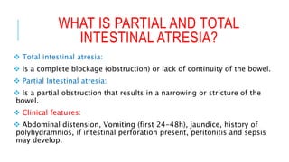
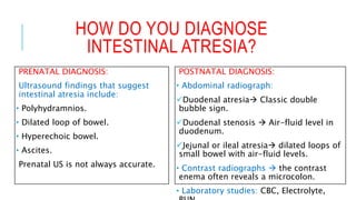
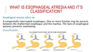
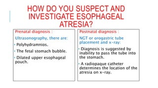
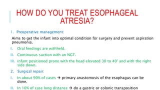
PROBLEM SOLVING:
VOMITING IN INFANTS AND
CHILDREN
Done by:
Arwa H. Al-
Onayzan.
ID: 215007943.
LEARNING OBJECTIVES:
 What is partial and total intestinal atresia? What are clinical
feature and how do you diagnose intestinal atresia?
 What is esophageal atresia? How do you classify? How do you
suspect, investigate and treat a case of esophageal atresia?
WHAT IS PARTIAL AND TOTAL
INTESTINAL ATRESIA?
 Total intestinal atresia:
 Is a complete blockage (obstruction) or lack of continuity of the bowel.
 Partial Intestinal atresia:
 Is a partial obstruction that results in a narrowing or stricture of the
bowel.
 Clinical features:
 Abdominal distension, Vomiting (first 24-48h), jaundice, history of
polyhydramnios, if intestinal perforation present, peritonitis and sepsis
may develop.
HOW DO YOU DIAGNOSE
INTESTINAL ATRESIA?
PRENATAL DIAGNOSIS:
Ultrasound findings that suggest
intestinal atresia include:
• Polyhydramnios.
• Dilated loop of bowel.
• Hyperechoic bowel.
• Ascites.
Prenatal US is not always accurate.
POSTNATAL DIAGNOSIS:
• Abdominal radiograph:
Duodenal atresia Classic double
bubble sign.
Duodenal stenosis  Air-fluid level in
duodenum.
Jejunal or ileal atresia dilated loops of
small bowel with air-fluid levels.
• Contrast radiographs  the contrast
enema often reveals a microcolon.
• Laboratory studies: CBC, Electrolyte,
WHAT IS ESOPHAGEAL ATRESIA AND IT’S
CLASSIFICATION?
Esophageal atresia refers to:
A congenitally interrupted esophagus. One or more fistulae may be present
between the malformed esophagus and the trachea. The lack of esophageal
patency prevents swallowing.
Classification:
HOW DO YOU SUSPECT AND
INVESTIGATE ESOPHAGEAL
ATRESIA?
Prenatal diagnosis :
Ultrasonography, there are:
• Polyhydramnios.
• The fetal stomach bubble.
• Dilated upper esophageal
pouch.
Postnatal diagnosis :
NGT or orogastric tube
placement and x-ray:
• Diagnosis is suggested by
inability to pass the tube into
the stomach.
• A radiopaque catheter
determines the location of the
atresia on x-ray.
HOW DO YOU TREAT ESOPHAGEAL
ATRESIA?
1. Preoperative management:
Aims to get the infant into optimal condition for surgery and prevent aspiration
pneumonia.
I. Oral feedings are withheld.
II. Continuous suction with an NGT.
III. infant positioned prone with the head elevated 30 to 40° and with the right
side down.
2. Surgical repair:
I. In about 90% of cases  primary anastomosis of the esophagus can be
done.
II. In 10% of case long distance  do a gastric or colonic transposition
REFERENCE
 Emedicine.medscape.com. (2018). Esophageal Atresia With or
Without Tracheoesophageal Fistula: Background, Pathophysiology,
Etiology. [online] Available at:
https://emedicine.medscape.com/article/935858-overview
[Accessed 24 Mar. 2018].
 MSD Manual Professional Edition. (2018). Esophageal Atresia -
Pediatrics - MSD Manual Professional Edition. [online] Available at:
https://www.msdmanuals.com/professional/pediatrics/congenital-
gastrointestinal-anomalies/esophageal-atresia [Accessed 24 Mar.
2018].
 Medup.ir. (2018). Uptodate Online. [online] Available at:
http://medup.ir/uptodate/contents/mobipreview.htm?30/55/31608.
[Accessed 24 Mar. 2018].

Vomiting in infants and children

  • 1.
    PROBLEM SOLVING: VOMITING ININFANTS AND CHILDREN Done by: Arwa H. Al- Onayzan. ID: 215007943.
  • 2.
    LEARNING OBJECTIVES:  Whatis partial and total intestinal atresia? What are clinical feature and how do you diagnose intestinal atresia?  What is esophageal atresia? How do you classify? How do you suspect, investigate and treat a case of esophageal atresia?
  • 3.
    WHAT IS PARTIALAND TOTAL INTESTINAL ATRESIA?  Total intestinal atresia:  Is a complete blockage (obstruction) or lack of continuity of the bowel.  Partial Intestinal atresia:  Is a partial obstruction that results in a narrowing or stricture of the bowel.  Clinical features:  Abdominal distension, Vomiting (first 24-48h), jaundice, history of polyhydramnios, if intestinal perforation present, peritonitis and sepsis may develop.
  • 4.
    HOW DO YOUDIAGNOSE INTESTINAL ATRESIA? PRENATAL DIAGNOSIS: Ultrasound findings that suggest intestinal atresia include: • Polyhydramnios. • Dilated loop of bowel. • Hyperechoic bowel. • Ascites. Prenatal US is not always accurate. POSTNATAL DIAGNOSIS: • Abdominal radiograph: Duodenal atresia Classic double bubble sign. Duodenal stenosis  Air-fluid level in duodenum. Jejunal or ileal atresia dilated loops of small bowel with air-fluid levels. • Contrast radiographs  the contrast enema often reveals a microcolon. • Laboratory studies: CBC, Electrolyte,
  • 5.
    WHAT IS ESOPHAGEALATRESIA AND IT’S CLASSIFICATION? Esophageal atresia refers to: A congenitally interrupted esophagus. One or more fistulae may be present between the malformed esophagus and the trachea. The lack of esophageal patency prevents swallowing. Classification:
  • 6.
    HOW DO YOUSUSPECT AND INVESTIGATE ESOPHAGEAL ATRESIA? Prenatal diagnosis : Ultrasonography, there are: • Polyhydramnios. • The fetal stomach bubble. • Dilated upper esophageal pouch. Postnatal diagnosis : NGT or orogastric tube placement and x-ray: • Diagnosis is suggested by inability to pass the tube into the stomach. • A radiopaque catheter determines the location of the atresia on x-ray.
  • 7.
    HOW DO YOUTREAT ESOPHAGEAL ATRESIA? 1. Preoperative management: Aims to get the infant into optimal condition for surgery and prevent aspiration pneumonia. I. Oral feedings are withheld. II. Continuous suction with an NGT. III. infant positioned prone with the head elevated 30 to 40° and with the right side down. 2. Surgical repair: I. In about 90% of cases  primary anastomosis of the esophagus can be done. II. In 10% of case long distance  do a gastric or colonic transposition
  • 8.
    REFERENCE  Emedicine.medscape.com. (2018).Esophageal Atresia With or Without Tracheoesophageal Fistula: Background, Pathophysiology, Etiology. [online] Available at: https://emedicine.medscape.com/article/935858-overview [Accessed 24 Mar. 2018].  MSD Manual Professional Edition. (2018). Esophageal Atresia - Pediatrics - MSD Manual Professional Edition. [online] Available at: https://www.msdmanuals.com/professional/pediatrics/congenital- gastrointestinal-anomalies/esophageal-atresia [Accessed 24 Mar. 2018].  Medup.ir. (2018). Uptodate Online. [online] Available at: http://medup.ir/uptodate/contents/mobipreview.htm?30/55/31608. [Accessed 24 Mar. 2018].

Editor's Notes

  • #4  An atresia is a congenital obstruction of the lumen of a hollow viscus. It can occur at any point in the gastrointestinal tract. The ileum is the most commonly affected site.
  • #5 Proximal atresias are more likely than distal lesions to be detected prenatally. Distal lesions usually are detected after birth.
  • #6 Tracheo-esophageal (TEF)
  • #8 infant should be positioned prone with the head elevated 30 to 40° and with the right side down to facilitate gastric emptying and minimize the risk of aspirating gastric acid through the fistula.