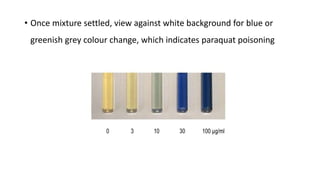
Paraquat Poisoning
• Introduction
• Methods of poisoning
• Kinetics
• Pharmacology and cellular toxicity
• Clinical feature
• Diagnosis
• Management
• Prognosis
Introduction
• Incidence of paraquat poisoning is 3.8/1,00,000 in a year
• A Bipyridyl compound, rapidly acting nonselective herbicide
• Mechanism of toxicity to plants includes inhibition of photosynthesis,
respiration, protein synthesis, growth stimulation
• Manufactured as liquid granules / aerosols
• Combined with Diquat – contains dye, emetic, stenching agent (to
reduce toxicity on ingestion)
• Mortality rates of 72.7% in a study conducted in 55 people
• 3,00,000 occur in Asian-pacific region due to paraquat poisoning
Methods of poisoning
• Ingestion – responsible for majority of deaths
• Transdermal absorption is minimal in the absence of skin lesions, but
deaths have been reported
• Inhalation / exposure to sprays – irritate conjunctiva and airway,
unlikely to cause systemic toxicity
Kinetics
• Highly polar and corrosive
• Not absorbed in significant amounts across intact skin or inhaled
droplets
• Rapidly but incompletely absorbed from gut (jejunum - 17.6%).
• Lethal oral dose of 20% concentrate solution is about
: 10-20 ml in adult
: 4-5 ml in children
• Plasma concentration peaks with in 2 hours of ingestion
• Tissue concentration peaks by 6 hours
• Elimination is mainly by kidneys unchanged form, most ingested
paraquat appears in the urine with in 24 hours in minor poisoning
• In severe poisoning kidney function is greatly reduced leading to
much slower elimination
Pharmacology and cellular toxicology
• Actively accumulate in many cells where it under goes redox cycling
and form superoxide radical, a highly reactive oxygen spices
• Superoxide causes direct cellular damage or react further to form
other ROS and nitrite radicals
• Redox cycling consumes NADPH (anti-oxidant)
• The resultant oxidative stress created by production of free radicals
and depletion of NADPH causes cell damage (lipid peroxidation,
mitochondrial dysfunction, necrosis and apoptosis) and triggers
secondary inflammation
• Over a period of hours to days these process lead to multiorgan
dysfunction
• Most organs affected are those with high blood flow and oxygen
tension and energy requirements – lungs, heart, kidneys and liver
• Brain is uncommonly affected as it doesn’t cross BBB
• Lung injury has two phases
• Initial destruction phase – loss of type 1 and 2 alveolar cells, infiltration
by inflammatory cells, haemorrhage. Changes are reversible
• Later proliferative phase – fibrosis of interstitium and alveolar spaces
• Paraquat and oxygen enhance each other toxicity sustaining redox cycle
• Myocardial injury and necrosis of adrenal gland
Clinical features
• Depends on quantity, route of exposure
• Skin exposure causes local skin irritation and ulceration of epithelial
surfaces due to severe caustic
• Eye exposure causes corneal injury due to severe corrosive action
• Upper respiratory tract exposure causes mucosal injury and epistaxis
due to corrosive action
• Inhalation – cough, dyspnoea, chest pain
• Ingestion – GI irritation and mucosal damage with ulceration, painful
mouth and pain with swallowing, nausea, vomiting and abdominal
pain
On examination
• Mouth and pharynx charred, ulcers
• Tongue dry due to vomiting, poor oral intake due to odynophagia
• Tachypnoea, tachycardia, hypotension
• Bilateral crepitation due to alveolitis
• Subcutaneous emphysema indicating mediastinitis
• Diffuse abdominal tenderness
Paraquat tongue early lesion, within 24
hours after ingestion
Paraquat tongue late lesion, 2 weeks after
ingestion with extensive ulceration
Classification based on clinical features
• Mild
• Severe
• Fulminant
Mild Asymptomatic
Nausea, vomiting, diarrhoea
Renal and hepatic injury
minimal/absent
Reduced pulmonary diffusion
capacity
Complete recovery expected
<20mg/kg
Or
<7.5ml of 20% concentration
solution in adult
Severe Initially nausea, vomiting,
diarrhoea
Abdominal pain
Mouth and throat ulcers
Positive colorimetric test for
paraquat in urine
1-4 days, renal failure, hepatic
impairment, hypotension
1-2 weeks cough, haemoptysis,
pleural fibrosis
Majority die within 2-3 weeks due
to pulmonary failure
20-40mg/kg
Or
7.5-15ml of 20% concentration in
adult
Fulminant Initially nausea, vomiting,
diarrhoea
Pain abdomen
Rapid development of renal and
hepatic failure
GI ulcers
Pancreatitis
Toxic myocarditis
Refractory hypotension
Convulsions
Coma
Death due to cardiogenic shock
and multiorgan failure with in 1-4
days
>40-50mg/kg
Or
>15-20ml of 20% concentration
solution
• Multisystem effects include GI tract corrosion, acute renal failure,
cardiac failure, hepatic failure, extensive pulmonary injury few hours
after ingestion
• Renal failure and hepatic cellular necrosis develop between 2nd – 5th day
• Progressive pulmonary fibrosis leading to refractory hypoxemia occur 5
days to several weeks
• Metabolic acidosis common due to hypoxia combined with multisystem
failure
Investigations
• Electrolytes
• RFT – acute kidney injury due to acute tubular necrosis, volume
depletion. Most commonly used to asses kidney function are
Creatinine and Cystatin C
• Rate of serum creatinine correlates with survival
• Amylase and lipase – acute pancreatitis in case patient develops pain
abdomen
• Increase in S . Creatinine
- <0.034mg/dL/hr over 5 hours – recovery expected
- >0.045mg/dL/hr over 6 hours
or
- increase in serum cystatin concentration of >0.09mg/L
over 6 hours – death
• Creatinine also increases due to muscle oxidative stress
• Therefore is not a marker of GFR in paraquat poisoning
• Alkalosis due to excessive vomiting
• Acidosis - respiratory acidosis (alveolitis or aspiration pneumonia) and
metabolic acidosis (diarrhoea, AKI, mitochondrial toxicity, hypotension)
• Lactic acidosis in severe poisoning due to multiorgan dysfunction,
hypotension, hypoxic acute respiratory distress syndrome
• Serum lactate above 4.4mmol/L or 3.35mmol/L associated with fatal
outcome ( sensitivity 82% and 74%, specificity 88% and 91%) and hence
aid in prognosis
• Chest X-Ray - diffuse pulmonary infiltrates bilaterally due to poison,
local infiltrates due to aspiration (more on the right)
pneumomediastinum, pneumothorax due to corrosive action
• Urinary dithionate test – in alkaline medium sodium dithionate
reduces paraquat to blue radical. Test should performed on fresh
urine sample around 6 hours after ingestion detect paraquat within
few hours after ingestion, semiquantitative test
• If urine paraquat concentration is more than 1mg/L the urine will turn
blue in the test (poor prognosis)
• The addition of sodium bicarbonate and sodium dithionite to
urine specimens containing decreasing concentrations of
paraquat shows characteristic color changes.
• Each number represents decreasing concentrations of
paraquat.
• Paraquat detection kits – 10ml urine to collected in a clear container,
add sachet A containing sodium bicarbonate, shake. Add sachet B
containing sodium dithionate wait for effervescence to subside and
shake again.
• Once mixture settled, view against white background for blue or
greenish grey colour change, which indicates paraquat poisoning
• Serum paraquat concentration – serum concentration levels to time
of poisoning predicts likelihood of death. A fatal outcome is likely if
the plasma concentration lies
above any line. (Sample to be drawn
at least after 4 hours of ingestion)
• Qualitative serum testing – conduct dithionate test in plasma with
positive urinary dithionate test. Equivocal colour change associated
with 50% mortality, definitive colour change associated with 100%
mortality
HRCT one year following a paraquat ingestion,
showing pulmonary fibrosis, more in the left lung.
Diagnosis
• History of ingestion
• Oropharyngeal burns and subsequent development of AKI, metabolic
acidosis, ARDS
• Confirmed with urinary or blood dithionate test
Management
• Survival rate is about 13% with no treatment and increases to 73% with
active treatment
• Resuscitation – assessment of airway, breathing and circulation
• Airway compromised due to mucosal damage or presence of vomitus
• Tachypnoea and/or hypoxia due to metabolic acidosis, aspiration, acute
alveolitis
• Mild to moderate hypoxia should not be treated with oxygen as worsens
oxidative stress
• Hypotension to be treated with crystalloids 15-20ml/kg over 15-30
minutes
• Maintaining high urine out put is desirable (how much?)
• Impaired consciousness indicates severe toxicity resulting in altered
consciousness from hypoxia, hypotension and severe acidosis or
co-ingestion of other agents like ethanol – intubated the patient
• Decontamination - gastric lavage with in 2 – 4hours, followed by
activated charcoal or Fuller’s earth. Lavage not indicated in as it is
contraindicated in caustic injury
• Nasogastric tube – pharyngeal or oesophageal burns, painful
swallowing
• IVF – hypotension
• Acute renal failure – daily fluid balance to be maintained with
ensuring good urine output
• Pain relief and sedation – opioids and benzodiazepines
• Hemoperfusion/haemodialysis – if presents wit in 2 hours of
ingestion in case of AKI without pneumonitis.
- Benefits are limited, endogenous clearance is high in first 6-12
hours and additional amount eliminated will be relatively modest.
- Subsequent elimination of accumulated paraquat from the lungs
is minimally dependent on plasma concentration
• Immunosuppression – suppressing acute inflammatory response, thus
reducing chances of lung fibrosis and death
- Cyclophosphamide, MESNA, Methylprednisolone and Dexamethasone
- Dexamethasone increases the expression of P-glycoprotein
- Significant reduction of paraquat accumulation in the lungs and increase
faecal excretion
- Has also shown to ameliorate histological and biochemical changes and
reduce lipid perioxidation
• 1g of cyclophosphamide daily for 2 days and 1g of
methylprednisolone daily for 3 days
Study Description Treatment Mortality
Addo & Poon-King [84] Uncontrolled 72
patients
CP, Dex, MP, vitamin B,
vitamin C
28%
Afzali & Gholyaf [87] RCT 20 patients CP, Dex, MP vs. conventional
treatment
33% vs. 81%
Perriens et al. [89] Uncontrolled 47 patients CP, Dex vs. conventional treatment 63% vs 61%
Lin et al. [86] RCT 23 patients CP, Dex, MP vs. conventional
treatment
31% vs 86%
Lin et al. [80] RCT 50 patients CP, Dex, MP vs. conventional
treatment
68% vs 82%
Lin et al. [85] 16 patients 17 historic
controls
CP, Dex, MP 25% vs 70%
Yasaka et al. [28] Uncontrolled 9 patients Vitamin E 100–4000 mg/day 78%
Hong et al. [99] Uncontrolled 5 patients Vitamin C escalating doses 0%
• Anti-oxidants
• Vitamin E – vitamin E stabilises membrane and scavenges ROS.
- Vitamin E deficiency rats had lower lethal dose and shorter
survival time, vitamin E treated rat lungs after 24 hours of exposure
showed less peroxidation
- human study only 2/9 patients survived
- 200-400mg/day
• Vitamin C – donate electron to free radicals and neutralise. Study
done on 10 patients, positive urine dithionate test and stable vitals
high dose of vitamin c and other oxidants were given, increased anti-
oxidants in the body
• N-acetylcysteine – replenishes cysteine, rate limiting in synthesis of
glutathione. Reduced paraquat induced apoptosis and inflammatory
response
- S - carboxymethycysteine 1500mg/day
• Deferoxamine – iron contributes to generation of free radicals by
Fenton’s reaction. Iron chelator used with no survival benefits
• Salicylic acid – inhibits cyclo-oxygenase. Scavenges hydroxyl radicals
and inhibit their production by Fenton’s reaction
- reduces inflammatory mediators IL4, oxidative stress, NF, lipid
peroxidation, platelet activation and histological lung damage
- single dose of salicylic acid 200mg/kg, showed no deaths when
compared to control group
Prognosis
• Details of exposure including time, route, accidental/intentional,
amount, concentration
• Quantity related to outcome
• >50 years and H/O renal disease worst prognosis
• S. Lactate aid prognosis
Thank you
Paraquat toxicity explained.pptx

Paraquat toxicity explained.pptx

  • 1.
  • 2.
    • Introduction • Methodsof poisoning • Kinetics • Pharmacology and cellular toxicity • Clinical feature • Diagnosis • Management • Prognosis
  • 3.
    Introduction • Incidence ofparaquat poisoning is 3.8/1,00,000 in a year • A Bipyridyl compound, rapidly acting nonselective herbicide • Mechanism of toxicity to plants includes inhibition of photosynthesis, respiration, protein synthesis, growth stimulation • Manufactured as liquid granules / aerosols • Combined with Diquat – contains dye, emetic, stenching agent (to reduce toxicity on ingestion)
  • 4.
    • Mortality ratesof 72.7% in a study conducted in 55 people • 3,00,000 occur in Asian-pacific region due to paraquat poisoning
  • 5.
    Methods of poisoning •Ingestion – responsible for majority of deaths • Transdermal absorption is minimal in the absence of skin lesions, but deaths have been reported • Inhalation / exposure to sprays – irritate conjunctiva and airway, unlikely to cause systemic toxicity
  • 6.
    Kinetics • Highly polarand corrosive • Not absorbed in significant amounts across intact skin or inhaled droplets • Rapidly but incompletely absorbed from gut (jejunum - 17.6%).
  • 7.
    • Lethal oraldose of 20% concentrate solution is about : 10-20 ml in adult : 4-5 ml in children • Plasma concentration peaks with in 2 hours of ingestion • Tissue concentration peaks by 6 hours • Elimination is mainly by kidneys unchanged form, most ingested paraquat appears in the urine with in 24 hours in minor poisoning
  • 8.
    • In severepoisoning kidney function is greatly reduced leading to much slower elimination
  • 9.
    Pharmacology and cellulartoxicology • Actively accumulate in many cells where it under goes redox cycling and form superoxide radical, a highly reactive oxygen spices • Superoxide causes direct cellular damage or react further to form other ROS and nitrite radicals • Redox cycling consumes NADPH (anti-oxidant)
  • 10.
    • The resultantoxidative stress created by production of free radicals and depletion of NADPH causes cell damage (lipid peroxidation, mitochondrial dysfunction, necrosis and apoptosis) and triggers secondary inflammation • Over a period of hours to days these process lead to multiorgan dysfunction
  • 11.
    • Most organsaffected are those with high blood flow and oxygen tension and energy requirements – lungs, heart, kidneys and liver • Brain is uncommonly affected as it doesn’t cross BBB
  • 12.
    • Lung injuryhas two phases • Initial destruction phase – loss of type 1 and 2 alveolar cells, infiltration by inflammatory cells, haemorrhage. Changes are reversible • Later proliferative phase – fibrosis of interstitium and alveolar spaces • Paraquat and oxygen enhance each other toxicity sustaining redox cycle • Myocardial injury and necrosis of adrenal gland
  • 13.
    Clinical features • Dependson quantity, route of exposure • Skin exposure causes local skin irritation and ulceration of epithelial surfaces due to severe caustic • Eye exposure causes corneal injury due to severe corrosive action • Upper respiratory tract exposure causes mucosal injury and epistaxis due to corrosive action
  • 14.
    • Inhalation –cough, dyspnoea, chest pain • Ingestion – GI irritation and mucosal damage with ulceration, painful mouth and pain with swallowing, nausea, vomiting and abdominal pain
  • 15.
    On examination • Mouthand pharynx charred, ulcers • Tongue dry due to vomiting, poor oral intake due to odynophagia • Tachypnoea, tachycardia, hypotension • Bilateral crepitation due to alveolitis • Subcutaneous emphysema indicating mediastinitis • Diffuse abdominal tenderness
  • 16.
    Paraquat tongue earlylesion, within 24 hours after ingestion Paraquat tongue late lesion, 2 weeks after ingestion with extensive ulceration
  • 17.
    Classification based onclinical features • Mild • Severe • Fulminant
  • 18.
    Mild Asymptomatic Nausea, vomiting,diarrhoea Renal and hepatic injury minimal/absent Reduced pulmonary diffusion capacity Complete recovery expected <20mg/kg Or <7.5ml of 20% concentration solution in adult Severe Initially nausea, vomiting, diarrhoea Abdominal pain Mouth and throat ulcers Positive colorimetric test for paraquat in urine 1-4 days, renal failure, hepatic impairment, hypotension 1-2 weeks cough, haemoptysis, pleural fibrosis Majority die within 2-3 weeks due to pulmonary failure 20-40mg/kg Or 7.5-15ml of 20% concentration in adult
  • 19.
    Fulminant Initially nausea,vomiting, diarrhoea Pain abdomen Rapid development of renal and hepatic failure GI ulcers Pancreatitis Toxic myocarditis Refractory hypotension Convulsions Coma Death due to cardiogenic shock and multiorgan failure with in 1-4 days >40-50mg/kg Or >15-20ml of 20% concentration solution
  • 20.
    • Multisystem effectsinclude GI tract corrosion, acute renal failure, cardiac failure, hepatic failure, extensive pulmonary injury few hours after ingestion • Renal failure and hepatic cellular necrosis develop between 2nd – 5th day • Progressive pulmonary fibrosis leading to refractory hypoxemia occur 5 days to several weeks • Metabolic acidosis common due to hypoxia combined with multisystem failure
  • 21.
    Investigations • Electrolytes • RFT– acute kidney injury due to acute tubular necrosis, volume depletion. Most commonly used to asses kidney function are Creatinine and Cystatin C • Rate of serum creatinine correlates with survival • Amylase and lipase – acute pancreatitis in case patient develops pain abdomen
  • 22.
    • Increase inS . Creatinine - <0.034mg/dL/hr over 5 hours – recovery expected - >0.045mg/dL/hr over 6 hours or - increase in serum cystatin concentration of >0.09mg/L over 6 hours – death • Creatinine also increases due to muscle oxidative stress • Therefore is not a marker of GFR in paraquat poisoning
  • 23.
    • Alkalosis dueto excessive vomiting • Acidosis - respiratory acidosis (alveolitis or aspiration pneumonia) and metabolic acidosis (diarrhoea, AKI, mitochondrial toxicity, hypotension) • Lactic acidosis in severe poisoning due to multiorgan dysfunction, hypotension, hypoxic acute respiratory distress syndrome • Serum lactate above 4.4mmol/L or 3.35mmol/L associated with fatal outcome ( sensitivity 82% and 74%, specificity 88% and 91%) and hence aid in prognosis
  • 24.
    • Chest X-Ray- diffuse pulmonary infiltrates bilaterally due to poison, local infiltrates due to aspiration (more on the right) pneumomediastinum, pneumothorax due to corrosive action
  • 25.
    • Urinary dithionatetest – in alkaline medium sodium dithionate reduces paraquat to blue radical. Test should performed on fresh urine sample around 6 hours after ingestion detect paraquat within few hours after ingestion, semiquantitative test • If urine paraquat concentration is more than 1mg/L the urine will turn blue in the test (poor prognosis)
  • 26.
    • The additionof sodium bicarbonate and sodium dithionite to urine specimens containing decreasing concentrations of paraquat shows characteristic color changes. • Each number represents decreasing concentrations of paraquat.
  • 27.
    • Paraquat detectionkits – 10ml urine to collected in a clear container, add sachet A containing sodium bicarbonate, shake. Add sachet B containing sodium dithionate wait for effervescence to subside and shake again.
  • 28.
    • Once mixturesettled, view against white background for blue or greenish grey colour change, which indicates paraquat poisoning
  • 29.
    • Serum paraquatconcentration – serum concentration levels to time of poisoning predicts likelihood of death. A fatal outcome is likely if the plasma concentration lies above any line. (Sample to be drawn at least after 4 hours of ingestion)
  • 30.
    • Qualitative serumtesting – conduct dithionate test in plasma with positive urinary dithionate test. Equivocal colour change associated with 50% mortality, definitive colour change associated with 100% mortality
  • 31.
    HRCT one yearfollowing a paraquat ingestion, showing pulmonary fibrosis, more in the left lung.
  • 32.
    Diagnosis • History ofingestion • Oropharyngeal burns and subsequent development of AKI, metabolic acidosis, ARDS • Confirmed with urinary or blood dithionate test
  • 33.
    Management • Survival rateis about 13% with no treatment and increases to 73% with active treatment • Resuscitation – assessment of airway, breathing and circulation • Airway compromised due to mucosal damage or presence of vomitus • Tachypnoea and/or hypoxia due to metabolic acidosis, aspiration, acute alveolitis • Mild to moderate hypoxia should not be treated with oxygen as worsens oxidative stress
  • 34.
    • Hypotension tobe treated with crystalloids 15-20ml/kg over 15-30 minutes • Maintaining high urine out put is desirable (how much?) • Impaired consciousness indicates severe toxicity resulting in altered consciousness from hypoxia, hypotension and severe acidosis or co-ingestion of other agents like ethanol – intubated the patient
  • 35.
    • Decontamination -gastric lavage with in 2 – 4hours, followed by activated charcoal or Fuller’s earth. Lavage not indicated in as it is contraindicated in caustic injury • Nasogastric tube – pharyngeal or oesophageal burns, painful swallowing • IVF – hypotension • Acute renal failure – daily fluid balance to be maintained with ensuring good urine output
  • 36.
    • Pain reliefand sedation – opioids and benzodiazepines • Hemoperfusion/haemodialysis – if presents wit in 2 hours of ingestion in case of AKI without pneumonitis. - Benefits are limited, endogenous clearance is high in first 6-12 hours and additional amount eliminated will be relatively modest. - Subsequent elimination of accumulated paraquat from the lungs is minimally dependent on plasma concentration
  • 37.
    • Immunosuppression –suppressing acute inflammatory response, thus reducing chances of lung fibrosis and death - Cyclophosphamide, MESNA, Methylprednisolone and Dexamethasone - Dexamethasone increases the expression of P-glycoprotein - Significant reduction of paraquat accumulation in the lungs and increase faecal excretion - Has also shown to ameliorate histological and biochemical changes and reduce lipid perioxidation
  • 38.
    • 1g ofcyclophosphamide daily for 2 days and 1g of methylprednisolone daily for 3 days
  • 39.
    Study Description TreatmentMortality Addo & Poon-King [84] Uncontrolled 72 patients CP, Dex, MP, vitamin B, vitamin C 28% Afzali & Gholyaf [87] RCT 20 patients CP, Dex, MP vs. conventional treatment 33% vs. 81% Perriens et al. [89] Uncontrolled 47 patients CP, Dex vs. conventional treatment 63% vs 61% Lin et al. [86] RCT 23 patients CP, Dex, MP vs. conventional treatment 31% vs 86% Lin et al. [80] RCT 50 patients CP, Dex, MP vs. conventional treatment 68% vs 82% Lin et al. [85] 16 patients 17 historic controls CP, Dex, MP 25% vs 70% Yasaka et al. [28] Uncontrolled 9 patients Vitamin E 100–4000 mg/day 78% Hong et al. [99] Uncontrolled 5 patients Vitamin C escalating doses 0%
  • 40.
    • Anti-oxidants • VitaminE – vitamin E stabilises membrane and scavenges ROS. - Vitamin E deficiency rats had lower lethal dose and shorter survival time, vitamin E treated rat lungs after 24 hours of exposure showed less peroxidation - human study only 2/9 patients survived - 200-400mg/day
  • 41.
    • Vitamin C– donate electron to free radicals and neutralise. Study done on 10 patients, positive urine dithionate test and stable vitals high dose of vitamin c and other oxidants were given, increased anti- oxidants in the body • N-acetylcysteine – replenishes cysteine, rate limiting in synthesis of glutathione. Reduced paraquat induced apoptosis and inflammatory response - S - carboxymethycysteine 1500mg/day
  • 42.
    • Deferoxamine –iron contributes to generation of free radicals by Fenton’s reaction. Iron chelator used with no survival benefits • Salicylic acid – inhibits cyclo-oxygenase. Scavenges hydroxyl radicals and inhibit their production by Fenton’s reaction - reduces inflammatory mediators IL4, oxidative stress, NF, lipid peroxidation, platelet activation and histological lung damage - single dose of salicylic acid 200mg/kg, showed no deaths when compared to control group
  • 43.
    Prognosis • Details ofexposure including time, route, accidental/intentional, amount, concentration • Quantity related to outcome • >50 years and H/O renal disease worst prognosis • S. Lactate aid prognosis
  • 44.