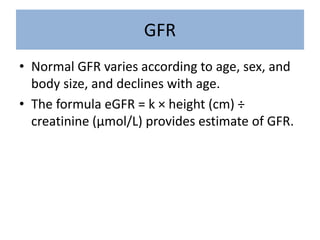
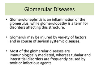
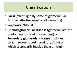
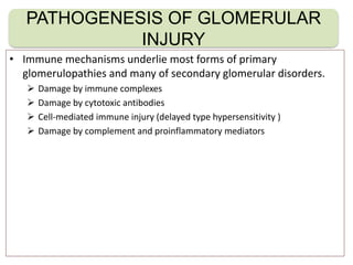
Glomerular Diseases
Natangwe Shimhanda
MBChB V
Glomerulus
• A tuft of capillaries situated within a
Bowman's capsule at the end of a renal tubule
in the vertebrate kidney that filters waste
products from the blood and thus initiates
urine formation.
• The basic filtration unit of the kidney.
Anatomy and Function
• Molecules <1.8nm are freely filtered e.g water,
sodium, insulin, glucose
• Molecules > 3.6nm are not filtered e.g
hemoglobin
Renal Corpuscle
3 Layers of the Glomerulus
Ultrafiltration Barrier
• Capillary Endothelium with fenestration pores
filters everything except blood cells
• Basement membrane prevents filtration of
large proteins
• Podocytes make the outer layer, and they
have pedicels that allow only small molecules
to pass through.
Filtration forces
• Filtration Depends on the Starling forces
– Oncotic pressure exerted by the plasma proteins in the
glomerular capillaries and bowman’s capsule
– Hydrostatic pressure exerted by the fluid in the capillary
walls
Glomerular Filtration Rate
• Total amount of filtrate in all the renal
corpuscles in both kidneys per minute.
• Assesses kidney function
• Takes into account Net Filtration pressure
(NFP), the surface area available for filtration
and the permeability of the glomeruli
Creatinine Clearance
• Males
{(140-age)*wt*1.22}/Cr
• Females
{(140-age)*wt*1.22}/Cr
multiply the answer by 0.85
• Ranges M 97-137
F 88-128
GFR
• Normal GFR varies according to age, sex, and
body size, and declines with age.
• The formula eGFR = k × height (cm) ÷
creatinine (μmol/L) provides estimate of GFR.
Glomerular Diseases
• Glomerulonephritis is an inflammation of the
glomerulus, while glomerulopathy is a term for
disorders affecting this structure.
• Glomeruli may be injured by variety of factors
and in course of several systemic diseases.
• Most of the glomerular diseases are
immunologically mediated, whereas tubular and
interstitial disorders are frequently caused by
toxic or infectious agents.
Classification
• Focal (affecting only some of glomeruli) or
Diffuse (affecting most or all glomeruli)
• Segmental/Global
• Primary glomerular disease (glomeruli are the
predominant site of involvement) or
Secondary glomerular disease (includes
certain systemic and hereditary diseases
which secondarily involve the glomeruli)
• Immune mechanisms underlie most forms of primary
glomerulopathies and many of secondary glomerular disorders.
 Damage by immune complexes
 Damage by cytotoxic antibodies
 Cell-mediated immune injury (delayed type hypersensitivity )
 Damage by complement and proinflammatory mediators
PATHOGENESIS OF GLOMERULAR
INJURY
METABOLIC GLOMERULAR INJURY
Diabetic nephropathy, Fabry’s disease
HAEMODYNAMIC GLOMERULAR INJURY
Systemic hypertension
DEPOSITION DISEASES
Amyloidosis, cryoglobulinaemia.
INFECTIOUS DISEASES
HBV, HCV, HIV, E.coli-derived nephrotoxin
INHERITED GLOMERULAR DISEASES
Alports syndrome
NON IMMUNE MECHANISM OF CELLULAR
INJURY IN GLOMERULUS
1. Proteinuria
• Transient proteinuria may occur during febrile
illnesses or after exercise and does not require
investigation.
• Persistent proteinuria is significant and should
be quantified by measuring the urine
protein/creatinine ratio in an early morning
sample (protein should not exceed 20
mg/mmol of creatinine).
Causes of proteinuria
• Orthostatic proteinuria
• Glomerular abnormalities
– Minimal change disease
– Glomerulonephritis
– Abnormal glomerular basement membrane (familial
nephritides)
• Increased glomerular filtration pressure
• Reduced renal mass
• Hypertension
• Tubular proteinuria.
(i) Nephrotic syndrome
A nonspecific disorder in which the kidneys are
damaged, causing them to leak large amounts
of protein from the blood into the urine.
Clinical state characterised by:
• Heavy proteinuria, 50mg/kg
• Hypoalubinaemia, 25gm/l
• Oedema
• Generalised hyperlipaedaemia
• Primary causes
– Minimal-change
nephropathy
– Focal glomerulosclerosis
– Membranous nephropathy
– Hereditary nephropathies
Nephrotic syndrome
• Secondary causes
– Diabetes mellitus
– Amyloidosis and paraproteinemias
– Post infectious-
– Group A beta haem strep and other bacteria eg.
Typhoid and syphilis
– Malaria
– Viral-chickenpox, HIV, Hep B, EBV
– Renal vein thrombosis
– Collagen vascular-SLE,
Nephrotic syndrome
Causes Cont’
• Hereditary nephritis- nail patella and Alport’s
• Sickle cell disease
• Malignancy: leukaemia, lymphoma, wilm’s
• Toxins: Bee stings, poison ivy, oak, snake
vernom
• Drugs: probrnicid, captopril, heroin, mercury,
gold, penicillamine etc
• Signs and symptoms
– Hypoalbuminemia
– Edema
– Hypercholesterolemia
– Hypoalbuminemia
– Tiredness
– Leukonychia
– Breathlessness
– fluid overload
– Dyslipidaemia
Nephrotic syndrome
• TREATMENT
–Supportive Treatment
• Monitoring and maintaining euvolemia
–Monitoring urine output, BP regularly
–Fluid restrict to 1L.
–Diuretics (IV furosemide).
Nephrotic syndrome
• TREATMENT
–Supportive Treatment
• Monitoring kidney function
–Do U&E daily and calculating GFR
–Treat hyperlipidemia to prevent further
atherosclerosis.
–Prevent and treat any complications
Nephrotic syndrome
• TREATMENT
–Specific Treatment
• Immunosuppression for the
glomerulonephritides
• Corticosteroid therapy: 60 mg/m2 per
day of prednisolone
Nephrotic syndrome
Treatment
• The steroid sensitive Nephrotic Syndrome resolves
spontaneously after a corticosteroid therapy course.
A renal biopsy must be done if unresponsive or
atypical features.
• Steroid-resistant nephrotic syndrome: Management
of the oedema is by diuretic therapy, salt restriction,
ACE inhibitors and sometimes NSAIDs which may
reduce proteinuria
(ii) Congenital nephrotic Syndrome
• Presents in utero, from birth up to 3/12
• Infantile: 4/12 up to 1 year of life.
Primary Causes
• Infantile microcytic disease
• Diffuse mesangial sclerosis (Drash)
• Minimal change lesion
• Focal Glomerulosclerosis
Secondary congenital and infantile:
• Infections: syphilis, Toxoplasmosis, CMV,
Rubella, Malaria
• Toxic: mercury
• SLE
• HUS
• Drug reaction
• Hepatoblastoma
• Syndromes: Nail patella, Lowe syndrome etc
Epidemiology:
• 2-7 children per 100,000 children/year
• 15 times more common in children than
adults
• Incidence higher in black children
• M:F=2:1
Pathophysiology:
• Protein: normal urine in children has 100mg
protein/24hrs (adults 150mg).
• Consists of albumin and Tamm-Horsfall
protein of tubular origin.
Increased protein loss is due to:
• Increased capillary permeability.
• Increased in size of pores of GBM
Pathophysiology continue:
• Selectivity: type of protein lost varies with the
underlying glomerular disease.
• Minimal change- highly selective, other
glomerular disease non selective.
• Hypoalbuminaemia: due to excessive urinary
loss of albumin.
• Oedema: cardinal feature of nephrotic due to
decreased plasma oncotic pressure
Clinical manifestations:
• Oedema usual and a predominant features
• Gastrointestinal disturbances e.g
• Dirrhoea (oedema of intestinal mucosa)
• hepatomegaly
• Abdominal pain- peritonitis, UTI
• Tachypnoea –pleural effusions, ascites, infection
• Poor appetite
• Psychological functional disturbances
Investigations:
• Proteinuria may be selective or non selective
• Urine Protein/creatinine ratio < 0,2 normal
• 0,1-0,5 minimal
• 0,2-2,0 moderate
• > 2,0 nephrotic
• FBC+ diff, ESR, u+e, Total protein, albumin,
cholesterol, INR, C3/C4, ASOT, Anti-DNASe B,
Hepatitis B, WR, HIV, ANA, CXR, TST, CMP
Complications:
• Renal failure: acute renal failure, HPT
• Infections: bacterial
• Thromboembolism: platelets, protein S&C def
• FTT and malnutrition
• Premature atherosclerosis from hyperlipemia
• Calcium and Vitamin D metabolism
2. Hematuria
• Urine that is red in colour or tests positive for
haemoglobin on urine sticks should be examined
under the microscope to confirm haematuria
(>10 red blood cells per high-power field).
• Glomerular haematuria is suggested by brown
urine, the presence of deformed red cells (which
occurs as they pass through the basement
membrane) and casts, and is often accompanied
by proteinuria.
Causes of Hematuria
Non-glomerular
• Infection (bacterial, viral,
TB, schistosomiasis)
• Trauma to genitalia, urinary
tract or kidneys
• Stones
• Tumours
• Sickle cell disease
• Bleeding disorders
• Renal vein thrombosis
• Hypercalciuria.
Glomerular
• Acute glomerulonephritis
(usually with proteinuria)
• Chronic glomerulonephritis
(usually with proteinuria)
• IgA nephropathy
• Familial nephritis, e.g.
Alport syndrome
• Thin basement membrane
disease.
Acute nephritis
• Increased glomerular cellularity restricts
glomerular blood flow and therefore filtration
is decreased. This leads to:
– decreased urine output and volume overload
– hypertension, which may cause seizures
– oedema, characteristically around the eyes
– haematuria and proteinuria.
Causes
• Post-infectious (including streptococcus)
• Vasculitis (Henoch–Schönlein purpura or,
rarely, SLE, Wegener granulomatosis,
microscopic polyarteritis, polyarteritis nodosa)
• IgA nephropathy and mesangiocapillary
glomerulonephritis
• Anti-glomerular basement membrane disease
(Goodpasture syndrome) – very rare.
Management
• Management is by attention to both water and
electrolyte balance and the use of diuretics when
necessary.
• Rarely, there may be a rapid deterioration in renal
function (rapidly progressive glomerulonephritis).
• This may occur with any cause of acute nephritis, but is
uncommon when the cause is post-streptococcal.
• If left untreated, irreversible renal failure may occur
over weeks or months, so renal biopsy and subsequent
treatment with immunosuppression and plasma
exchange may be necessary.
Post-streptococcal and
post-infectious nephritis
• Usually follows a streptococcal sore throat or skin
infection and is diagnosed by evidence of a recent
streptococcal infection (culture of the organism,
raised ASO/ anti-DNAse B titres) and low
complement C3 levels that return to normal after
3–4 weeks.
• Streptococcal nephritis is a common condition in
developing countries, but has become
uncommon in developed countries.
• Longterm prognosis is good.
Treatment
• Phenoxymethylpenicillin 250mg/5ml P.O Q.I.D
5/7 or
• Azithromycin 10mg/kg P.O 3/7
Henoch–Schönlein purpura
• It usually occurs between the ages of 3 and 10 years, is
twice as common in boys, peaks during the winter
months and is often preceded by an upper
respiratoryinfection.
• Despite much research, the cause is unknown.
• It is postulated that genetic predisposition and antigen
exposure increase circulating IgA levels and disrupt IgG
synthesis.
• The IgA and IgG interact to produce complexes that
activate complement and are deposited in affected
organs, precipitating an inflammatory response with
vasculitis.
Henoch–Schönlein purpura
Henoch–Schönlein purpura is
the combination of some of
the following features:
• Characteristic skin rash
• Arthralgia
• Periarticular oedema
• Abdominal pain
• Glomerulonephritis.
Clinical Findings
• At presentation, affected children often have a
fever.
• The rash is the most obvious feature.
• It is symmetrically distributed over the buttocks,
the extensor surfaces of the arms and legs, and
the ankles.
• The trunk is spared unless lesions are induced by
trauma.
• The rash may initially be urticarial, rapidly
becoming maculopapular and purpuric.
Management
• if severe, can be treated with corticosteroids
• All children with renal involvement are
followed for a year to detect those with
persisting urinary abnormalities (5–10%), who
require long-term follow-up.
• This is necessary as hypertension and
declining renal function may develop after an
interval of several years.
IgA nephropathy
• This may present with episodes of
macroscopic haematuria, commonly in
association with upper respiratory tract
infections.
• Histological findings and management are as
for Henoch–Schönlein purpura, which may be
a variant of the same pathological process but
not restricted to the kidney.
Vasculitis
• The commonest vasculitis to involve the kidney is Henoch–Schönlein
purpura (see above).
• However, renal involvement may occur in rarer vasculitides such as
polyarteritis nodosa, microscopic polyarteritis and Wegener
granulomatosis.
• Characteristic symptoms are fever, malaise, weight loss, skin rash and
arthropathy with prominent involvement of the respiratory tract in
Wegener disease.
• ANCA (antineutrophil cytoplasm antibodies) are present and diagnostic in
these diseases.
• Renal arteriography, to demonstrate the presence of aneurysms, will
diagnose polyarteritis nodosa.
• Renal involvement may be severe and rapidly progressive.
• Treatment is with steroids, plasma exchange and intravenous
cyclophosphamide, which may need to be continued for many months.
Systemic lupus erythematosus (SLE)
• SLE is a disease that presents mainly in adolescent girls
and young women.
• It is much commoner in Asians and Afro-Caribbeans
than Caucasians. It is characterized by the presence of
multiple autoantibodies, including antibodies to
double-stranded DNA.
• The C3 and C4 components of complement may be
low, particularly during active phases of the disease.
• Haematuria and proteinuria are indications for renal
biopsy, as immunosuppression is always necessary and
its intensity will depend on the severity of renal
involvement.
Glomerulonephritis
• Also known as glomerular
nephritis (GN) or glomerular disease
• It is a disease of the kidney,
characterized by inflammation of the
glomeruli
• Acute Glomerulonephritis
–Cause
• infections such as strep throat
• Lupus erythematous
• Goodpasture's syndrome
• Wegener's disease
• polyarteritis nodosa
Glomerulonephritis
• Acute Glomerulonephritis
–Symptoms
• Puffy face in the morning
• hematuria (or brown urine)
• oliguria
Glomerulonephritis
• Chronic Glomerulonephritis
–Cause
• This condition may develop after survival
of the acute phase of rapidly progressive
glomerulonephritis
Glomerulonephritis
• Chronic Glomerulonephritis
–Signs and symptoms
• Proteinuria/hematuria
• High blood pressure
• oedema
• nocturia
• Very bubbly or foamy urine
Glomerulonephritis
• Diagnosis
–Urine test
–Blood test
–Throat swab
–Renal function tests
–Kidney biopsy
–Imaging tests
Glomerulonephritis
• Treatment
–Diet salts restrictions and adequate fluid
intake
–The following medications may also be
prescribed to treat possible underlying
causes:
• Bacterial infections - a targeted antibiotic
• Lupus or vasculitis - corticosteroids and
immunosuppressants
Glomerulonephritis
• Treatment
• IgA - possibly fish oil supplements.
• Goodpasture's syndrome -
plasmapheresis is a procedure designed
to reduce blood plasma levels without
depleting the body of its blood cells.
Antibodies are removed and donated
plasma replaces the depleted plasma
Glomerulonephritis
Investigations of Hematuria
All patients
• Urine microscopy (with phase contrast) and culture
• Protein and calcium excretion
• Kidney and urinary tract ultrasound
• Plasma urea, electrolytes, creatinine, calcium, phosphate, albumin
• Full blood count, platelets, clotting screen, sickle cell screen.
If suggestive of glomerular haematuria
• ESR, complement levels and anti-DNA antibodies
• Throat swab and antistreptolysin O/anti-DNAse B titres
• Hepatitis B and C screen
• Renal biopsy if indicated
• Test mother’s urine for blood (if Alport syndrome suspected)
• Hearing test (if Alport syndrome suspected).
Hemolytic Uraemic Syndrome
• Hemolytic-uremic syndrome (HUS) is a clinical
syndrome characterized by progressive renal failure
that is associated with microangiopathic (nonimmune,
Coombs-negative) hemolytic anemia and
thrombocytopenia.
• It predominantly, but not exclusively, affects children.
Most cases are preceded by an episode of infectious,
sometimes bloody, diarrhea acquired as a foodborne
illness or from a contaminated water supply caused by
E. coli O157:H7, other non-o157:H7 E. coli serotypes,
Shigella, and Campylobacter.
• The typical pathophysiology of HUS involves
the binding of Shiga-toxin to the
globotriaosylceramide (Gb3; also called
ceramide trihexoside which accumulates in
Fabry disease) receptor on the surface of the
glomerular endothelium.
Hereditary Glomerulopathies
• Predominant hematuria
– Alport’s syndrome
– Benign Familial hematuria
– Hemolytic Uraemic Syndrome
Alport’s syndrome
• Genetically heterogeneous disease arising
from mutations of the gene coding for type IV
collagen, a major constituent of the basement
membrane.
• Persistent hematuria often progressing
proteinuria, hypertension and renal failure.
• Often associated with high frequency sensori-
neural deafness and ocular defects
• Benign Familial hematuria is usually due to the
thin basement membrane. No associated
deafness and the condition is inherited as an
autosomal dominant.
References
• Lissauer T, Clayden G, 2012. Illustrated
Textbook Of Paediatrics. Fourth edition. ©
2012 Elsevier Ltd
• Wittenberg D.F. (2009), Coovadia’s Paediatrics
and Child Health. Oxford University Press
Southern Africa (pty) Ltd

Glomerular diseases

  • 1.
  • 2.
    Glomerulus • A tuftof capillaries situated within a Bowman's capsule at the end of a renal tubule in the vertebrate kidney that filters waste products from the blood and thus initiates urine formation. • The basic filtration unit of the kidney.
  • 3.
  • 4.
    • Molecules <1.8nmare freely filtered e.g water, sodium, insulin, glucose • Molecules > 3.6nm are not filtered e.g hemoglobin
  • 5.
  • 6.
    3 Layers ofthe Glomerulus
  • 7.
    Ultrafiltration Barrier • CapillaryEndothelium with fenestration pores filters everything except blood cells • Basement membrane prevents filtration of large proteins • Podocytes make the outer layer, and they have pedicels that allow only small molecules to pass through.
  • 8.
    Filtration forces • FiltrationDepends on the Starling forces – Oncotic pressure exerted by the plasma proteins in the glomerular capillaries and bowman’s capsule – Hydrostatic pressure exerted by the fluid in the capillary walls
  • 10.
    Glomerular Filtration Rate •Total amount of filtrate in all the renal corpuscles in both kidneys per minute. • Assesses kidney function • Takes into account Net Filtration pressure (NFP), the surface area available for filtration and the permeability of the glomeruli
  • 11.
    Creatinine Clearance • Males {(140-age)*wt*1.22}/Cr •Females {(140-age)*wt*1.22}/Cr multiply the answer by 0.85 • Ranges M 97-137 F 88-128
  • 12.
    GFR • Normal GFRvaries according to age, sex, and body size, and declines with age. • The formula eGFR = k × height (cm) ÷ creatinine (μmol/L) provides estimate of GFR.
  • 13.
    Glomerular Diseases • Glomerulonephritisis an inflammation of the glomerulus, while glomerulopathy is a term for disorders affecting this structure. • Glomeruli may be injured by variety of factors and in course of several systemic diseases. • Most of the glomerular diseases are immunologically mediated, whereas tubular and interstitial disorders are frequently caused by toxic or infectious agents.
  • 14.
    Classification • Focal (affectingonly some of glomeruli) or Diffuse (affecting most or all glomeruli) • Segmental/Global • Primary glomerular disease (glomeruli are the predominant site of involvement) or Secondary glomerular disease (includes certain systemic and hereditary diseases which secondarily involve the glomeruli)
  • 15.
    • Immune mechanismsunderlie most forms of primary glomerulopathies and many of secondary glomerular disorders.  Damage by immune complexes  Damage by cytotoxic antibodies  Cell-mediated immune injury (delayed type hypersensitivity )  Damage by complement and proinflammatory mediators PATHOGENESIS OF GLOMERULAR INJURY
  • 16.
    METABOLIC GLOMERULAR INJURY Diabeticnephropathy, Fabry’s disease HAEMODYNAMIC GLOMERULAR INJURY Systemic hypertension DEPOSITION DISEASES Amyloidosis, cryoglobulinaemia. INFECTIOUS DISEASES HBV, HCV, HIV, E.coli-derived nephrotoxin INHERITED GLOMERULAR DISEASES Alports syndrome NON IMMUNE MECHANISM OF CELLULAR INJURY IN GLOMERULUS
  • 18.
    1. Proteinuria • Transientproteinuria may occur during febrile illnesses or after exercise and does not require investigation. • Persistent proteinuria is significant and should be quantified by measuring the urine protein/creatinine ratio in an early morning sample (protein should not exceed 20 mg/mmol of creatinine).
  • 19.
    Causes of proteinuria •Orthostatic proteinuria • Glomerular abnormalities – Minimal change disease – Glomerulonephritis – Abnormal glomerular basement membrane (familial nephritides) • Increased glomerular filtration pressure • Reduced renal mass • Hypertension • Tubular proteinuria.
  • 20.
    (i) Nephrotic syndrome Anonspecific disorder in which the kidneys are damaged, causing them to leak large amounts of protein from the blood into the urine. Clinical state characterised by: • Heavy proteinuria, 50mg/kg • Hypoalubinaemia, 25gm/l • Oedema • Generalised hyperlipaedaemia
  • 21.
    • Primary causes –Minimal-change nephropathy – Focal glomerulosclerosis – Membranous nephropathy – Hereditary nephropathies Nephrotic syndrome
  • 22.
    • Secondary causes –Diabetes mellitus – Amyloidosis and paraproteinemias – Post infectious- – Group A beta haem strep and other bacteria eg. Typhoid and syphilis – Malaria – Viral-chickenpox, HIV, Hep B, EBV – Renal vein thrombosis – Collagen vascular-SLE, Nephrotic syndrome
  • 23.
    Causes Cont’ • Hereditarynephritis- nail patella and Alport’s • Sickle cell disease • Malignancy: leukaemia, lymphoma, wilm’s • Toxins: Bee stings, poison ivy, oak, snake vernom • Drugs: probrnicid, captopril, heroin, mercury, gold, penicillamine etc
  • 24.
    • Signs andsymptoms – Hypoalbuminemia – Edema – Hypercholesterolemia – Hypoalbuminemia – Tiredness – Leukonychia – Breathlessness – fluid overload – Dyslipidaemia Nephrotic syndrome
  • 25.
    • TREATMENT –Supportive Treatment •Monitoring and maintaining euvolemia –Monitoring urine output, BP regularly –Fluid restrict to 1L. –Diuretics (IV furosemide). Nephrotic syndrome
  • 26.
    • TREATMENT –Supportive Treatment •Monitoring kidney function –Do U&E daily and calculating GFR –Treat hyperlipidemia to prevent further atherosclerosis. –Prevent and treat any complications Nephrotic syndrome
  • 27.
    • TREATMENT –Specific Treatment •Immunosuppression for the glomerulonephritides • Corticosteroid therapy: 60 mg/m2 per day of prednisolone Nephrotic syndrome
  • 28.
    Treatment • The steroidsensitive Nephrotic Syndrome resolves spontaneously after a corticosteroid therapy course. A renal biopsy must be done if unresponsive or atypical features. • Steroid-resistant nephrotic syndrome: Management of the oedema is by diuretic therapy, salt restriction, ACE inhibitors and sometimes NSAIDs which may reduce proteinuria
  • 29.
    (ii) Congenital nephroticSyndrome • Presents in utero, from birth up to 3/12 • Infantile: 4/12 up to 1 year of life. Primary Causes • Infantile microcytic disease • Diffuse mesangial sclerosis (Drash) • Minimal change lesion • Focal Glomerulosclerosis
  • 30.
    Secondary congenital andinfantile: • Infections: syphilis, Toxoplasmosis, CMV, Rubella, Malaria • Toxic: mercury • SLE • HUS • Drug reaction • Hepatoblastoma • Syndromes: Nail patella, Lowe syndrome etc
  • 31.
    Epidemiology: • 2-7 childrenper 100,000 children/year • 15 times more common in children than adults • Incidence higher in black children • M:F=2:1
  • 32.
    Pathophysiology: • Protein: normalurine in children has 100mg protein/24hrs (adults 150mg). • Consists of albumin and Tamm-Horsfall protein of tubular origin. Increased protein loss is due to: • Increased capillary permeability. • Increased in size of pores of GBM
  • 33.
    Pathophysiology continue: • Selectivity:type of protein lost varies with the underlying glomerular disease. • Minimal change- highly selective, other glomerular disease non selective. • Hypoalbuminaemia: due to excessive urinary loss of albumin. • Oedema: cardinal feature of nephrotic due to decreased plasma oncotic pressure
  • 34.
    Clinical manifestations: • Oedemausual and a predominant features • Gastrointestinal disturbances e.g • Dirrhoea (oedema of intestinal mucosa) • hepatomegaly • Abdominal pain- peritonitis, UTI • Tachypnoea –pleural effusions, ascites, infection • Poor appetite • Psychological functional disturbances
  • 35.
    Investigations: • Proteinuria maybe selective or non selective • Urine Protein/creatinine ratio < 0,2 normal • 0,1-0,5 minimal • 0,2-2,0 moderate • > 2,0 nephrotic • FBC+ diff, ESR, u+e, Total protein, albumin, cholesterol, INR, C3/C4, ASOT, Anti-DNASe B, Hepatitis B, WR, HIV, ANA, CXR, TST, CMP
  • 36.
    Complications: • Renal failure:acute renal failure, HPT • Infections: bacterial • Thromboembolism: platelets, protein S&C def • FTT and malnutrition • Premature atherosclerosis from hyperlipemia • Calcium and Vitamin D metabolism
  • 37.
    2. Hematuria • Urinethat is red in colour or tests positive for haemoglobin on urine sticks should be examined under the microscope to confirm haematuria (>10 red blood cells per high-power field). • Glomerular haematuria is suggested by brown urine, the presence of deformed red cells (which occurs as they pass through the basement membrane) and casts, and is often accompanied by proteinuria.
  • 38.
    Causes of Hematuria Non-glomerular •Infection (bacterial, viral, TB, schistosomiasis) • Trauma to genitalia, urinary tract or kidneys • Stones • Tumours • Sickle cell disease • Bleeding disorders • Renal vein thrombosis • Hypercalciuria. Glomerular • Acute glomerulonephritis (usually with proteinuria) • Chronic glomerulonephritis (usually with proteinuria) • IgA nephropathy • Familial nephritis, e.g. Alport syndrome • Thin basement membrane disease.
  • 39.
    Acute nephritis • Increasedglomerular cellularity restricts glomerular blood flow and therefore filtration is decreased. This leads to: – decreased urine output and volume overload – hypertension, which may cause seizures – oedema, characteristically around the eyes – haematuria and proteinuria.
  • 40.
    Causes • Post-infectious (includingstreptococcus) • Vasculitis (Henoch–Schönlein purpura or, rarely, SLE, Wegener granulomatosis, microscopic polyarteritis, polyarteritis nodosa) • IgA nephropathy and mesangiocapillary glomerulonephritis • Anti-glomerular basement membrane disease (Goodpasture syndrome) – very rare.
  • 41.
    Management • Management isby attention to both water and electrolyte balance and the use of diuretics when necessary. • Rarely, there may be a rapid deterioration in renal function (rapidly progressive glomerulonephritis). • This may occur with any cause of acute nephritis, but is uncommon when the cause is post-streptococcal. • If left untreated, irreversible renal failure may occur over weeks or months, so renal biopsy and subsequent treatment with immunosuppression and plasma exchange may be necessary.
  • 42.
    Post-streptococcal and post-infectious nephritis •Usually follows a streptococcal sore throat or skin infection and is diagnosed by evidence of a recent streptococcal infection (culture of the organism, raised ASO/ anti-DNAse B titres) and low complement C3 levels that return to normal after 3–4 weeks. • Streptococcal nephritis is a common condition in developing countries, but has become uncommon in developed countries. • Longterm prognosis is good.
  • 43.
    Treatment • Phenoxymethylpenicillin 250mg/5mlP.O Q.I.D 5/7 or • Azithromycin 10mg/kg P.O 3/7
  • 44.
    Henoch–Schönlein purpura • Itusually occurs between the ages of 3 and 10 years, is twice as common in boys, peaks during the winter months and is often preceded by an upper respiratoryinfection. • Despite much research, the cause is unknown. • It is postulated that genetic predisposition and antigen exposure increase circulating IgA levels and disrupt IgG synthesis. • The IgA and IgG interact to produce complexes that activate complement and are deposited in affected organs, precipitating an inflammatory response with vasculitis.
  • 45.
    Henoch–Schönlein purpura Henoch–Schönlein purpurais the combination of some of the following features: • Characteristic skin rash • Arthralgia • Periarticular oedema • Abdominal pain • Glomerulonephritis.
  • 46.
    Clinical Findings • Atpresentation, affected children often have a fever. • The rash is the most obvious feature. • It is symmetrically distributed over the buttocks, the extensor surfaces of the arms and legs, and the ankles. • The trunk is spared unless lesions are induced by trauma. • The rash may initially be urticarial, rapidly becoming maculopapular and purpuric.
  • 47.
    Management • if severe,can be treated with corticosteroids • All children with renal involvement are followed for a year to detect those with persisting urinary abnormalities (5–10%), who require long-term follow-up. • This is necessary as hypertension and declining renal function may develop after an interval of several years.
  • 48.
    IgA nephropathy • Thismay present with episodes of macroscopic haematuria, commonly in association with upper respiratory tract infections. • Histological findings and management are as for Henoch–Schönlein purpura, which may be a variant of the same pathological process but not restricted to the kidney.
  • 49.
    Vasculitis • The commonestvasculitis to involve the kidney is Henoch–Schönlein purpura (see above). • However, renal involvement may occur in rarer vasculitides such as polyarteritis nodosa, microscopic polyarteritis and Wegener granulomatosis. • Characteristic symptoms are fever, malaise, weight loss, skin rash and arthropathy with prominent involvement of the respiratory tract in Wegener disease. • ANCA (antineutrophil cytoplasm antibodies) are present and diagnostic in these diseases. • Renal arteriography, to demonstrate the presence of aneurysms, will diagnose polyarteritis nodosa. • Renal involvement may be severe and rapidly progressive. • Treatment is with steroids, plasma exchange and intravenous cyclophosphamide, which may need to be continued for many months.
  • 50.
    Systemic lupus erythematosus(SLE) • SLE is a disease that presents mainly in adolescent girls and young women. • It is much commoner in Asians and Afro-Caribbeans than Caucasians. It is characterized by the presence of multiple autoantibodies, including antibodies to double-stranded DNA. • The C3 and C4 components of complement may be low, particularly during active phases of the disease. • Haematuria and proteinuria are indications for renal biopsy, as immunosuppression is always necessary and its intensity will depend on the severity of renal involvement.
  • 51.
    Glomerulonephritis • Also knownas glomerular nephritis (GN) or glomerular disease • It is a disease of the kidney, characterized by inflammation of the glomeruli
  • 52.
    • Acute Glomerulonephritis –Cause •infections such as strep throat • Lupus erythematous • Goodpasture's syndrome • Wegener's disease • polyarteritis nodosa Glomerulonephritis
  • 53.
    • Acute Glomerulonephritis –Symptoms •Puffy face in the morning • hematuria (or brown urine) • oliguria Glomerulonephritis
  • 54.
    • Chronic Glomerulonephritis –Cause •This condition may develop after survival of the acute phase of rapidly progressive glomerulonephritis Glomerulonephritis
  • 55.
    • Chronic Glomerulonephritis –Signsand symptoms • Proteinuria/hematuria • High blood pressure • oedema • nocturia • Very bubbly or foamy urine Glomerulonephritis
  • 56.
    • Diagnosis –Urine test –Bloodtest –Throat swab –Renal function tests –Kidney biopsy –Imaging tests Glomerulonephritis
  • 57.
    • Treatment –Diet saltsrestrictions and adequate fluid intake –The following medications may also be prescribed to treat possible underlying causes: • Bacterial infections - a targeted antibiotic • Lupus or vasculitis - corticosteroids and immunosuppressants Glomerulonephritis
  • 58.
    • Treatment • IgA- possibly fish oil supplements. • Goodpasture's syndrome - plasmapheresis is a procedure designed to reduce blood plasma levels without depleting the body of its blood cells. Antibodies are removed and donated plasma replaces the depleted plasma Glomerulonephritis
  • 59.
    Investigations of Hematuria Allpatients • Urine microscopy (with phase contrast) and culture • Protein and calcium excretion • Kidney and urinary tract ultrasound • Plasma urea, electrolytes, creatinine, calcium, phosphate, albumin • Full blood count, platelets, clotting screen, sickle cell screen. If suggestive of glomerular haematuria • ESR, complement levels and anti-DNA antibodies • Throat swab and antistreptolysin O/anti-DNAse B titres • Hepatitis B and C screen • Renal biopsy if indicated • Test mother’s urine for blood (if Alport syndrome suspected) • Hearing test (if Alport syndrome suspected).
  • 60.
    Hemolytic Uraemic Syndrome •Hemolytic-uremic syndrome (HUS) is a clinical syndrome characterized by progressive renal failure that is associated with microangiopathic (nonimmune, Coombs-negative) hemolytic anemia and thrombocytopenia. • It predominantly, but not exclusively, affects children. Most cases are preceded by an episode of infectious, sometimes bloody, diarrhea acquired as a foodborne illness or from a contaminated water supply caused by E. coli O157:H7, other non-o157:H7 E. coli serotypes, Shigella, and Campylobacter.
  • 61.
    • The typicalpathophysiology of HUS involves the binding of Shiga-toxin to the globotriaosylceramide (Gb3; also called ceramide trihexoside which accumulates in Fabry disease) receptor on the surface of the glomerular endothelium.
  • 62.
    Hereditary Glomerulopathies • Predominanthematuria – Alport’s syndrome – Benign Familial hematuria – Hemolytic Uraemic Syndrome
  • 63.
    Alport’s syndrome • Geneticallyheterogeneous disease arising from mutations of the gene coding for type IV collagen, a major constituent of the basement membrane. • Persistent hematuria often progressing proteinuria, hypertension and renal failure. • Often associated with high frequency sensori- neural deafness and ocular defects
  • 64.
    • Benign Familialhematuria is usually due to the thin basement membrane. No associated deafness and the condition is inherited as an autosomal dominant.
  • 65.
    References • Lissauer T,Clayden G, 2012. Illustrated Textbook Of Paediatrics. Fourth edition. © 2012 Elsevier Ltd • Wittenberg D.F. (2009), Coovadia’s Paediatrics and Child Health. Oxford University Press Southern Africa (pty) Ltd

Editor's Notes

  • #8 All 3 layers contain negatively charged glycoproteins which makes it difficult for negatively charged molecules to pass through e.g. Albumin is not filtered despite being in the filtration range