Downloaded 147 times

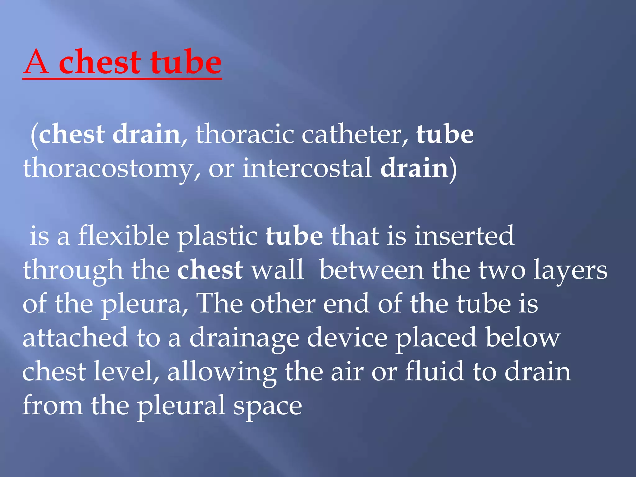
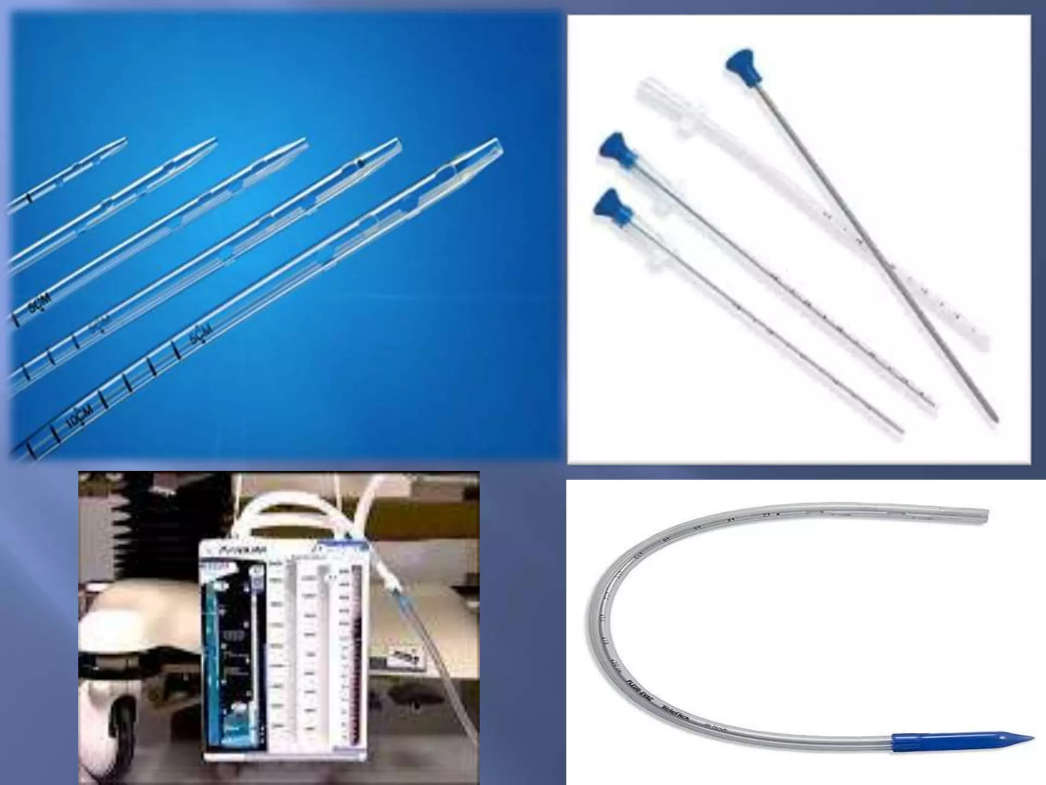


A chest tube is a flexible plastic tube inserted between the pleural layers of the chest wall to drain air or fluid from the pleural space. It is used to treat conditions where air or fluid has accumulated in the pleural space such as pneumothorax, pleural effusions, or hemothorax. The tube is inserted using local anesthesia in the intercostal space and attached to a drainage system placed below chest level. It is secured with sutures and the insertion site is dressed. The tube remains in place until drainage stops and a chest x-ray confirms its proper position and resolution of the underlying condition.


