GLAUCOMA is a chronic eye disease that can damage optic nerve. According to WHO It
is the second leading cause of blindness, and is predicted to affect around 80 million people by 2020.
Development of the disease leads to loss of vision, which occurs increasingly over a long period of
time. As the symptoms only occur when the disease is quite advanced so that glaucoma is called the
silent thief of sight. Glaucoma cannot be cured, but its development can be slowed down by
treatment. Therefore, detecting glaucoma in time is critical. However, many glaucoma patients are
unaware of the disease until it has reached its advanced stage. In this paper, some manual and
automatic methods are discussed to detect glaucoma. Manual analysis of the eye is time consuming
and the accuracy of the parameter measurements also varies with different clinicians. To overcome
these problems with manual analysis, the objective of this survey is to introduce a method to
automatically analyze the ultrasound images of the eye. Automatic analysis of this disease is much
more effective than manual analysis.
Glaucoma is the most leading cause of irreversible blindness with the population of Africa and Asia ranking the highest over the rate of glaucoma affected regions around the world. The defect will damage eyes irreversibly by affecting the optic cup and optic disc of an eye. The early detection of glaucoma is an unavoidable need in the medical field. The widely used technique to detect glaucoma is an invasive method that may lead to other effects on the eye. This reason led to the introduction of a non invasive method that follows image processing for the detection of glaucoma. Retinal image based detection is the best way to choose as it comes under non invasive methods of detection. Detection of glaucoma using retinal images requires various medical features of the eyes such as optic cup diameter, optic disc diameter and optic cup to disc ratio are used. Glaucoma disease detection from retinal images supports convolutional neural networks CNN . The textual features obtained from retinal images such as the optic cup to optic disc measures are used for this classification. Convolutional Neural Networks use little pre processing techniques that can be implemented relatively uncomplicated compared to other image classification techniques. The implementation of this project follows the traditional CNN architecture, applying filter layers such as Convolution layer and Pooling layer and also activation functions such as ReLu function and sigmoid function to pre process as well as to update weights respectively on the hidden layers of the CNN followed by classifying the image. Vishnubhotla Poornasree | Vijayagiri Ashritha | Venumula Deeksha Reddy | J. Srilatha ""Glaucoma Detection from Retinal Images"" Published in International Journal of Trend in Scientific Research and Development (ijtsrd), ISSN: 2456-6470, Volume-3 | Issue-4 , June 2019, URL: https://www.ijtsrd.com/papers/ijtsrd23732.pdf
Paper URL: https://www.ijtsrd.com/computer-science/other/23732/glaucoma-detection-from-retinal-images/vishnubhotla-poornasree
Glaucoma Detection in Retinal Images Using Image Processing Techniques: A SurveyEswar Publications
Glaucoma is a disease associated with human eyes and second conducting movement o fblindness across the globe if
eyes are not treated at preliminary stage. Glaucoma normally occurs with increased intra-ocular pressure (IOP) in eyes and gradually damagesthe vision field of eyes. The term ocular-hypertension is related to those people in whom IOP increases consistently and does not damage the optic nerve. Glaucoma has different types such as open-angle, close-angle, congenital, normal tension and etcetera. Normal tension glaucoma affects vision field and damages optic nerve as well. The term angle means the distance between iris and cornea; if this distance is large it is referred to as open-angle glaucoma and similarly if the distance between iris and cornea is short than this is called close-angle glaucoma. Open-angle glaucoma is common as compared to close-angle glaucoma. Close-angle glaucoma is very painful and affects vision field of eyes quickly as compared to open-angle glaucoma. In this
paper, the state of the art CAD systems and image processing methods are studied and compared systematically in terms of their classification accuracy, methodology approach, sensitivity and specificity. The comparison results indicate that the accuracy of these CAD systems and image processing methods is not up to the mark.
FUZZY CLUSTERING BASED GLAUCOMA DETECTION USING THE CDR sipij
Glaucoma is a serious eye disease, overtime it will result in gradual blindness. Early detection of thedisease will help prevent against developing a more serious condition. A vertical cup-to-disc ratio which isthe ratio of the vertical diameter of the optic cup to that of the optic disc, of the fundus eye image is an important clinical indicator for glaucoma diagnosis. This paper presents an automated method for the extraction of optic disc and optic cup using Fuzzy C Means clustering technique combined with
thresholding. Using the extracted optic disc and optic cup the vertical cup-to-disc ratio was calculated.
The validity of this new method has been tested on 365 colour fundus images from two different publicly
available databases DRION, DIARATDB0 and images from an ophthalmologist. The result of the method
seems to be promising and useful for clinical work.
The International Journal of Engineering & Science is aimed at providing a platform for researchers, engineers, scientists, or educators to publish their original research results, to exchange new ideas, to disseminate information in innovative designs, engineering experiences and technological skills. It is also the Journal's objective to promote engineering and technology education. All papers submitted to the Journal will be blind peer-reviewed. Only original articles will be published.
Detection of Glaucoma using Optic Disk and Incremental Cup Segmentation from ...theijes
Medical researchers, detection of eye disease is very important because it may causes blindness. Glaucoma is one of the diseases that cause blindness. Standard procedure for detection glaucoma is to analysis of optic disk (OD) and cup region in retinal image. In this paper, introduce an automatic OD parameterized technique which is based on segmentation and Incremental Cup segmentation. The incremental cup segmentation method is based on anatomical evidence such as vessel bends at the cup boundary, considered relevant by glaucoma experts. Bends in a vessel are robustly detected using a region of support concept, which automatically selects the right scale for analysis. A multi-stage strategy is applied to derive a reliable subset of vessel bends called r-bends followed by a local 2-D spline fitting to derive the desired cup boundary. The results are compared with existing methods using different retinal images.
Development of novel BMIP algorithms for human eyes affected with glaucoma an...Premier Publishers
Glaucoma is one of the second driving eye maladies on the planet, if not treated legitimately may prompt lasting visual impairment. There are no particular side effects when the glaucoma disease is considered, especially for this type of eye disease, the effect of which is the vision loss in the human eyes. Because of measuring, the container zone increments, which will result in the vision impairment in the human eyes. Normally exceptionally prepared opthalmogists physically review eye pictures as tedious way. In this unique circumstance, we are attempting to build up some novel calculations for programmed recognition of eyes influenced with glaucoma utilizing picture preparing separating and change strategies and actualize the same on equipment utilizing micro-controller framework. The product that will be created by us could be implanted on the equipment to test the sound and undesirable fundus pictures for the recognition of glaucoma. The calculations that could be created can be actualized wrt the eye pictures in HDL language utilizing Xilinx ISE, MATLAB and MODELSIM, TI based unit or NI based pack (any one) is the equipment apparatus that is considered for execution purposes.
International Journal of Computational Engineering Research(IJCER) is an intentional online Journal in English monthly publishing journal. This Journal publish original research work that contributes significantly to further the scientific knowledge in engineering and Technology.
Glaucoma Screening Test By Segmentation of Optical Disc& Cup Segmentation Usi...IJERA Editor
Glaucoma is one of the most common causes of blindness and it is becoming even more important considering
the ageing society. Because healing of died retinal nerve fibers is not possible early detection and prevention is
essential. Robust, automated mass-screening will help to extend the symptom-free life of affected patients. We
devised a novel, automated, appearance based glaucoma classification system that does not depend on
segmentation based measurements. Our purely data-driven approach is applicable in large-scale screening
examinations. The proposed segmentation methods have been evaluated in a database of 650 images with optic
disc and optic cup boundaries manually marked by trained professionals. Our expected Experimental results
may be average overlapping error of 9.5% and 24.1% in optic disc and optic cup segmentation, respectively.
Diabetic retinopathy is a disease, caused by alternation in the retinal blood vessels. It is a strong sign of early blindness and if it is not treated may tend to complete blindness and the vision lost once cannot be restored once again. In this paper different image processing techniques are used to differentiate between the normal and the diseased image. The attempt is made to see where the problem actually lies so that proper diagnosis of patient can be done. Pre processing of an image, optic disk detection, Blood vessels extraction, Exudates detection are some of the methods that are applied here. Other algorithms are designed to obtain the desired result. A large number of populations are affected by this disease around the world.
An Approach for the Detection of Vascular Abnormalities in Diabetic Retinopathyijdmtaiir
Diabetic Retinopathy is a common complication of
diabetes that is caused by changes in the blood vessels of the
retina. The blood vessels in the retina get altered. Exudates are
secreted, micro-aneurysms and hemorrhages occur in the
retina. The appearance of these features represents the degree
of severity of the disease. In this paper the proposed approach
detects the presence of abnormalities in the retina using image
processing techniques by applying morphological processing
techniques to the fundus images to extract features such as
blood vessels, micro aneurysms and exudates. These features
are used for the detection of severity of Diabetic Retinopathy.
It can quickly process a large number of fundus images
obtained from mass screening to help reduce the cost, increase
productivity and efficiency for ophthalmologists.
Segmentation of the Blood Vessel and Optic Disc in Retinal Images Using EM Al...iosrjce
IOSR Journal of Computer Engineering (IOSR-JCE) is a double blind peer reviewed International Journal that provides rapid publication (within a month) of articles in all areas of computer engineering and its applications. The journal welcomes publications of high quality papers on theoretical developments and practical applications in computer technology. Original research papers, state-of-the-art reviews, and high quality technical notes are invited for publications.
Review of methods for diabetic retinopathy detection and severity classificationeSAT Journals
Abstract Diabetic Retinopathy is a serious vascular disorder that might lead to complete blindness. Therefore, the early detection and the treatment are necessary to prevent major vision loss. Though the Manual screening methods are available, they are time consuming and inefficient on a large image database of patients. Moreover, it demands skilled professionals for the diagnosis. Automatic Diabetic Retinopathy diagnosis systems can replace manual methods as they can significantly reduce the manual labor involved in the screening process. Screening conducted over a larger population can become efficient if the system can separate normal and abnormal cases, instead of the manual examination of all images. Therefore, Automatic Retinopathy detection systems have attracted large popularity in the recent times. Automatic retinopathy detection systems employ image processing and computer vision techniques to detect different anomalies associated with retinopathy. This paper reviews various methods of diabetic retinopathy detection and classification into different stages based on severity levels and also, various image databases used for the research purpose are discussed. Keywords— Automatic Diabetic Retinopathy detection, computer vision, Diabetic Retinopathy, image databases, image processing, manual screening
Automatic identification and classification of microaneurysms for detection o...eSAT Journals
Abstract Headlights of vehicles pose a great danger during night driving. The drivers of most vehicles use high, bright beam while driving at night. This causes a discomfort to the person travelling from the opposite direction. He experiences a sudden glare for a short period of time. This is caused due to the high intense headlight beam from the other vehicle coming towards him from the opposite direction. We are expected to dim the headlight to avoid this glare. This glare causes a temporary blindness to a person resulting in road accidents during the night. To avoid such incidents, we have fabricated a prototype of automatic headlight dimmer. This automatically switches the high beam into low beam thus reducing the glare effect by sensing the approaching vehicle. It also eliminates the requirement of manual switching by the driver which is not done at all times. The construction, working and the advantages of this prototype model is discussed in detail in this paper. Keywords: Headlight, automatic, dimmer, control, high beam, low beam, Kelvin (K).
Automatic Detection of Diabetic Maculopathy from Funduas Images Using Image A...Eman Al-dhaher
Diabetic retinopathy is a severe eye disease that affects many diabetic patients. It changes the small blood vessels in the retina resulting in loss of vision. Early detection and diagnosis have been identified as one of the ways to achieve a reduction in the percentage of visual impairment and blindness caused by diabetic retinopathy with emphasis on regular screening for detection and monitoring of this disease.
The work focuses on developing a fundus image analysis system that extracts the fundal features of the retina such as optic disk, macula (i.e., fovea) and exudates lesions (hard and soft exudates), which are the fundamental steps in an automated analyzing system to display and diagnosis diabetic retinopathy.
Performance analysis of retinal image blood vessel segmentationacijjournal
The retinal image diagnosis
is an important methodology for diabetic retinopathy detection and analysis. in
this paper, the morphological operations and svm classifier are used to detect and segment the blood
vessels from the retinal image. the proposed system consists of three stage
s
-
first is preprocessing of retinal
image to separate the green channel and second stage is retinal image enhancement and third stage is
blood vessel segmentation using morphological operations and svm classifier. the performance of the
proposed system is
analyzed using publicly available dataset
Optic Disc and Macula Localization from Retinal Optical Coherence Tomography ...IJECEIAES
This research used images from Optical Coherence Tomography (OCT) examination as well as fundus images to localize the optical disc and macular layer of retina. The researchers utilized the OCT and fundus image to interpret the distance between macular center and optic disc in the image. The distance will express the area of macula that can be employed for further research. This distance could recognize the thickness of macula parameters diameter that will be used in localizing process of optic disc and macula. The parameters are the circle radius, the size of window’s filter, the constant value and the size of optic disc element structure as well as the size of macula. The results of this study are expected to improve the accuracy of macula detection that experience the edema.
Glaucoma is a chronic eye disease in which the optic nerve head is progressively damaged which leads to loss of
vision. Early diagnosis and treatment is the key to preserving sight in people with glaucoma. Current tests using
intraocular pressure (IOP) are not sensitive enough for population based glaucoma screening. Assessment of the
damaged optic nerve head is both more promising, and superior to IOP measurement or visual field testing. This paper
presents superpixel classification based optic disc and optic cup segmentation for glaucoma screening. In optic disc
segmentation, histograms and centre surround statistics are used to classify each superpixel as disc or non-disc. For optic
cup segmentation, in addition to the histograms and centre surround statistics, the location information is also included
into the feature space to boost the performance. The segmented optic disc and optic cup are used to compute the CDR
for glaucoma screening. The Cup to Disc Ratio (CDR) of the color retinal fundus camera image is the primary identifier
to confirm Glaucoma given patient.
Keywords — IOP measurement, optic cup segmentation, optic disc segmentation, CDR.
Automatic detection of optic disc and blood vessels from retinal images using...eSAT Journals
Abstract Diabetic retinopathy is the common cause of blindness. This paper presents the mathematical morphology method to detect and eliminate the optic disc (OD) and the blood vessels. Detection of optic disc and the blood vessels are the necessary steps in the detection of diabetic retinopathy because the blood vessels and the optic disc are the normal features of the retinal image. And also, the optic disc and the exudates are the brightest portion of the image. Detection of optic disc and the blood vessels can help the ophthalmologists to detect the diseases earlier and faster. Optic disc and the blood vessels are detected and eliminated by using mathematical morphology methods such as closing, filling, morphological reconstruction and Otsu algorithm. The objective of this paper is to detect the normal features of the image. By using the result, the ophthalmologists can detect the diseases easily. Keywords: Blood vessels, Diabetic retinopathy, mathematical morphology, Otsu algorithm, optic disc (OD)
Automatic detection of optic disc and blood vessels from retinal images using...eSAT Publishing House
IJRET : International Journal of Research in Engineering and Technology is an international peer reviewed, online journal published by eSAT Publishing House for the enhancement of research in various disciplines of Engineering and Technology. The aim and scope of the journal is to provide an academic medium and an important reference for the advancement and dissemination of research results that support high-level learning, teaching and research in the fields of Engineering and Technology. We bring together Scientists, Academician, Field Engineers, Scholars and Students of related fields of Engineering and Technology
The legal cause of blindness for the workingage
population in western countries is Diabetic Retinopathy - a
complication of diabetes mellitus - is a severe and wide- spread
eye disease. Digital color fundus images are becoming
increasingly important for the diagnosis of Diabetic Retinopathy.
In order to facilitate and improve diagnosis in different ways, this
fact opens the possibility of applying image processing techniques
.Microaneurysms is the earliest sign of DR, therefore an
algorithm able to automatically detect the microaneurysms in
fundus image captured. Since microaneurysms is a necessary
preprocessing step for a correct diagnosis. Some methods that
address this problem can be found in the literature but they have
some drawbacks like accuracy or speed. The aim of this thesis is
to develop and test a new method for detecting the
microaneurysms in retina images. To do so preprocessing, gray
level 2D feature based vessel extraction is done using neural
network by using extra neurons which is evaluated on DRIVE
database which is superior than rulebased methods. To identify
microaneurysms in an image morphological opening and image
enhancement is performed. The complete algorithm is developed
by using a MATLAB implementation and the diagnosis in an
image can be estimated with the better accuracy and in shorter
time than previous techniques
Diabetic retinopathy is the cause for
blindness in the human society. Early detection of it prevents
blindness. Image processing techniques can reduce the work of
ophthalmologists and the tools used to detect Diabetic
Retinopathy Patients. Proliferative diabetic retinopathy is the
most advanced stage of diabetic retinopathy, and is classified by
the growth of new blood vessels. These blood vessels are
abnormal and fragile, and are susceptible to leaking blood and
fluid onto the retina, which can cause severe vision loss. First,
vessel-like patterns are segmented by using Ridge Strength
Measurement and Watershed lines. The second step is measuring
the vessel pattern obtained [5][10]. Many features that are
extracted from the blood vessels such as shape, position,
orientation, brightness, contrast and line density have been used
to quantitative patterns in retinal vasculature. Based on the seven
features extracted, the segment is classified as normal or
abnormal by using Support Vector Machine Classifier [6][8]. The
obtained accuracy may be sufficient to reduce the workload of an
ophthalmologist and to prioritize the patient grading queues.
Automated Screening of Diabetic Retinopathy Using Image Processingiosrjce
IOSR Journal of Pharmacy and Biological Sciences(IOSR-JPBS) is a double blind peer reviewed International Journal that provides rapid publication (within a month) of articles in all areas of Pharmacy and Biological Science. The journal welcomes publications of high quality papers on theoretical developments and practical applications in Pharmacy and Biological Science. Original research papers, state-of-the-art reviews, and high quality technical notes are invited for publications.
Automatic Detection of Non-Proliferative Diabetic Retinopathy Using Fundus Im...iosrjce
To diagnosis of Diabetic Retinopathy (DR) it is the prime cause of blindness in the working age
population of the world. Detection method is proposed to detect dark or red lesions such as microaneurysms
and hemorrhages in fundus images.Developed during this work, this first is for collection of lesion data
information and was used by the ophthalmologist in marking images for database while the automatic
diagnosing and displaying the diagnosis result in a more friendly user interface and is as shown in chapter
three of this report. The primary aim of this project is to develop a system that will be able to identify patients
with BDR and PDR from either colour image or grey level image obtained from the retina of the patient. The
algorithm was tested fundus images. The Operating Characteristics (ROC) was determined for red spot lesion
and bleeding, while cross over points were only detected leaving further classification as part of future work
needed to complete this global project. Sensitivity and specificity was calculated for the algorithm is given
respectively as 96.3% and 95.1%
Detection and Grading of Diabetic Maculopathy Automatically in Digital Retina...paperpublications3
Abstract: Diabetic Retinopathy (DR) is a critical eye disease which can be regarded as manifestation of diabetes on the retina the symptoms can blur or distort the patient’s vision and are a main cause of blindness. Exudates are one of the signs of Diabetic Retinopathy. If the disease is detected early and treated promptly many of the visual loss can be prevented. This paper explains the development of an automatic fundus image processing and analytic system to facilitate diagnosis of the ophthalmologists. The algorithms to detect the optic disc; blood vessels and exudates are investigated. The proposed system extracts macula from digital retinal image using the optic disc location. Many common features such as intensity, geometric and correlations are used to distinguish between them. The system uses GLCM for feature extraction. The system uses a SVM based classifier to differentiate the retinal images in different stages of maculopathy by using the macula co-ordinates and exudates feature set.
An Efficient Integrated Approach for the Detection of Exudates and Diabetic M...acijjournal
Diabetic Retinopathy (DR) is a major cause of blindness. Exudates are one of the primary signs of diabetic retinopathy which is a main cause of blindness that could be prevented with an early screening process In this approach, the process and knowledge of digital image processing to diagnose exudates
from images of retina is applied. An automated method to detect and localize the presence of exudates and Maculopathy from low-contrast digital images of Retinopathy patient’s with non-dilated pupils is proposed. First, the image is segmented using colour K-means Clustering algorithm. The segmented image along with Optic Disc (OD) is chosen. To Classify these segmented region, features based on colour and texture are extracted. The selected feature vector are then classified into exudates and nonexudates using a Support Vector Machine (SVM) Classifier. Also the detection of Diabetic Maculopathy,
which is the severe stage of Diabetic Retinopathy is performed using Morphological Operation. Using a clinical reference standard, images with exudates were detected with 96% success rate. This method appears promising as it can detect the very small areas of exudates.
A UTOMATIC C OMPUTATION OF CDR U SING F UZZY C LUSTERING T ECHNIQUEScsandit
Eye disease identification techniques are highly im
portant in the field of ophthalmology. A
vertical Cup-to-Disc Ratio which is the ratio of th
e vertical diameter of the optic cup to that of
the optic disc, of the fundus eye image is one of t
he important signs of glaucoma. This paper
presents an automated method for the extraction of
optic disc and optic cup using Fuzzy C
Means clustering technique. The validity of this ne
w method has been tested on 454 colour
fundus images from
three different publicly available databases DRION,
DIARATDB0 and
DIARETDB1 and, images from an ophthalmologist. The
average success rate of optic disc and
optic cup segmentation is 94.26percentage. The scat
ter plot depicts high positive correlation
between clinical CDR and the CDR obtained using the
new method. The result of the system
seems to be promising and useful for clinical work
A UTOMATIC C OMPUTATION OF CDR U SING F UZZY C LUSTERING T ECHNIQUEScsandit
Eye disease identification techniques are highly im
portant in the field of ophthalmology. A
vertical Cup-to-Disc Ratio which is the ratio of th
e vertical diameter of the optic cup to that of
the optic disc, of the fundus eye image is one of t
he important signs of glaucoma. This paper
presents an automated method for the extraction of
optic disc and optic cup using Fuzzy C
Means clustering technique. The validity of this ne
w method has been tested on 454 colour
fundus images from
three different publicly available databases DRION,
DIARATDB0 and
DIARETDB1 and, images from an ophthalmologist. The
average success rate of optic disc and
optic cup segmentation is 94.26percentage. The scat
ter plot depicts high positive correlation
between clinical CDR and the CDR obtained using the
new method. The result of the system
seems to be promising and useful for clinical work.
Similar to SURVEY OF GLAUCOMA DETECTION METHODS (20)
A NEW DATA ENCODER AND DECODER SCHEME FOR NETWORK ON CHIPEditor IJMTER
System-on-chip (soc) based system has so many disadvantages in power-dissipation as
well as clock rate while the data transfer from one system to another system in on-chip. At the same
time, a higher operated system does not support the lower operated bus network for data transfer.
However an alternative scheme is proposed for high speed data transfer. But this scheme is limited to
SOCs. Unlike soc, network-on-chip (NOC) has so many advantages for data transfer. It has a special
feature to transfer the data in on-chip named as transitional encoder. Its operation is based on input
transitions. At the same time it supports systems which are higher operated frequencies. In this
project, a low-power encoding scheme is proposed. The proposed system yields lower dynamic
power dissipation due to the reduction of switching activity and coupling switching activity when
compared to existing system. Even-though many factors which is based on power dissipation, the
dynamic power dissipation is only considerable for reasonable advantage. The proposed system is
synthesized using quartus II 9.1 software. Besides, the proposed system will be extended up to
interlink PE communication with help of routers and PE’s which are performed by various
operations. To implement this system in real NOC’s contains the proposed encoders and decoders for
data transfer with regular traffic scenarios should be considered.
A RESEARCH - DEVELOP AN EFFICIENT ALGORITHM TO RECOGNIZE, SEPARATE AND COUNT ...Editor IJMTER
Coins are important part of our life. We use coins in a places like stores, banks, buses, trains
etc. So it becomes a basic need that coins can be sorted, counted automatically. For this, there is
necessary that the coins can be recognized automatically. Automated Coin Recognition System for the
Indian Coins of Rs. 1, 2, 5 and 10 with the rotation invariance. We have taken images from the both
sides of coin. So this system is capable to recognizing coins from both sides. Features are taken from the
images using techniques as a Hough Transformation, Pattern Averaging etc.
Analysis of VoIP Traffic in WiMAX EnvironmentEditor IJMTER
Worldwide Interoperability for Microwave Access (WiMAX) is currently one of the
hottest technologies in wireless communication. It is a standard based on the IEEE 802.16 wireless
technology that provides a very high throughput broadband connections over long distances. In
parallel, Voice Over Internet Protocol (VoIP) is a new technology which provides access to voice
communication over internet protocol and hence it is becomes an alternative to public switched
telephone networks (PSTN) due to its capability of transmission of voice as packets over IP
networks. A lot of research has been done in analyzing the performances of VoIP traffic over
WiMAX network. In this paper we review the analysis carried out by several authors for the most
common VoIP codec’s which are G.711, G.723.1 and G.729 over a WiMAX network using various
service classes. The objective is to compare the results for different types of service classes with
respect to the QoS parameters such as throughput, average delay and average jitter.
A Hybrid Cloud Approach for Secure Authorized De-DuplicationEditor IJMTER
The cloud backup is used for the personal storage of the people in terms of reducing the
mainlining process and managing the structure and storage space managing process. The challenging
process is the deduplication process in both the local and global backup de-duplications. In the prior
work they only provide the local storage de-duplication or vice versa global storage de-duplication in
terms of improving the storage capacity and the processing time. In this paper, the proposed system
is called as the ALG- Dedupe. It means the Application aware Local-Global Source De-duplication
proposed system to provide the efficient de-duplication process. It can provide the efficient deduplication process with the low system load, shortened backup window, and increased power
efficiency in the user’s personal storage. In the proposed system the large data is partitioned into
smaller part which is called as chunks of data. Here the data may contain the redundancy it will be
avoided before storing into the storage area.
Aging protocols that could incapacitate the InternetEditor IJMTER
The biggest threat to the Internet is the fact that it was never really designed. For e.g., the
BGP protocol is used by Internet routers to exchange information about changes to the Internet's
network topologies. However, it also is among the most fundamentally broken; as Internet routing
information can be poisoned with bogus routing information. Instead, it evolved in fits and start,
thanks to various protocols that have been cobbled together to fulfill the needs of the moment. Few
of protocols from them were designed with security in mind. or if they were sported no more than
was needed to keep out a nosy neighbor, not a malicious attacker. The result is a welter of aging
protocols susceptible to exploit on an Internet scale. Here are six Internet protocols that could stand
to be replaced sooner rather than later or are (mercifully) on the way out.
A Cloud Computing design with Wireless Sensor Networks For Agricultural Appli...Editor IJMTER
The emergence of exactitude agriculture has been promoted by the numerous developments within
the field of wireless sensing element actor networks (WSAN). These WSANs offer important data
for gathering, work management, development of crops, and limitation of crop diseases. Goals of
this paper to introducing cloud computing as a brand new way (technique) to be utilized in addition
to WSANs to any enhance their application and benefits to the area of agriculture.
A CAR POOLING MODEL WITH CMGV AND CMGNV STOCHASTIC VEHICLE TRAVEL TIMESEditor IJMTER
Carpooling (also car-sharing, ride-sharing, lift-sharing), is the sharing of car journeys so
that more than one person travels in a car. It helps to resolve a variety of problems that continue to
plague urban areas, ranging from energy demands and traffic congestion to environmental pollution.
Most of the existing method used stochastic disturbances arising from variations in vehicle travel
times for carpooling. However it doesn’t deal with the unmet demand with uncertain demand of the
vehicle for car pooling. To deal with this the proposed system uses Chance constrained
formulation/Programming (CCP) approach of the problem with stochastic demand and travel time
parameters, under mild assumptions on the distribution of stochastic parameters; and relates it with a
robust optimization approach. Since real problem sizes can be large, it could be difficult to find
optimal solutions within a reasonable period of time. Therefore solution algorithm using tabu
heuristic solution approach is developed to solve the model. Therefore, we constructed a stochastic
carpooling model that considers the in- fluence of stochastic travel times. The model is formulated as
an integer multiple commodity network flow problem. Since real problem sizes can be large, it could
be difficult to find optimal solutions within a reasonable period of time.
Sustainable Construction With Foam Concrete As A Green Green Building MaterialEditor IJMTER
A green building is an environmentally sustainable building, designed, constructed and
operated to minimise the total environmental impacts. Carbon dioxide (CO2) is the primary
greenhouse gas emitted through human activities. It is claimed that 5% of the world’s carbon dioxide
emission is attributed to cement industry, which is the vital constituent of concrete. Due to the
significant contribution to the environmental pollution, there is a need for finding an optimal solution
along with satisfying the civil construction needs. Apart from normal concrete bricks, a clay brick,
Foam concrete is a new innovative technology for sustainable building and civil construction which
fulfills the criteria of being a Green Material. This paper concludes that Foam Concrete can be an
effective sustainable material for construction and also focuses on the cost effectiveness in using
Foam Concrete as a building material in replacement with Clay Brick or other bricks.
USE OF ICT IN EDUCATION ONLINE COMPUTER BASED TESTEditor IJMTER
A good education system is required for overall prosperity of a nation. A tremendous
growth in the education sector had made the administration of education institutions complex. Any
researches reveal that the integration of ICT helps to reduce the complexity and enhance the overall
administration of education. This study has been undertaken to identify the various functional areas
to which ICT is deployed for information administration in education institutions and to find the
current extent of usage of ICT in all these functional areas pertaining to information administration.
The various factors that contribute to these functional areas were identified. A theoretical model was
derived and validated.
Textual Data Partitioning with Relationship and Discriminative AnalysisEditor IJMTER
Data partitioning methods are used to partition the data values with similarity. Similarity
measures are used to estimate transaction relationships. Hierarchical clustering model produces tree
structured results. Partitioned clustering produces results in grid format. Text documents are
unstructured data values with high dimensional attributes. Document clustering group ups unlabeled text
documents into meaningful clusters. Traditional clustering methods require cluster count (K) for the
document grouping process. Clustering accuracy degrades drastically with reference to the unsuitable
cluster count.
Textual data elements are divided into two types’ discriminative words and nondiscriminative
words. Only discriminative words are useful for grouping documents. The involvement of
nondiscriminative words confuses the clustering process and leads to poor clustering solution in return.
A variation inference algorithm is used to infer the document collection structure and partition of
document words at the same time. Dirichlet Process Mixture (DPM) model is used to partition
documents. DPM clustering model uses both the data likelihood and the clustering property of the
Dirichlet Process (DP). Dirichlet Process Mixture Model for Feature Partition (DPMFP) is used to
discover the latent cluster structure based on the DPM model. DPMFP clustering is performed without
requiring the number of clusters as input.
Document labels are used to estimate the discriminative word identification process. Concept
relationships are analyzed with Ontology support. Semantic weight model is used for the document
similarity analysis. The system improves the scalability with the support of labels and concept relations
for dimensionality reduction process.
Testing of Matrices Multiplication Methods on Different ProcessorsEditor IJMTER
There are many algorithms we found for matrices multiplication. Until now it has been
found that complexity of matrix multiplication is O(n3). Though Further research found that this
complexity can be decreased. This paper focus on the algorithm and its complexity of matrices
multiplication methods.
Malware is a worldwide pandemic. It is designed to damage computer systems without
the knowledge of the owner using the system. Software‟s from reputable vendors also contain
malicious code that affects the system or leaks information‟s to remote servers. Malware‟s includes
computer viruses, spyware, dishonest ad-ware, rootkits, Trojans, dialers etc. Malware detectors are
the primary tools in defense against malware. The quality of such a detector is determined by the
techniques it uses. It is therefore imperative that we study malware detection techniques and
understand their strengths and limitations. This survey examines different types of Malware and
malware detection methods.
SURVEY OF TRUST BASED BLUETOOTH AUTHENTICATION FOR MOBILE DEVICEEditor IJMTER
Practical requirements for securely demonstrating identities between two handheld
devices are an important concern. The adversary can inject a Man-In- The-Middle (MITM) attack to
intrude the protocol. Protocols that employ secret keys require the devices to share private
information in advance, in which it is not feasible in the above scenario. Apart from insecurely
typing passwords into handheld devices or comparing long hexadecimal keys displayed on the
devices’ screen, many other human-verifiable protocols have been proposed in the literature to solve
the problem. Unfortunately, most of these schemes are unsalable to more users. Even when there are
only three entities attempt to agree a session key, these protocols need to be rerun for three times.
So, in the existing method a bipartite and a tripartite authentication protocol is presented using a
temporary confidential channel. Besides, further extend the system into a transitive authentication
protocol that allows multiple handheld devices to establish a conference key securely and efficiently.
But this method detects only the outsider attacks. Method does not consider the insider attacks. So,
in the proposed method trust score based method is introduced which computes the trust values for
the nodes and provide the security. The trust score is computed has a positive influence on the
confidence with which an entity conducts transactions with that node. Network the behavior of the
node will be monitored periodically and its trust value is also updated .So depending on the behavior
of the node in the network trust relation will be established between two nodes.
Survey: Multipath routing for Wireless Sensor NetworkEditor IJMTER
Reliability is playing very vital role in some application of Wireless Sensor Networks
and multipath routing is one of the ways to increase the probability of reliability. More over energy
consumption is constraint. In this paper, we provide a survey of the state-of-the-art of proposed
multipath routing algorithm for Wireless Sensor Networks. We study the design, analyze the tradeoff
of each design, and overview several presenting algorithms.
Step up DC-DC Impedance source network based PMDC Motor DriveEditor IJMTER
This paper is devoted to the Quasi Z source network based DC Drive. The cascaded
(two-stage) Quasi Z Source network could be derived by the adding of one diode, one inductor,
and two capacitors to the traditional quasi-Z-source inverter The proposed cascaded qZSI inherits all
the advantages of the traditional solution (voltage boost and buck functions in a single stage,
continuous input current, and improved reliability). Moreover, as compared to the conventional qZSI,
the proposed solution reduces the shoot-through duty cycle by over 30% at the same voltage boost
factor. Theoretical analysis of the two-stage qZSI in the shoot-through and non-shoot-through
operating modes is described. The proposed and traditional qZSI-networks are compared. A
prototype of a Quasi Z Source network based DC Drive was built to verify the theoretical
assumptions. The experimental results are presented and analyzed.
SPIRITUAL PERSPECTIVE OF AUROBINDO GHOSH’S PHILOSOPHY IN TODAY’S EDUCATIONEditor IJMTER
The paper reflects the spiritual philosophy of Aurobindo Ghosh which is helpful in today’s
education. In 19th century he wrote about spirituality, in accordance with that it is a core and vital part
of today’s education. It is very much essential for today’s kid. Here I propose the overview of that
philosophy.At the utmost regeneration of those values in today’s generation is the great deal with
education system. To develop the values and spiritual education in the youngers is the great moto of
mine. It is the materialistic world and without value redefinition among them is the harder task but not
difficult.
Software Quality Analysis Using Mutation Testing SchemeEditor IJMTER
The software test coverage is used measure the safety measures. The safety critical analysis is
carried out for the source code designed in Java language. Testing provides a primary means for
assuring software in safety-critical systems. To demonstrate, particularly to a certification authority, that
sufficient testing has been performed, it is necessary to achieve the test coverage levels recommended or
mandated by safety standards and industry guidelines. Mutation testing provides an alternative or
complementary method of measuring test sufficiency, but has not been widely adopted in the safetycritical industry. The system provides an empirical evaluation of the application of mutation testing to
airborne software systems which have already satisfied the coverage requirements for certification.
The system mutation testing to safety-critical software developed using high-integrity subsets of
C and Ada, identify the most effective mutant types and analyze the root causes of failures in test cases.
Mutation testing could be effective where traditional structural coverage analysis and manual peer
review have failed. They also show that several testing issues have origins beyond the test activity and
this suggests improvements to the requirements definition and coding process. The system also
examines the relationship between program characteristics and mutation survival and considers how
program size can provide a means for targeting test areas most likely to have dormant faults. Industry
feedback is also provided, particularly on how mutation testing can be integrated into a typical
verification life cycle of airborne software. The system also covers the safety and criticality levels of
Java source code.
Software Defect Prediction Using Local and Global AnalysisEditor IJMTER
The software defect factors are used to measure the quality of the software. The software
effort estimation is used to measure the effort required for the software development process. The defect
factor makes an impact on the software development effort. Software development and cost factors are
also decided with reference to the defect and effort factors. The software defects are predicted with
reference to the module information. Module link information are used in the effort estimation process.
Data mining techniques are used in the software analysis process. Clustering techniques are used
in the property grouping process. Rule mining methods are used to learn rules from clustered data
values. The “WHERE” clustering scheme and “WHICH” rule mining scheme are used in the defect
prediction and effort estimation process. The system uses the module information for the defect
prediction and effort estimation process.
The proposed system is designed to improve the defect prediction and effort estimation process.
The Single Objective Genetic Algorithm (SOGA) is used in the clustering process. The rule learning
operations are carried out sing the Apriori algorithm. The system improves the cluster accuracy levels.
The defect prediction and effort estimation accuracy is also improved by the system. The system is
developed using the Java language and Oracle relation database environment.
Software Cost Estimation Using Clustering and Ranking SchemeEditor IJMTER
Software cost estimation is an important task in the software design and development process.
Planning and budgeting tasks are carried out with reference to the software cost values. A variety of
software properties are used in the cost estimation process. Hardware, products, technology and
methodology factors are used in the cost estimation process. The software cost estimation quality is
measured with reference to the accuracy levels.
Software cost estimation is carried out using three types of techniques. They are regression based
model, anology based model and machine learning model. Each model has a set of technique for the
software cost estimation process. 11 cost estimation techniques fewer than 3 different categories are
used in the system. The Attribute Relational File Format (ARFF) is used maintain the software product
property values. The ARFF file is used as the main input for the system.
The proposed system is designed to perform the clustering and ranking of software cost
estimation methods. Non overlapped clustering technique is enhanced with optimal centroid estimation
mechanism. The system improves the clustering and ranking process accuracy. The system produces
efficient ranking results on software cost estimation methods.
Single Phase Thirteen-Level Inverter using Seven Switches for Photovoltaic sy...Editor IJMTER
This paper proposes a single-phase thirteen-level inverter using seven switches, with a
novel pulse width-modulated (PWM) control scheme. The Proposed multilevel inverter output
voltage level increasing by using less number of switches driven by the multicarrier modulation
techniques. The inverter is capable of producing thirteen levels of output-voltage (Vdc, 5/6Vdc,
4/6Vdc, 3/6Vdc, 2/6Vdc, 1/6Vdc, 0, -5/6Vdc, -4/6Vdc, -3/6Vdc, -2/6Vdc, -1/6Vdc,-Vdc) from the
dc supply voltage. A digital multi carrier PWM algorithm was implemented in a Spartan 3E FPGA.
The proposed system was verified through simulation and implemented in a prototype.
Explore the innovative world of trenchless pipe repair with our comprehensive guide, "The Benefits and Techniques of Trenchless Pipe Repair." This document delves into the modern methods of repairing underground pipes without the need for extensive excavation, highlighting the numerous advantages and the latest techniques used in the industry.
Learn about the cost savings, reduced environmental impact, and minimal disruption associated with trenchless technology. Discover detailed explanations of popular techniques such as pipe bursting, cured-in-place pipe (CIPP) lining, and directional drilling. Understand how these methods can be applied to various types of infrastructure, from residential plumbing to large-scale municipal systems.
Ideal for homeowners, contractors, engineers, and anyone interested in modern plumbing solutions, this guide provides valuable insights into why trenchless pipe repair is becoming the preferred choice for pipe rehabilitation. Stay informed about the latest advancements and best practices in the field.
Democratizing Fuzzing at Scale by Abhishek Aryaabh.arya
Presented at NUS: Fuzzing and Software Security Summer School 2024
This keynote talks about the democratization of fuzzing at scale, highlighting the collaboration between open source communities, academia, and industry to advance the field of fuzzing. It delves into the history of fuzzing, the development of scalable fuzzing platforms, and the empowerment of community-driven research. The talk will further discuss recent advancements leveraging AI/ML and offer insights into the future evolution of the fuzzing landscape.
Event Management System Vb Net Project Report.pdfKamal Acharya
In present era, the scopes of information technology growing with a very fast .We do not see any are untouched from this industry. The scope of information technology has become wider includes: Business and industry. Household Business, Communication, Education, Entertainment, Science, Medicine, Engineering, Distance Learning, Weather Forecasting. Carrier Searching and so on.
My project named “Event Management System” is software that store and maintained all events coordinated in college. It also helpful to print related reports. My project will help to record the events coordinated by faculties with their Name, Event subject, date & details in an efficient & effective ways.
In my system we have to make a system by which a user can record all events coordinated by a particular faculty. In our proposed system some more featured are added which differs it from the existing system such as security.
Sachpazis:Terzaghi Bearing Capacity Estimation in simple terms with Calculati...Dr.Costas Sachpazis
Terzaghi's soil bearing capacity theory, developed by Karl Terzaghi, is a fundamental principle in geotechnical engineering used to determine the bearing capacity of shallow foundations. This theory provides a method to calculate the ultimate bearing capacity of soil, which is the maximum load per unit area that the soil can support without undergoing shear failure. The Calculation HTML Code included.
Automobile Management System Project Report.pdfKamal Acharya
The proposed project is developed to manage the automobile in the automobile dealer company. The main module in this project is login, automobile management, customer management, sales, complaints and reports. The first module is the login. The automobile showroom owner should login to the project for usage. The username and password are verified and if it is correct, next form opens. If the username and password are not correct, it shows the error message.
When a customer search for a automobile, if the automobile is available, they will be taken to a page that shows the details of the automobile including automobile name, automobile ID, quantity, price etc. “Automobile Management System” is useful for maintaining automobiles, customers effectively and hence helps for establishing good relation between customer and automobile organization. It contains various customized modules for effectively maintaining automobiles and stock information accurately and safely.
When the automobile is sold to the customer, stock will be reduced automatically. When a new purchase is made, stock will be increased automatically. While selecting automobiles for sale, the proposed software will automatically check for total number of available stock of that particular item, if the total stock of that particular item is less than 5, software will notify the user to purchase the particular item.
Also when the user tries to sale items which are not in stock, the system will prompt the user that the stock is not enough. Customers of this system can search for a automobile; can purchase a automobile easily by selecting fast. On the other hand the stock of automobiles can be maintained perfectly by the automobile shop manager overcoming the drawbacks of existing system.
Overview of the fundamental roles in Hydropower generation and the components involved in wider Electrical Engineering.
This paper presents the design and construction of hydroelectric dams from the hydrologist’s survey of the valley before construction, all aspects and involved disciplines, fluid dynamics, structural engineering, generation and mains frequency regulation to the very transmission of power through the network in the United Kingdom.
Author: Robbie Edward Sayers
Collaborators and co editors: Charlie Sims and Connor Healey.
(C) 2024 Robbie E. Sayers
Courier management system project report.pdfKamal Acharya
It is now-a-days very important for the people to send or receive articles like imported furniture, electronic items, gifts, business goods and the like. People depend vastly on different transport systems which mostly use the manual way of receiving and delivering the articles. There is no way to track the articles till they are received and there is no way to let the customer know what happened in transit, once he booked some articles. In such a situation, we need a system which completely computerizes the cargo activities including time to time tracking of the articles sent. This need is fulfilled by Courier Management System software which is online software for the cargo management people that enables them to receive the goods from a source and send them to a required destination and track their status from time to time.
Water scarcity is the lack of fresh water resources to meet the standard water demand. There are two type of water scarcity. One is physical. The other is economic water scarcity.
Vaccine management system project report documentation..pdfKamal Acharya
The Division of Vaccine and Immunization is facing increasing difficulty monitoring vaccines and other commodities distribution once they have been distributed from the national stores. With the introduction of new vaccines, more challenges have been anticipated with this additions posing serious threat to the already over strained vaccine supply chain system in Kenya.
Industrial Training at Shahjalal Fertilizer Company Limited (SFCL)MdTanvirMahtab2
This presentation is about the working procedure of Shahjalal Fertilizer Company Limited (SFCL). A Govt. owned Company of Bangladesh Chemical Industries Corporation under Ministry of Industries.
COLLEGE BUS MANAGEMENT SYSTEM PROJECT REPORT.pdfKamal Acharya
The College Bus Management system is completely developed by Visual Basic .NET Version. The application is connect with most secured database language MS SQL Server. The application is develop by using best combination of front-end and back-end languages. The application is totally design like flat user interface. This flat user interface is more attractive user interface in 2017. The application is gives more important to the system functionality. The application is to manage the student’s details, driver’s details, bus details, bus route details, bus fees details and more. The application has only one unit for admin. The admin can manage the entire application. The admin can login into the application by using username and password of the admin. The application is develop for big and small colleges. It is more user friendly for non-computer person. Even they can easily learn how to manage the application within hours. The application is more secure by the admin. The system will give an effective output for the VB.Net and SQL Server given as input to the system. The compiled java program given as input to the system, after scanning the program will generate different reports. The application generates the report for users. The admin can view and download the report of the data. The application deliver the excel format reports. Because, excel formatted reports is very easy to understand the income and expense of the college bus. This application is mainly develop for windows operating system users. In 2017, 73% of people enterprises are using windows operating system. So the application will easily install for all the windows operating system users. The application-developed size is very low. The application consumes very low space in disk. Therefore, the user can allocate very minimum local disk space for this application.
AKS UNIVERSITY Satna Final Year Project By OM Hardaha.pdf
SURVEY OF GLAUCOMA DETECTION METHODS
1. Scientific Journal Impact Factor (SJIF): 1.711
International Journal of Modern Trends in Engineering
and Research
www.ijmter.com
@IJMTER-2014, All rights Reserved 197
e-ISSN: 2349-9745
p-ISSN: 2393-8161
SURVEY OF GLAUCOMA DETECTION METHODS
Pranchal Chaudhari1
, Rupali Nikhare2
1
Information Technology,PIIT,Panvel
2
Assistant Professor, Information Technology, PIIT,Panvel
Abstract- GLAUCOMA is a chronic eye disease that can damage optic nerve. According to WHO It
is the second leading cause of blindness, and is predicted to affect around 80 million people by 2020.
Development of the disease leads to loss of vision, which occurs increasingly over a long period of
time. As the symptoms only occur when the disease is quite advanced so that glaucoma is called the
silent thief of sight. Glaucoma cannot be cured, but its development can be slowed down by
treatment. Therefore, detecting glaucoma in time is critical. However, many glaucoma patients are
unaware of the disease until it has reached its advanced stage. In this paper, some manual and
automatic methods are discussed to detect glaucoma. Manual analysis of the eye is time consuming
and the accuracy of the parameter measurements also varies with different clinicians. To overcome
these problems with manual analysis, the objective of this survey is to introduce a method to
automatically analyze the ultrasound images of the eye. Automatic analysis of this disease is much
more effective than manual analysis.
Keywords-Glaucoma, Optic cup segmentation, Optic disc segmentation, cup-to-disc ratio, active
contour, vessel bend.
I. INTRODUCTION
Glaucoma is the second leading cause of blindness that can damage the eye’s optic nerve, resulting
in loss of vision and thereby cause permanent blindness. Though there is no cure, early diagnosis
with adequate medication and care, it is possible to stop further impact on vision to a patient.With
the predicted increase in life expectancy, the anticipated number of people becoming blind from the
disease will rise substantially in the near future. Despite increasing public health awareness and the
availability of advanced technology diagnostic tests in developed countries.
Figure 1. Normal eye and glaucoma effected eye
2. International Journal of Modern Trends in Engineering and Research (IJMTER)
Volume 02, Issue 01, [January - 2015] e-ISSN: 2349-9745, p-ISSN: 2393-8161
@IJMTER-2014, All rights Reserved 198
There are three methods to detect glaucoma
1) Assessment of raised intraocular pressure (IOP).
2) Assessment of abnormal visual field.
3) Assessment of damaged optic nerve head.
The IOP measurement using noncontact tonometry is neither specific nor sensitive enough to be an
effective screening tool because glaucoma can be present with or without increased IOP.Visual field
testing requires special equipment that is usually present only in hospitals. It is a subjective examination
as it assumes that patients fully understand the testing instructions, cooperate and complete the test.
Moreover, the test is usually time consuming. Thus, the information obtained may not be reliable.
The assessment of optic nerve damage is superior to the other two methods. Optic nerve can be
assessed by trained specialists or through 3D imaging techniques such as Heidelberg Retinal
Tomography (HRT) and Ocular Computing Tomography (OCT). However, optic nerve assessment
by specialists is subjective and the availability of HRT and OCT equipment is limited due to the high
cost involved.
An automatic and economic system is highly desirable for detection of glaucoma in large-scale
screening programs. The digital color fundus image is a more cost effective imaging modality to
assess optic nerve damage compared to HRT and OCT, and it has been widely used in recent years to
diagnose various ocular diseases, including glaucoma. An ophthalmologist will diagnose Glaucoma
by measuring the CDR (Cup to Disc Ratio) which is the ratio of the vertical height of the optic cup
and optic disc.
II. GLAUCOMA DETECTION METHODS
Glaucoma is a disease characterized by degeneration of optic nerves. So the fall in blood flow to the
optic nerve give to the visual field defects associated with glaucoma. Drug therapy to control the
elevated intraocular pressure and serial evaluation of the optical nerves are the principal method of
curing the disease. Standard methods of evaluation of the optic nerve using ophthalmology or stereo
photography or evaluation of visual fields. There are manual and automatic detection methods
available. The survey is conducted on different glaucoma detection methods in image processing.
This section briefly describes some of the techniques that are used for the detection of glaucoma.
Figure 2. Classification of Glaucoma Detection Methods
Quantitative
Glaucoma Detection Methods
Qualitative
Confocal Scanning Laser
Ophthalmology
Scanning Laser Polarimetry
Optical Coherence Tomography
Automated Glaucoma Detection by using
CDR
Optic Disc and Optic Cup Segmentation
from Monocular color Retinal Images For
Glaucoma Assessment
3. International Journal of Modern Trends in Engineering and Research (IJMTER)
Volume 02, Issue 01, [January - 2015] e-ISSN: 2349-9745, p-ISSN: 2393-8161
@IJMTER-2014, All rights Reserved 199
A. Automated Glaucoma Detection by using CDR
The diagnosis of glaucoma can be done through measurement of CDR (cup-to-disc ratio). Currently,
CDR evaluation is manually performed by trained ophthalmologists or expensive equipment such as
Heidelberg Retinal Tomography (HRT). However, CDR evaluation by an ophthalmologist is
subjective and the availability of HRT is very limited.
In [7] this method CDR is calculated automatically from nonsterographic retinal fundus photographs.
To automatically extract the disc, two methods making use of an edge detection method and
variational level-set method are proposed. For the cup, color component analysis and threshold level-
set method are evaluated. To reshape the obtained disc and cup boundary from above mentationed
methods, ellipse fitting is applied to the obtained image.
Methodology
To calculate the vertical cup to disc ratio (CDR), the optic cup and disc first have to be segmented
from the retinal images. Figure 3 shows the framework for building the glaucoma detection system.
Figure 3. Framework for building the glaucoma detection system.
In order to extract the optic disc and cup, each retinal fundus image has been captured using a high
resolution retinal fundus camera and saved as a 3072 x 2048 high-resolution digital image, as shown
in Figure (a). Thus, the region of interest (ROI) around the optic disc must first be delineated.
The set of fundus images are firstly examined, and it is found that the optic disc region is usually of a
brighter pallor or higher color intensity than the surrounding retinal area. The fundus images with the
highest intensity are selected as potential candidates for the optic disc center, as shown in Figure (b).
The intensity-weighted centroid method is proposed to find an approximate ROI centre. The
boundary of the ROI is defined as a rectangle around the ROI centre with dimensions of twice the
typical optic disc diameter, and is used as the initial boundary for the optic disc segmentation, as
shown in Figure (c). The ROI is returned as an image of size 480x750 pixels as shown in Figure (d).
Optic nerve image
ROI Detection
Disc segmentation Cup Segmentation
Disc boundary smoothing Cup boundary smoothing
Calculate CDR
4. International Journal of Modern Trends in Engineering and Research (IJMTER)
Volume 02, Issue 01, [January - 2015] e-ISSN: 2349-9745, p-ISSN: 2393-8161
@IJMTER-2014, All rights Reserved 200
Figure (a) Figure (b)
Figure (c) Figure (d)
Figure 4. a) Input image of size 3072 x 2048 pixels b) A brighter pallor detected (blue area) c) ROI localization d)
ROI image of size 480 x 750 pixels.
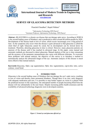
1. Optic Disc Segmentation
To automatically extract an optic disc boundary, image pre-processing is introduced. Figure 5 shows
a simplified workflow of optic disc segmentation.
Figure 5. Optic Disc Segmentation
A coarse localization of optic disc region is presented using the red channel. The red component is
utilized as it is found to have higher contrast between the optic disc and non-optic disc area than for
Input Image
Image Pre-processing
Edge Detection by Canny Algorithm Variational Level-Set
K-mean Clustering
Ellipse Fitting
Ellipse Fitting
Compared Result
5. International Journal of Modern Trends in Engineering and Research (IJMTER)
Volume 02, Issue 01, [January - 2015] e-ISSN: 2349-9745, p-ISSN: 2393-8161
@IJMTER-2014, All rights Reserved 201
other channels. To remove the blood vessels, a morphological closing operation is performed. After
performing the closing operation, a median filter is applied to further smoothen the obtained image.
The outputs of the image pre-processing are shown in Figure 6
(a) (b) (c) (d)
Figure 6. a) Input Image b) Red channel c) Closing operation and d) Median filter
1.2 Edge Detection Approach
The Canny method is specified for edge detection because the Canny algorithm can detect edges
with noise suppressed at the same time. This method uses two thresholds, to detect strong and weak
edges, and it includes the weak edges in the output only if they are connected to strong edges. The
optimum threshold of the each input retinal image is found to be different due to the variant
intensities in each image.
In order to extract only a disc boundary, the edge image has to be classified into three groups based
on the distance from the center of the optic disc as shown in below Figure 7(a). This classification is
achieved by performing k-means clustering to the edge of the image 7(b). K-means clustering is a
method of cluster analysis which aims to partition n observations into k clusters in which each
observation belongs to the cluster with the nearest mean. Then, the group that contains only edge
detection of a disc boundary is selected, and the noise can be rejected as shown in Figure 7(c).After
that, the application of direct ellipse fitting is used to obtain boundaries in order to find a smoother
contour. The outputs are shown in Figure 7(d).
(a) (b) (c) (d)
Figure 7. a) Input image after performing the image pre-processing with the center of an optic disc b) Edge detection
c) The result after noise is rejected d) Disc boundary smoothing.
1.3 Variational Level-Set Approach
The variational level- set algorithm is used as a global approach for the optimization of active
contours for the segmentation of objects of interest from the background. This method is employed
by initializing a curve centered at the detected optic disc location. The curve is evolved based on the
average intensity value inside and outside the curve. The curve evolution always converges to the
optic disc boundary irrespective of the shape or size of the initial contour.
6. International Journal of Modern Trends in Engineering and Research (IJMTER)
Volume 02, Issue 01, [January - 2015] e-ISSN: 2349-9745, p-ISSN: 2393-8161
@IJMTER-2014, All rights Reserved 202
(a) (b) (c) (d)
Figure 8. Sample Evolution Results at Different Iterations
2. Optic Cup Segmentation
Optic Cup Segmentation, two approaches are used .The color component analysis approach and
threshold level set approach.
2.1 Color Component Analysis Approach.
In the color component analysis method, RGB components of the input images are analyzed, and it is
found that the optic cup is more easily discriminated in the green image because the visibility and
contrast of the optic cup is superior and its pixels are of higher intensities, while the neuroretinal rim
and the retinal vessels are often of lower intensities. The next step is to use morphological opening
operation in order to remove noise around the cup region. Then the edge detection and ellipse fitting
is proposed to obtain the cup boundary smoothing.
(a) (b) (c) (d) (e)
Figure 9. a) Input Image b) Color Green Detected c) Morphological Opening Operation d) Edge Detection
e) Cup Boundary Smoothing
2.2 Threshold Level Set Approach
In this approach, the green channel of the input image is selected as the basis for further
segmentation due to the optimum observed contrast between the cup and disc boundaries in this
channel. The method to select the top 1/3 of the grayscale intensity is to find the threshold value
from the normalized cumulative histogram and then compare it with all the intensity values of the
input image. Then an intensity value is that is greater than the threshold value is selected.Text step is
to use a morphological opening operation in order to remove noise around the cup region.Next, the
intensity-weighted centroid method is proposed to find an approximate initial point. This is found to
give a good initial approximation for the initial cup region. Then, a threshold level-set algorithm is
applied to segment the optic cup.
(a) (b) (c) (d) (e)
Figure 10. A) Input Image B) Color Green Detected C) Morphological Opening Operation D) The Initial Point Of
Cup Region E) The Initial Cup Contour.
7. International Journal of Modern Trends in Engineering and Research (IJMTER)
Volume 02, Issue 01, [January - 2015] e-ISSN: 2349-9745, p-ISSN: 2393-8161
@IJMTER-2014, All rights Reserved 203
3. Ellipse Fitting for Optic Disc and Cup
Ellipse fitting algorithm can be used to smooth the optic cup and disc boundary. Ellipse fitting is
usually based on the least square fitting algorithm which assumes that the best-fit curve of a given
type is the curve that has the minimal sum of the deviations squared from, given data points. Direct
Least Square Fitting Algorithm is chosen to fit the optic disc from popular ellipse fitting algorithms.
It is ellipse specific, thus the effect of noise (ocular blood vessel, hemorrhage, etc.) around the disc
area can be minimized while forming the ellipse. It can also be easily solved naturally by a
generalized Eigen system.
4. Calculate CDR
The ratio of the size of the optic cup to the optic disc, also known as the cup-to-disc ratio (CDR), the
value of CDR which is more than 0.65 is used to assess a patient as a possible glaucoma case.
B. Optic Disc and Optic Cup Segmentation from Monocular Color
Retinal Images for Glaucoma Assessment
Interest in OD segmentation is not limited to glaucoma detection. It is a fundamental task for
automatic processing of retinal images such as image sequence registration, and automatic
measurements for treatment evaluation or for diabetic retinopathy diagnosis. Initial attempts have
been made with shape-based template matching in which OD is modelled as a circular or elliptical
object. This matching is performed on an edge map extracted from the underlying image. This
approach suffers due to vessel edges present in and around the OD region. To handle this,
morphological-based pre-processing step is employed to suppress the vessel prior to template
matching[1].
1. Localised and vector-valued C-V active contour model
This model assumes that an image consists of statistically homogeneous regions and therefore lacks
the ability to deal with objects having intensity inhomogeneity. Intensity inhomogeneity is very
common in natural images, especially in OD region it is a frequently occurring phenomena. In
computer vision, there have been some attempts to improve C-V model for such situations. Here, the
basic idea is to use local instead of global image intensity into the region-based active contour
model.
These methods report significant improvement in the segmentation over original C-V model for
segmenting objects with heterogeneous intensity statistics. However other than intensity
heterogeneity within OD, smooth region transition at boundary locations and occurrence of similar
characteristic regions near the OD boundaries (atrophy) make OD segmentation a much more
difficult case altogether. The local intensity based statistics is not sufficient to discriminate between
the OD and atrophy regions. We propose a region-based active contour model which uses local
image information at a support domain around each point of interest (POI) inspired by localised C-V
models by using a richer form of local image information gathered over a multi-dimensional feature
space. The intention is to represent the POI more holistically by including descriptions of the
intensity, colour, texture, etc. This approach should yield a better representation of image regions and
make the proposed model robust to the distractions found near the OD boundaries.
2. OD localisation and contour initialisation
The first step is to localise the OD region and extract a region of interest for further processing. The
red colour plane of CFI gives good definition of OD region and thus is a good choice for the OD
localisation task. The contour initialization is the next essential step to initiate the active contour
evolution. In our method, we perform localisation and initialisation steps together by performing
circular Hough transform on the gradient map.
8. International Journal of Modern Trends in Engineering and Research (IJMTER)
Volume 02, Issue 01, [January - 2015] e-ISSN: 2349-9745, p-ISSN: 2393-8161
@IJMTER-2014, All rights Reserved 204
The vessel segments are identified using a curvature-based technique. These regions are suppressed
and inpainted by performing selective morphological closing in 8 directions and retaining maximum
response for each vessel pixel. Next, a Canny edge detector at a very low threshold is applied on the
pre-processed (vessel-free) image to get edge points. On these points, a circular Hough transform is
applied for a range of expected OD radius (rmin to rmax). This range is chosen based on the retinal
image resolution. We select the OD center which has maximum value in the accumulator matrix
while performing Circular Hough transform. Next, the edges near the identified center location in the
image domain are used to estimate the radius of the circle. The circle points are identified using
estimated radius and used to initialise the active contour mentioned in section
Figure 11. Sample Results of C-V Active Contour.
2.1 Segmentation in multi-dimensional feature space
A multi-dimensional image representation is obtained from colour and texture feature space. In
normal image conditions, red colour plane gives a better contrast of the OD region. To better
characterise OD in pathological situations, two different texture representations are derived.
3. CUP Segmentation
The objective is to segment the cup region by using both vessel bends and pallor information. vessel
bends can occur at many places within the OD region. However, only a subset of these points
define the cup boundary. It refer to this as relevant vessel bends or r-bends. The first problem at hand
is to find this subset. We use multiple sources of information for this purpose: the pallor region
which spatially defines the inner limit of r-bends, bending angle and location in the OD region.
3.1 Cup segmentation using r-bends information
The objective is to segment the cup region by using both vessel bends and pallor information. cyan
points in figure 13, vessel bends can occur at many places within the OD region. However, only a
subset of these points defines the cup boundary. this is relevant vessel bends or r-bends. The first
problem at hand is to find this subset, multiple sources of information is use for this purpose: the
pallor region which spatially defines the inner limit of r-bends, bending angle and location in the OD
region.
A second problem is that the anatomy of the OD region is such that the r-bends are non-uniformly
distributed across a cup boundary with more points on the top and bottom; they are mostly absent in
the nasal side and very few in number in the temporal side. We propose a local interpolating spline to
naturally approximate the cup boundary in regions where r- bends are absent.
9. International Journal of Modern Trends in Engineering and Research (IJMTER)
Volume 02, Issue 01, [January - 2015] e-ISSN: 2349-9745, p-ISSN: 2393-8161
@IJMTER-2014, All rights Reserved 205
Figure 12. The Proposed Cup Segmentation Method
Figure 13. a) Angle of a vessel bend, b) uniform pallor samples(red),
bend points(cyan) and c) fitted circle(red) and potential r-bends
3.1.1 Medial axis detection
The OD region has both thick and thin vessels. Detecting both reliably is difficult in the presence of
inter-image intensity variations. This method formulates the blood vessel detection as a problem of
trench detection in the intensity surface. The selection of this space gives robustness to the image
variations and detection is solely driven by the shape of trench and directional continuity associated
with a vessel structure. Trenches are regions characterized by high curvature, oriented in a particular
direction.
3.1.2 Vessel Bend detection
The amount of bending in vessels varies according to the caliber of vessel. Thin vessels show
significant bending compared to a thick vessel. This is due to the fact that thick vessels are more
rigid. The selection of appropriate scale for detecting bends in both types of vessels is crucial
because bend in a thick vessel is apparent only at a larger scale compared to a bend in thin vessel.
We employ a scheme based on the concept of dynamic region of support (ROS) which has been
proposed for corner detection to find the appropriate scale to analyse a candidate point. This is
explained below.
First, we extract vessel segments terminated by end and/or junction points. For each segment, we
compute 1D shape (curvature) profile and locate the local maxima. These local maxima constitute a
candidate set of bends bi. A ROS for any bi is defined as a segment of vessel around bi and bound on
either side by the nearest curvature minimum. Choosing the bounds to be based on curvature minima
automatically ensures the size of the ROS to be large for thick vessels and small for thin vessels.
The angle of bend θ is then computed as the angle between the lines joining a bend point and the
center of mass on both sides of the ROS. The center of mass of an arm is defined by the mean
position of pixels on the arm. Since only vessels bending into the cup are of interest, bends above θ =
10. International Journal of Modern Trends in Engineering and Research (IJMTER)
Volume 02, Issue 01, [January - 2015] e-ISSN: 2349-9745, p-ISSN: 2393-8161
@IJMTER-2014, All rights Reserved 206
170◦ are eliminated from the candidate set. The detected vessel bends in a sample image are
highlighted in figure(13) with cyan markers.
3.1.3 Multi-stage selection of r-bends
The task of identifying the r-bends from bi is performed in two stages to reduce the required
analysis, by utilizing anatomical knowledge associated with r-bends. In the first stage, a coarse
selection is done based on a bend’s proximity to the pallor region. In the second stage, the spatial
position and bending information are used to identify the set of r-bends.
(a) (b)
Figure 14 a)Estimated Cup Boundry b)Final Cup Boundry
3.1.4 2D spline interpolation
Typically, r-bends are sparse and not uniformly distributed across the sectors. In their absence,
experts use their clinical knowledge (experience of direct 3D cup examination) to approximate a cup
boundary. Hence, it is difficult to get the cup boundary in the regions with no r-bends. We choose a
local, cubic cardinal spline, which is a generalisation of Catmull-Rom spline, with a shape parameter
t. The parameter t helps control the bending behaviour and thus the shape according to the sector.
The value of t is kept high in sectors 2&4 as they usually have low vessel density (r-bends)
compared to sector 1&3. A closed-form 2D spline curve is obtained by considering, sequentially a
subset of r-bends. Figure 7(a) shows the interpolated cup boundary passing through the r-bends and
Fig. 7(b) shows final obtained boundaries for a sample OD region.
C. Confocal Scanning Laser Ophthalmology
In [6] Confocal scanning laser ophthalmoscopy (CSLO), a laser based image gaining which is
proposed to improve the quality of the examination compared to ordinary ophthalmologic
examination. A laser id scanned across the retina along with a detector system. Once a single spot on
the retina is illuminated at any time, ensuing in a high-contrast image of great reproducibility that
can be used to estimate the width of the RNFL. In addition, this technique does not need maximal
mydriasis, which may be a problem in patients having glaucoma. The Heidelberg Retinal
Tomography is possibly the most common example of Confocal scanning laser ophthalmoscopy
(CSLO).
Figure 15. HRT Machine
11. International Journal of Modern Trends in Engineering and Research (IJMTER)
Volume 02, Issue 01, [January - 2015] e-ISSN: 2349-9745, p-ISSN: 2393-8161
@IJMTER-2014, All rights Reserved 207
D. Scanning Laser Polarimetry
RNFL is birefringent, which causes a change in the state of divergence of a laser beam as it passes. It
uses a 780-nm diode to illuminate optic nerve. The polarization state of the light emerging from the
eye is then evaluated and linked with RNFL thickness. Unlike CSLO, scanning laser polarimetry
(SLP) can unswervingly measure the thickness of the RNFL[6]. GDx® is an ordinary example of a
scanning laser polarimeter. GDx®contain a normative database and statistical software package to
permit comparison to age-matched normal subjects of the same racial origin.
The advantages of this system are that images can be obtained without pupil dilation, and evaluation
can be done roughly in 10 minutes. Modern instruments have added improved and erratic corneal
compensation technology to account for corneal polarization.
Figure 16. GDx VCC
E. Optical Coherence Tomography
Optical coherence tomography (OCT) uses near-infrared light to provide direct crosssectional
measurement of the RNFL [6]. The principles employed are alike to those used in B-mode
ultrasound except light, not sound, is used to create the 2-dimensional images.
The light source can be directed into the eye through a conservative slit-lamp biomicroscope and
focused on the retina through a distinctive 78-diopter lens. This system requires dilation of the
patient’s pupil. OCT is an example of this technology.
Figure 17. OCT
III. CONCLUSION
Glaucoma is a silent disease that comes with no symptoms and warning. Initially no one can say that
the patient is having any sort of problem either by looking and touching the eye. Hence its detection
and diagnosis are very essential.