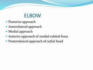
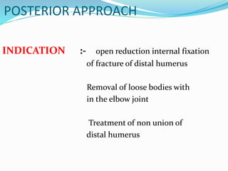
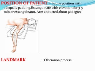
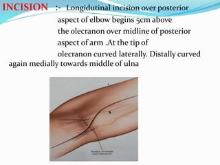
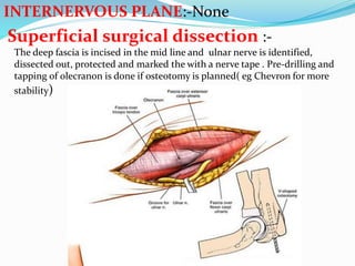
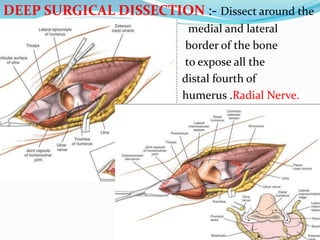
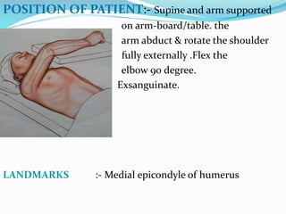
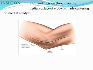
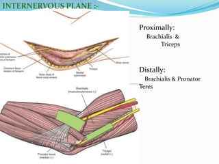
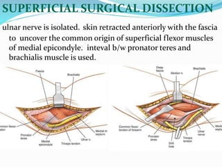
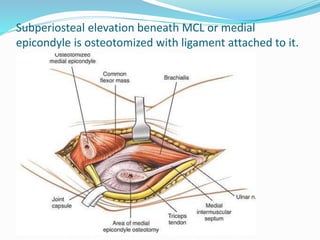
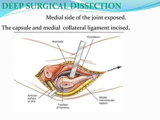
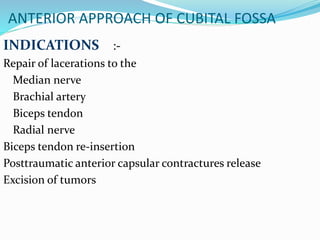
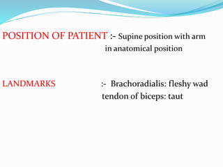
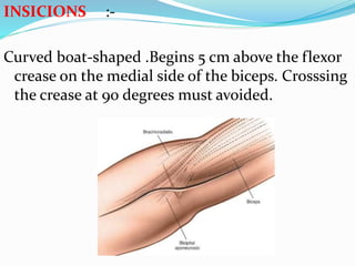
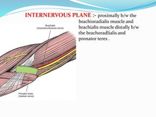
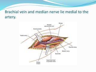
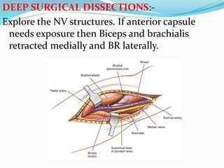
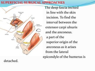
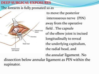
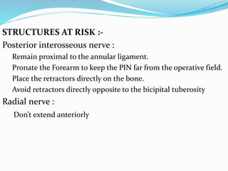
This document describes various surgical approaches to the elbow, including the posterior, medial, anterolateral, anterior of cubital fossa, and posterolateral approaches. For each approach, it provides the indications, patient positioning, landmarks, incision details, internervous planes, superficial and deep dissections, and structures at risk. The posterior approach is used for fractures of the distal humerus. The medial approach provides access to the medial condyle and coronoid process. The anterolateral approach is used for fractures of the radial head and capitellum.
this ppt provides a comprehensive review & exam oriented details
compiled from journals & old edition textbooks. because ITB contracture has become a rare presentation. & new edition books doesnt speak about it much...
conventional plates including different functions of screws, modes of plate application, Compression Mode.
Neutralization Mode.
Buttress plate.
Antiglide plate.
Bridge plating or span plating.
Tension band.
prebending precountouring
working length
lag screw
AO principles
biological fixation
MIPO
Intramedullary nailing of fractures.dr mohamed ashraf.HOD.govt TD medical co...drashraf369
presentation of biology,biomechanics and practice of intramedullary nailing of long bone fractures by dr mohamed ashraf,govt TD medical college,alleppey,kerala,india
Intramedullary nailing of fractures.dr mohamed ashraf.HOD.govt TD medical co...drashraf369
presentation of biology,biomechanics and practice of intramedullary nailing of long bone fractures by dr mohamed ashraf,govt TD medical college,alleppey,kerala,india
Osseous anatomy, Types of approaches(Position,landmarks,Incision,Superficial and Deep surgical dissection) , structures at risk, Extensile approaches with diagrams and eponymous .
Macroeconomics- Movie Location
This will be used as part of your Personal Professional Portfolio once graded.
Objective:
Prepare a presentation or a paper using research, basic comparative analysis, data organization and application of economic information. You will make an informed assessment of an economic climate outside of the United States to accomplish an entertainment industry objective.
How to Build a Module in Odoo 17 Using the Scaffold MethodCeline George
Odoo provides an option for creating a module by using a single line command. By using this command the user can make a whole structure of a module. It is very easy for a beginner to make a module. There is no need to make each file manually. This slide will show how to create a module using the scaffold method.
Biological screening of herbal drugs: Introduction and Need for
Phyto-Pharmacological Screening, New Strategies for evaluating
Natural Products, In vitro evaluation techniques for Antioxidants, Antimicrobial and Anticancer drugs. In vivo evaluation techniques
for Anti-inflammatory, Antiulcer, Anticancer, Wound healing, Antidiabetic, Hepatoprotective, Cardio protective, Diuretics and
Antifertility, Toxicity studies as per OECD guidelines
Safalta Digital marketing institute in Noida, provide complete applications that encompass a huge range of virtual advertising and marketing additives, which includes search engine optimization, virtual communication advertising, pay-per-click on marketing, content material advertising, internet analytics, and greater. These university courses are designed for students who possess a comprehensive understanding of virtual marketing strategies and attributes.Safalta Digital Marketing Institute in Noida is a first choice for young individuals or students who are looking to start their careers in the field of digital advertising. The institute gives specialized courses designed and certification.
for beginners, providing thorough training in areas such as SEO, digital communication marketing, and PPC training in Noida. After finishing the program, students receive the certifications recognised by top different universitie, setting a strong foundation for a successful career in digital marketing.
Exploiting Artificial Intelligence for Empowering Researchers and Faculty, In...Dr. Vinod Kumar Kanvaria
Exploiting Artificial Intelligence for Empowering Researchers and Faculty,
International FDP on Fundamentals of Research in Social Sciences
at Integral University, Lucknow, 06.06.2024
By Dr. Vinod Kumar Kanvaria
Unit 8 - Information and Communication Technology (Paper I).pdfThiyagu K
This slides describes the basic concepts of ICT, basics of Email, Emerging Technology and Digital Initiatives in Education. This presentations aligns with the UGC Paper I syllabus.
This presentation includes basic of PCOS their pathology and treatment and also Ayurveda correlation of PCOS and Ayurvedic line of treatment mentioned in classics.
How to Add Chatter in the odoo 17 ERP ModuleCeline George
In Odoo, the chatter is like a chat tool that helps you work together on records. You can leave notes and track things, making it easier to talk with your team and partners. Inside chatter, all communication history, activity, and changes will be displayed.
Introduction to AI for Nonprofits with Tapp NetworkTechSoup
Dive into the world of AI! Experts Jon Hill and Tareq Monaur will guide you through AI's role in enhancing nonprofit websites and basic marketing strategies, making it easy to understand and apply.
2. ELBOW
Posterior approach
Anterolateral approach
Medial approach
Anterior approach of medial cubital fossa
Posterolateral approach of radial head
3. POSTERIOR APPROACH
INDICATION :- open reduction internal fixation
of fracture of distal humerus
Removal of loose bodies with
in the elbow joint
Treatment of non union of
distal humerus
4. POSITION OF PATIENT :- Prone position with
adequate padding.Exsanguinate with elevation for 3-5
min or exsanguinator. Arm abducted about 90degree
LANDMARK :- Olecranon process
5. INCISION :- Longidutinal incision over posterior
aspect of elbow begins 5cm above
the olecranon over midline of posterior
aspect of arm .At the tip of
olecranon curved laterally. Distally curved
again medially towards middle of ulna
6. INTERNERVOUS PLANE:-None
Superficial surgical dissection :-
The deep fascia is incised in the mid line and ulnar nerve is identified,
dissected out, protected and marked the with a nerve tape . Pre-drilling and
tapping of olecranon is done if osteotomy is planned( eg Chevron for more
stability)
7. DEEP SURGICAL DISSECTION :- Dissect around the
medial and lateral
border of the bone
to expose all the
distal fourth of
humerus .Radial Nerve.
8. STRUCTURES AT RISK :-
Ulnar nerve : Identify and protect.
Median nerve : Always remain in subperiosteal plane
Radial nerve: Don’t dissect too proximal at lateral intermuscular
septum
Brachial artery: Anteriorly located
Extension:-
Proximally– Not possible than the distal third of humerus
Distally - can be extended along the subcutaneous border of ulna
9. MEDIAL APPROACH
INDICATIONS :- Removal of loose bodies
Open reduction and internal
fixation of fractures of the
corocoid process of the ulna
Open reduction and internal
fixation of fractures of the
medial humeral condyle and
epicondyle.
Contraindications:
1. Exploration of elbow as poor access to
the lateral side
10. POSITION OF PATIENT:- Supine and arm supported
on arm-board/table. the
arm abduct & rotate the shoulder
fully externally .Flex the
elbow 90 degree.
Exsanguinate.
LANDMARKS :- Medial epicondyle of humerus
11. INSICION :- Curved incision 8-10cm on the
medial surface of elbow is made centering
on medial condyle.
13. SUPERFICIAL SURGICAL DISSECTION
ulnar nerve is isolated. skin retracted anteriorly with the fascia
to uncover the common origin of superficial flexor muscles
of medial epicondyle. inteval b/w pronator teres and
brachialis muscle is used.
16. STRUCTURES AT RISK
Ulnar nerve.
Median nerve and its main branch AIN with vigorous traction
of medial epicondyle or superficial flexor muscles
EXTENSION
Proximally :
b/w triceps and brachialis muscle subperiosteally
Distally:
exposure provides adequate view of the
brachialis inserting into coronoid. it cannot offer a more
distal exposure but only upto the branching off of the
median nerve.
17. ANTEROLATERAL APPROACH
INDICATIONS :- Open reduction and internal fixation
of the capitulum #
Excision of proximal radius tumors
Treatment of aseptic necrosis of the capitulum
Drainage of septic elbow arthritis
Neural decompression :lesions of the proximal
half of the PIN and of the proximal part of the
superficial radial nerve access to the arcade Frohse, as well as
treatment of radial head fractures with paralysis of this nerve
Biceps tendon avulsion re-attachment to radial
tuberosity
TEA
18. POSITION OF PATIENT :- Supine with arm on arm-
board
LANDMARKS :- Brachioradialis :palpable thick wad
Biceps tendon: easily palpable taut
structure
19. INCISION :- curved S incision given around the
anterior aspect of the elbow. Begins at 5
cm above flexor crease along lateral border of the
biceps muscle. The lower portion
curves over the medial border of
the brachioradialis muscle
21. SUPERFICIAL SURGICAL DISSECTION :- Deep
fascia is incised along the medial border of the
brachioradialis. The lateral antebrachial cutaneous nerve
(LCNFA) is identified and preserved.
22. Deeper blunt dissection with finger. Radial nerve between
BR and Brachialis. PIN enters supinator. SupercialRadialN is beneath the
Brachioradialis Motor branch to ECRB.
23. DEEP SURGICAL DISSECTION :-
longitudinal incision is made in the anterior
capsule of the joint between the
radial nerve laterally and the brachialis
muscle medially to expose the radial head
and capitulum. To expose the
radius further, forearm is fully supinated
& supinator muscle removed distally in a
subperiosteal manner
24.
25. STRUCTURES AT RISK :-
Radial nerve : in brachioradialis and brachialis interval
Posterior interosseous nerve : Winding around the radial
neck.
Lateral cutaneous nerve of forearm: LCNFA emerging
from brachioradialis and biceps brachii interval
Reccurent branch of radial artery: Ligation decreases
post-op bleed and chance of VIC
EXTENSION:-
Proximally: BR/Triceps
Distally: Along entire anterior surface of the radius between
BR/PT and further distally BR/FCR.
26. ANTERIOR APPROACH OF CUBITAL FOSSA
INDICATIONS :-
Repair of lacerations to the
Median nerve
Brachial artery
Biceps tendon
Radial nerve
Biceps tendon re-insertion
Posttraumatic anterior capsular contractures release
Excision of tumors
27. POSITION OF PATIENT :- Supine position with arm
in anatomical position
LANDMARKS :- Brachoradialis: fleshy wad
tendon of biceps: taut
28. INSICIONS :-
Curved boat-shaped .Begins 5 cm above the flexor
crease on the medial side of the biceps. Crosssing
the crease at 90 degrees must avoided.
29. INTERNERVOUS PLANE :- proximally b/w the
brachioradialis muscle and
brachialis muscle distally b/w
the brachoradlialis and
pronator teres .
30. SUPERFICIAL SURGICAL DISSECTIONS :-
The deep fascia is incised in line with the skin incision and the numerous veins
that cross the elbow in this area are ligated.Lateral cutaneous nerve of the
forearm (LCNFA) in the interval between the biceps tendon and the brachialis,
is identified and preserved
Lacertus fibrosus is identified as the brachial artery is immediately under it.
32. DEEP SURGICAL DISSECTIONS:-
Explore the NV structures. If anterior capsule
needs exposure then Biceps and brachialis
retracted medially and BR laterally.
33. STRUCTURES AT RISK
1. LCNFA a sensory branch of musculocutaneous nerve
at distal ¼ of the arm. Emerges between biceps &
brachialis.
2. Radial artery
3.PIN
34. EXTENSION
1. For Median Nerve
Proximally: Medial border of biceps
Distally: Pronator Teres simple retraction between humeral
and ulnar heads
2. For Brachilal Artery
As above
35. EXTENSION
3. For Radial Artery
Proximally: Plane between PT & BR
Distally : Between FCR & BR to the wrist
36. POSTEROLATERAL APPROACH OF
RADIAL HEAD
Indications: Radial head excision or prosthetic
replacement
POSITION OF PATIENT
- Supine on operating table with
affected arm over
chest ,pronate the forearm
37. LANDMARK :- Lateral humeral epicondyle.
Olecranon tip
INCISION :- gentle curve beginning over the posterior
surface of the lateral humeral epicondyle
and continuing downward and
medially over the posterior
border of the ulna, at about 6 cm distal
to the tip of the olecranon.
39. SUPERFICIAL SURGICAL APPROACHES
The deep fascia incised
in line with the skin
incision. To find the
interval between the
extensor carpi ulnaris
and the anconeus.
a part of the
superior origin of the
anconeus as it arises
from the lateral
epicondyle of the humerus is
detached.
40. DEEP SURGICAL EXPOSURES
The forearm is fully pronated so as
to move the posterior
interosseous nerve (PIN)
away from the operative
field . The capsule
of the elbow joint is incised
longitudinally to reveal
the underlying capitulum,
the radial head, and
the annular ligament. No
dissection below annular ligament as PIN within the
supinator.
41. STRUCTURES AT RISK :-
Posterior interosseous nerve :
Remain proximal to the annular ligament.
Pronate the Forearm to keep the PIN far from the operative field.
Place the retractors directly on the bone.
Avoid retractors directly opposite to the bicipital tuberosity
Radial nerve :
Don’t extend anteriorly