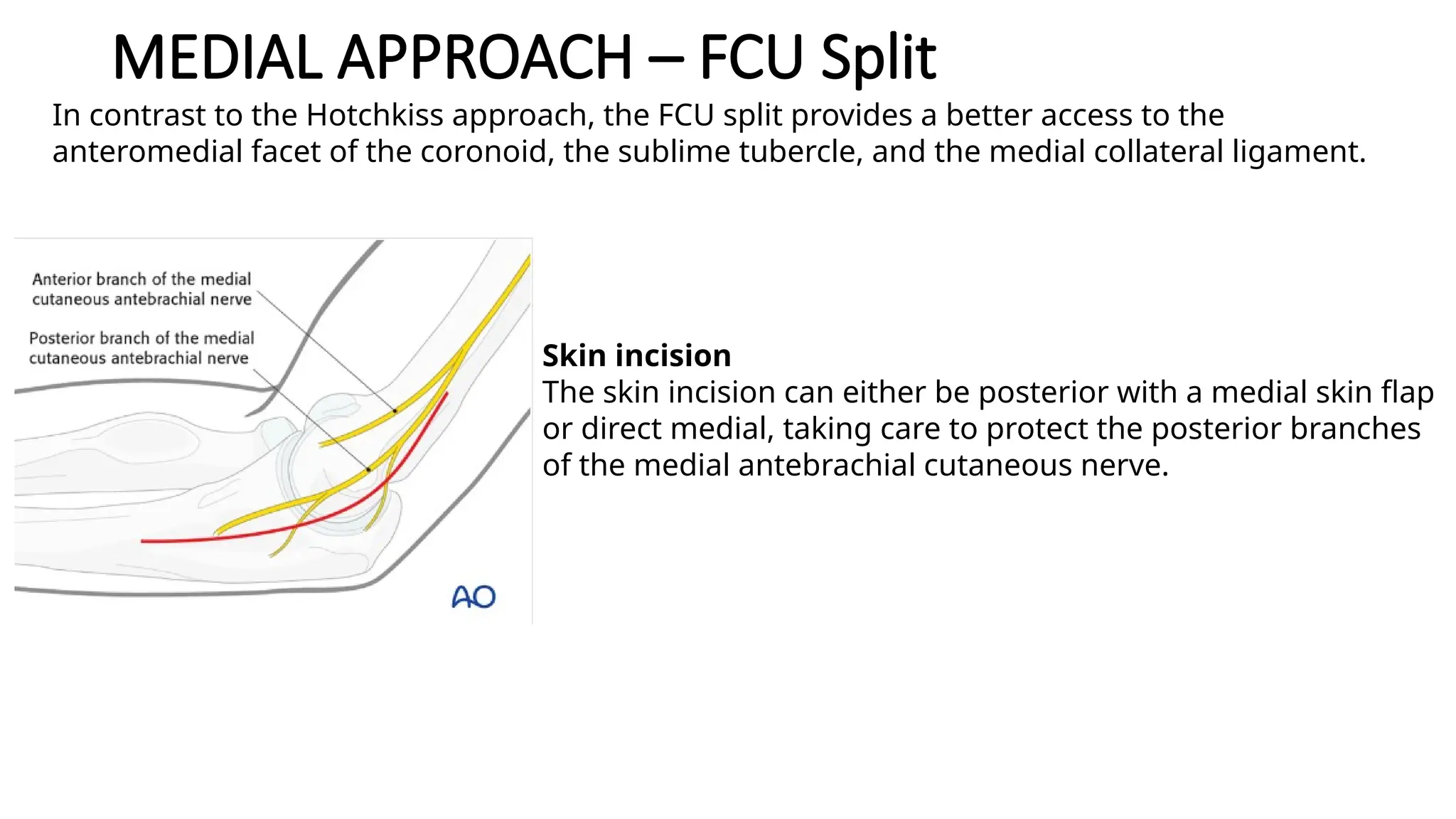
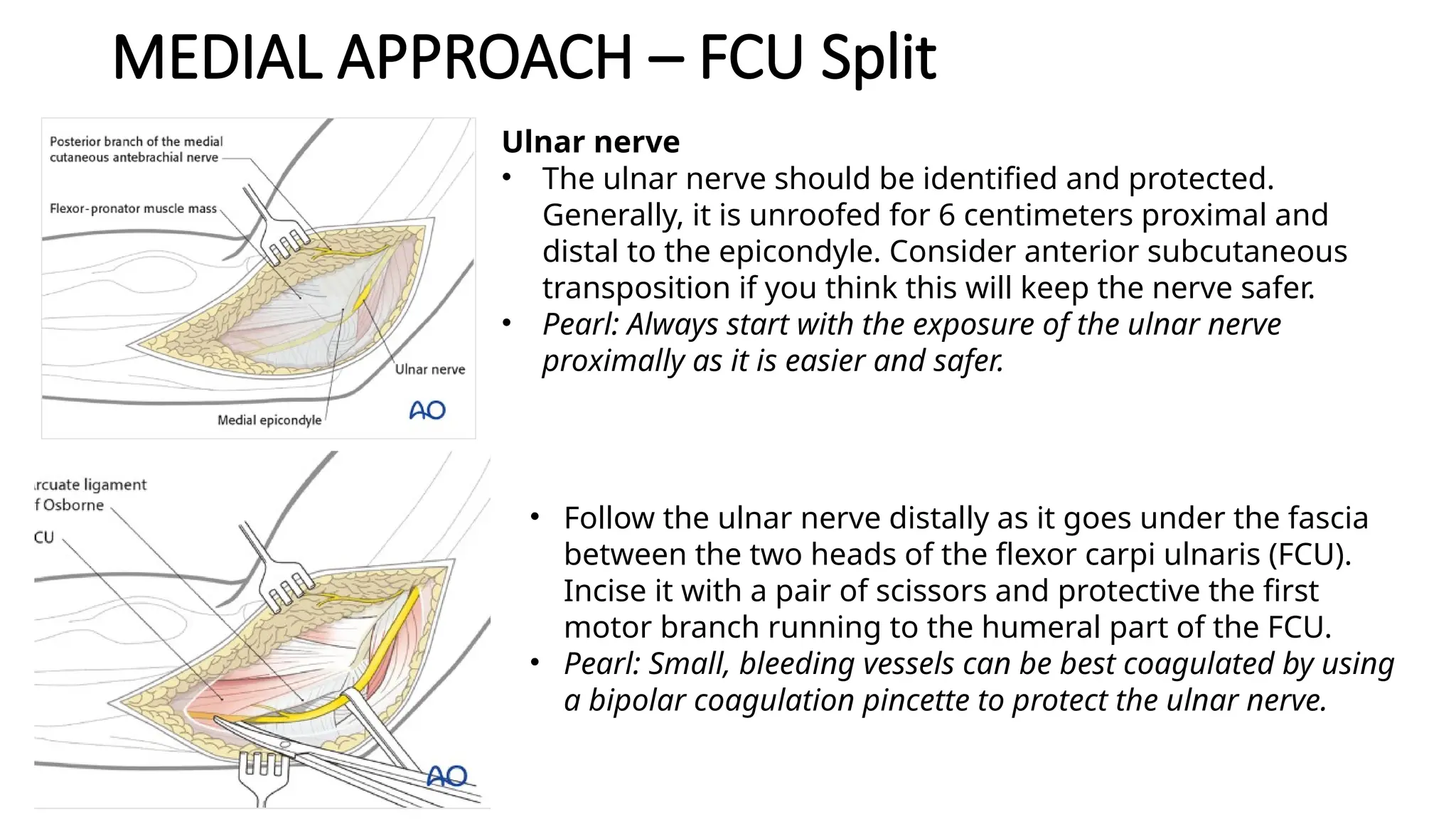
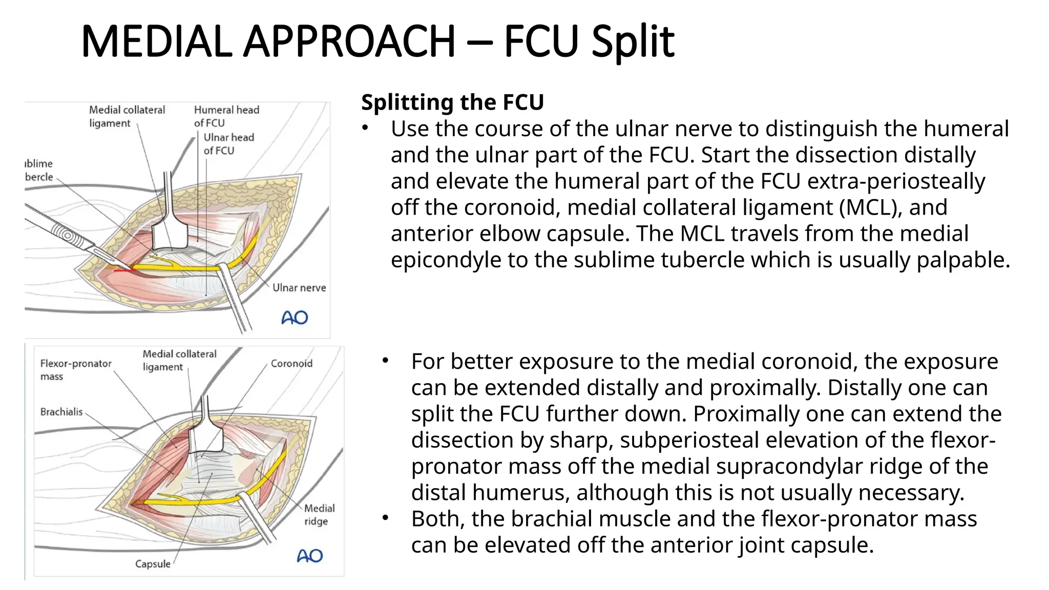
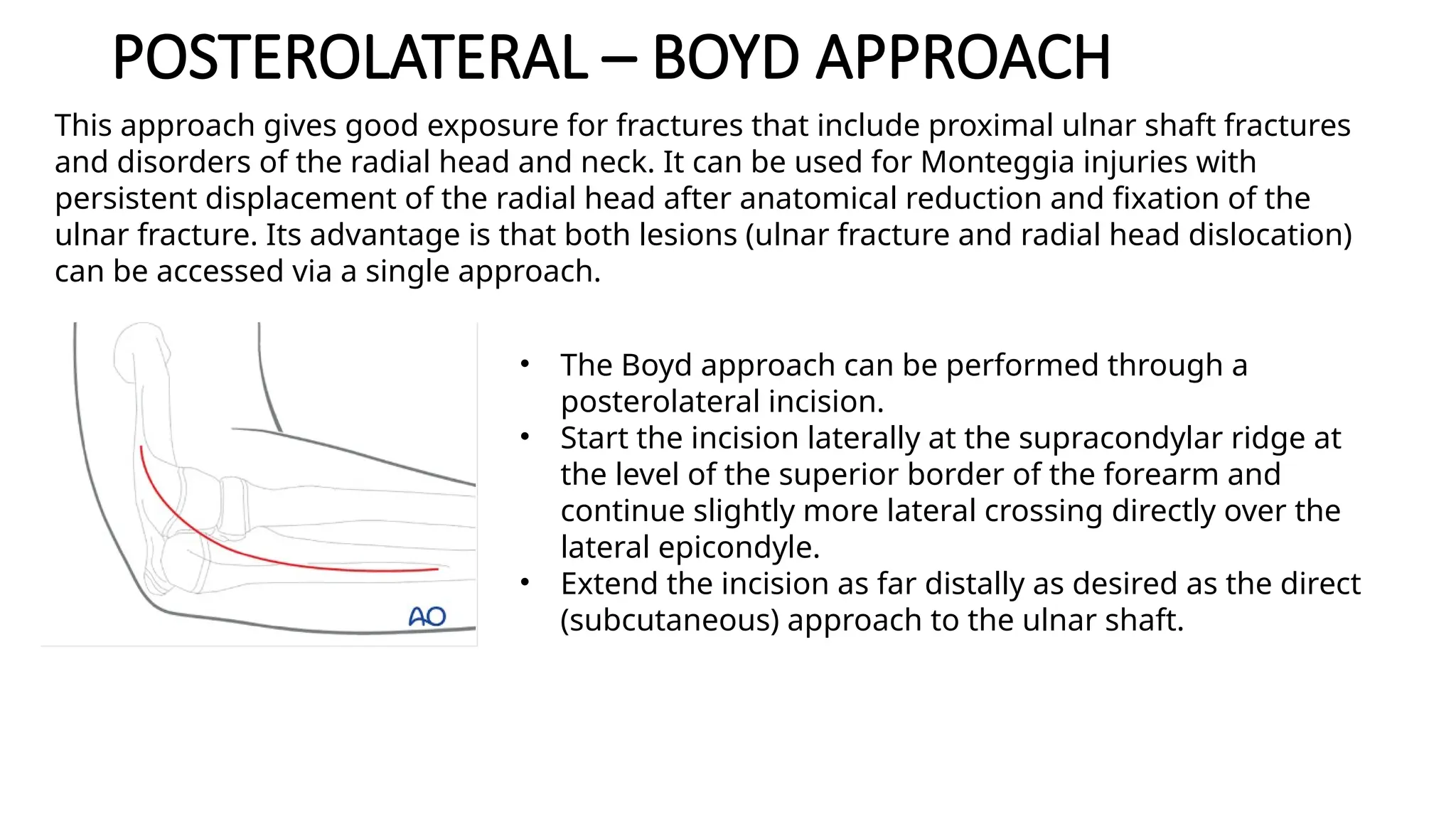
UPPER EXTREMITY
APPROACH- ELBOW
Elbow surgery is all about picking up the right surgical approach
POSTERIOR APPROACH
• Most common for distal humeral fracture
• 10 posterior approach depends on how we manage the triceps
• Olecranon osteotomy
(+Anconeus flap)
• Triceps on technique
• Triceps reflecting (Medial 
Lateral) and (Lateral 
Medial)
• Paraolecranon approach
(Lateral and Medial)
• Triceps split
• Triceps tongue
• TRAP approach
• Diamond “pup up”
• Decide your priority  save the triceps, avoid ulnar nerve problem,
what do I need to see, risk of late complication, personal preferences
CASE
• Articular comminution
• I need to see well the articular surface
• Ulnar nerve and skin healing are of
minor important
Olecranon
Osteotomy
POSTERIOR APPROACH WITH OLECRANON OSTEOTOMY
Positioning
Indications :
1. Open reduction and internal fixation of fractures of the distal humerus
2. Removal of loose bodies within the elbow joint
3. Treatment of nonunions of the distal humerus
90 degree
Apply torniquet
Padding
Landmark : Olecranon process
Incision :
• begin 5cm proximal to the olecranon in the midline of the posterior
distal humerus
• curve laterally proximal to the tip of the of the olecranon along the
lateral aspect of the olecranon process
• then curve medially over the middle of the posterior aspect of the
subcutaneous ulna
Internervous plane  None
• the extensor mechanism is either split or detached
• the radial nerve innervates the triceps muscle more proximally
Superficial Dissection
• first, palpate the ulnar nerve and fully dissect it out
• is helpful to pass tape or penrose for identification at all times
• incise deep posterior fascia in the midline
• can either split triceps fascia, or continue with olecranon osteotomy
• if performing olecranon osteotomy, drill and tap olecranon prior to
osteotomy
• score the olecranon with an osteotome to allow perfect reduction when
the osteotomy is repaired
• V-shaped osteotomy of the olecranon 2 cm from the tip using an
oscillating saw
• Dissect the ulnar nerve from its bed and hold it free with tape. Predrill
• the olecranon before performing an osteotomy for easy reattachment.
A V-shaped
• osteotomy is inherently more stable than a transverse osteotomy.
Deep Dissection
• strip soft tissue from the edges of the osteotomy site and retract the
olecranon fragment proximally, elevating triceps rfom the back of the
humerus
• subperiosteal dissection of the medial and lateral borders of the
humerus allows exposure of entire distal fourth of the humerus
• Be careful not to extend the dissection proximally above the distal
fourth of the humerus, because the radial nerve, which passes from the
posterior to the anterior compartment of the arm through the lateral
intermuscular septum, may be damaged. Flex the elbow to relax the
anterior structures if they need to be elevated off the front of the
humerus
Perform a V-shaped osteotomy of the olecranon
and retract it proximally, with the triceps muscle
attached. Strip a portion of the joint capsule with
an osteotome.
DANGER
•Ulnar nerve
•should initially be indentified and protected during the
approach
•can usually be palpated 2cm proximal to medial epicondyle
•transposition of the ulnar nerve has shown no benefit to
reducing the incidence of ulnar neuritis
•It is at most risk when transverse K wires are inserted from
the lateral side that may overpenetrate the medial cortex.
•Median nerve
•strict subperiosteal dissection off the anterior surface of the
humerus protects the nerve
•flexion of the elbow relaxes the anterior structures.
•Radial nerve
•in danger proximally as it travels from the posterior to
anterior brachial compartments through lateral
intermuscular septum
•can usually be found at the lateral border of the humerus
near distal 1/3 junction
•Brachial artery
•runs with the median nerve
POSTERIOR APPROACH – TRICEPS
PRESERVING
Advantages
• Fast Rehab
• Less post op pain
• Don’t cut bridges
Disadvantages
• Difficult to address articular
fragment
POSTERIOR APPROACH – TRICEPS ON
• Triceps on / paratriceps / Alonso Liames
• All the triceps is left intact
• Difficult to see the trochlea and ulna
• Risk of malreduction
• Risk of rotational malalignment of the ulna component during TEA
• Some comminution of the throclea (seems to
be fixable)
• Not severe comminution of the coloumn of
the trochle
POSTERIOR APPROACH – TRICEPS ON
Skin incision :
• Incise the skin, beginning at the tip of the
olecranon.
• The incision runs proximally in a straight
line from the olecranon along the
posterior midline of the arm
ULNAR WINDOW RADIAL WINDOW
As a first step, identify and mobilize the ulnar nerve. It
may be helpful to protect it with a vessel loop.
Next, mobilize the triceps muscle and retract it
laterally. This may be achieved by bluntly dissecting
the medial head of the triceps from the posterior
aspect of the humerus.
The entire triceps muscle is isolated with a gauze wrap
This permits the whole triceps muscle to be moved
towards either the lateral or medial side, to provide
access to the humerus (“triceps flip”).
POSTERIOR APPROACH – PARAOLECRANON
• Triceps on / paratriceps / Alonso Liames
• All the triceps is left intact
• Difficult to see the trochlea and ulna
• Risk of malreduction
• Risk of rotational malalignment of the ulna component during TEA
• Some comminution of the throclea (seems to
be fixable)
• Not severe comminution of the coloumn of
the trochlea
• Primarily approach for arthroplasty
Triceps on Vs Paraolecranon
• Paraolecranon Medial
• Better view of the ulna compare to
triceps on
• Gentle with the ulnar nerve
• Triceps on
• Paraolecranon Lateral
• Better view of the ulna compare to
triceps on
• Triceps can be strongly repaired
POSTERIOR APPROACH –
TRICEP REFLECTING (BRYAN MOREY)
• This is an approach primarily used for arthroplasty.
• A disadvantage of this approach is the need to repair and protect the triceps at the end of the
procedure.
• Consequently, there is a risk for triceps insufficiency
Make an incision centered on the junction of the middle
and distal thirds of the humeral shaft.
Avoid placing the incision over the tip of the olecranon.
make a straight incision slightly medial, lateral or a curved
incision. The incision ends over the ulnar diaphysis.
Elevate full-thickness fasciocutaneous flaps to protect the
cutaneous nerves.
POSTERIOR APPROACH –
TRICEP REFLECTING (BRYAN MOREY)
Identify the ulnar nerve proximally along the medial border of the
triceps. Release the ulnar nerve
Note: If the ulnar nerve has been mobilized, it is essential that the
OR report should clearly describe how the ulnar nerve has been
protected and the location of the nerve at the end of the
operation.
Detach the triceps insertion subperiosteally from the proximal
ulna towards the radial side.
POSTERIOR APPROACH –
TRICEP REFLECTING (BRYAN MOREY)
• Incise the posterior capsule proximal to the olecranon.
• At the level of the olecranon, detach the extensor apparatus
subperiosteally or with a sliver of bone using a fine osteotome.
• Release the extensor muscles from the lateral epicondyle of the
humerus and the anconeus from the posterolateral humerus
and ulna. Now the entire extensor apparatus flap can be
retracted to the radial side
• Flex the elbow beyond 100° to enhance visualization of the
articular surface.
• Optionally, the tip of the olecranon may be removed
POSTERIOR APPROACH –
TRICEP REFLECTING (BRYAN MOREY)
Place heavy
nonabsorbable
braided sutures
through these
tunnels.
Pull the extensor
apparatus into
place using a
Kocher forceps.
• Reattach the triceps tendon
insertion to the olecranon
with transosseous sutures.
• Start by drilling the
transosseous tunnels in the
olecranon using a 2–2.5 mm
drill bit
• Secure the triceps tendon
and fascia with locking
sutures and surgical knots.
This provides compression of
the triceps tendon to the
olecranon.
• The patient should avoid
resisted extension activities
while the tendon heals to
POSTERIOR APPROACH – TRICEP SPLIT
• This approach is most commonly used for fractures involving the distal half of the
humerus. However, it can be extended for more proximal fractures once the radial
nerve has been identified.
Incise the skin,
beginning at the tip
of the olecranon
Incise the deep fascia in
line with the skin incision
By palpation with a finger, identify the
interval between the lateral and long heads
of the triceps. The opening of this interval
will be developed from proximal to distal,
remembering that the radial nerve lies
beneath the triceps as it crosses the
POSTERIOR APPROACH – TRICEP SPLIT
• Then develop the proximal interval between the two heads by
dissection, retracting the lateral head laterally and the long head
medially.
• Within the spiral groove, identify the radial nerve and the
accompanying profunda brachii artery.
• Distally, split the common triceps tendon, along the line of the
skin incision, by sharp dissection.
• Release the lateral head of the triceps from the humerus proximally,
and incise it distally, in line with the humeral shaft. Release the
muscle from the bone only as much as needed and protect the ulnar
nerve medially. The ulnar nerve is constrained at the point where it
passes distally through the medial intermuscular septum, from the
anterior to the posterior compartment of the arm.
• Release the medial head of the triceps proximally until the axillary
nerve is found.
• Now the posterior humerus, crossed by radial nerve and its
accompanying vessels, lies exposed from the axillary nerve and
posterior circumflex humeral artery proximally to the capitellum
POSTERIOR APPROACH – TRICEP SPLIT
Distal extension
Splitting the triceps tendon limits distal exposure, which can be
improved by approaching the humerus from the lateral side of this
muscle. For more details see the distal posterior approach.
Proximal extension
Limited proximal extension, even beyond the axillary nerve, is
possible with careful mobilization and retraction of both radial and
axillary nerves and their accompanying vessels.
LATERAL APPROACH
• For radial head, coronoid, terrible triad ( combination of fractures of
the radial head and ulnar coronoid process and dislocation of the
elbow joint)
• Kocher
• Kaplan
• Proximally – extended
lateral approach
• Extensile postero-
lateral approach
LATERAL APPROACH
KOCHER KAPLAN
Anatomy Kocher Interval More anterior : common extensor tendon
(ECRB – EDC), better view for terrible triad
PIN Not at risk At risk
What you see See the radial head from below (easier
lesser sigmoid notch)  landmark for
radial head replacement
See the radial head from above / lateral
Instability Not clear Preserve stability
Extendable Yes (Proximal – distal) Yes (Proximal)
LATERAL APPROACH
• Either a posterior skin incision with a lateral skin flap or a lateral
skin incision can be used.
• For a lateral skin incision, place the elbow at 90 degrees and try to
pinch the lateral condyle (easier in thin patients). Make a straight
skin incision directly over the middle of the lateral condyle. Start
with a small incision (6-8 cm or so) and extend proximal or distal as
needed.
• Note: The posterior interosseous nerve, within the supinator muscle,
crosses the posterior radius, from anteriorly, three finger-breadths
distal to the radial head. It must be protected during this approach.
• Incise the subcutaneous tissue in line with the incision
and raise flaps to expose the fascia over the muscles.
LATERAL APPROACH
• lies in the interval between the extensor carpi radialis brevis and
extensor digitorum communis (Kaplan interval). The associated
ligament and muscle injury will make the rest of the exposure very
easy.
• The Kaplan interval can also be identified by elevating the origin of
the extensor carpi radialis brevis from the supracondylar ridge of
the distal humerus, elevating the brachialis from the anterior
humerus, then continuing distally until the joint is entered and the
capitellum is visualized. Elevating these muscles is necessary to
exposure the coronoid from the lateral side. Split the common wrist
and digital extensor musculature at the point that divides the
capitellum in half anterior/posterior.
• The interval between the anconeus and extensor carpi
ulnaris (Kocher interval) is relatively more posterior and
thus risks injuring the lateral collateral ligament complex.
LATERAL APPROACH
Deep surgical incision
• The annular ligament is divided in line with the muscle interval.
• If the lateral collateral
ligament is intact and
better exposure of the
radial head and neck are
desired, one can consider
osteotomy of the lateral
humeral epicondyle. The
osteotomy line in the
illustration is marked in
• The soft tissues and
osteotomized lateral
epicondyle are reflected
anteriorly to provide
access to the proximal
radius and ulna.
• Screw fixation of the
osteotomy can be
difficult because the
fragment is small and
metaphyseal. A tension
band wire is an
alternative, using the
muscle origin as a more
reliable point of fixation.
MEDIAL APPROACH
• Medial approaches to the elbow :
• 1. Hotchkiss – Over the top
• 2. Medial Coronoid approach (Split FCU)
• 3. Taylor and Scham (Posteromedial approach)
Hotchkiss - Over the Top Split FCU
Anatomy Anterior to the FCU In between the FCU
Ulnar nerve More protected At risk
What you see Capsule, tip of coronoid Tip of coronoid and base of the coronoid
MCL Difficult to see posterior MCL Easier to identify
Extendable Yes (Proximal) Yes (Proximal – distal)
• Indication :
• Open fixation of medial epicondyle fracture
• For medial epicondyle safe k wire insertion bilateral
crossed
• Ulnar nerve exploration
MEDIAL APPROACH- HOTCHKISS
Skin incision
The skin incision can either be posterior with a medial skin
flap or direct medial, taking care to protect branches of
the medial antebrachial cutaneous nerve which travels
more anteriorly.
• Indication :
• The Hotchkiss “over the top” approach is the most anterior of the medial approaches and
provides good access to the tip of the coronoid process and the whole anterior elbow joint.
MEDIAL APPROACH- HOTCHKISS
Ulnar nerve
The ulnar nerve should be identified and protected. Generally, it is
unroofed for 6 centimeters proximal and distal. Consider anterior
subcutaneous transposition if you think this will keep the nerve safer.
Pearl: Always start with the exposure of the ulnar nerve proximally. It is
easier and safer to identify the ulnar nerve proximally.
Follow the ulnar nerve distally as it goes under the fascia between the
two heads of the flexor carpi ulnaris (FCU). Incise it with a pair of
scissors and protective the first motor branch running to the humeral
part of the FCU.
Pearl: Small, bleeding vessels can be best coagulated by using a bipolar
coagulation pincette to protect the ulnar nerve.
MEDIAL APPROACH- HOTCHKISS
Splitting the flexor-pronator mass
Use blunt dissection to identify the anterior edge of the flexor-pronator
mass, over the top of the brachialis and near to where the median
nerve and brachial artery lie. With the ulnar nerve identified posteriorly,
the flexor-pronator mass can be split, usually in the middle of the
anterior-posterior width, but sometimes more posteriorly to get better
access to the anteromedial coronoid. Elevate the palmaris longus,
flexor carpi radialis and pronator teres origins off the medial
epicondyle.
Extend this dissection proximally by extra-periosteal dissection of the
brachialis muscle and the flexor-pronator mass off the medial
supracondylar ridge of the distal humerus and the anterior elbow
capsule.
MEDIAL APPROACH- HOTCHKISS
• Using blunt Hohmann retractors, the exposure can be
improved across the entire anterior elbow joint.
• One can split the pronator mass more distally for better
exposure to the medial coronoid, for instance if a longer plate
fixation is planned.
Capsulotomy
The elbow joint can be entered with an anterior capsulotomy or
capsulectomy depending on the circumstances.
MEDIAL APPROACH – TAYLOR SCHAM
Skin incision
The skin incision can either be posterior with a medial skin flap or
direct medial, taking care to protect the posterior branches of the
medial antebrachial cutaneous nerve
The Taylor and Scham approach is a good choice for medial plate fixation of large, basilar
fractures of the coronoid
MEDIAL APPROACH – TAYLOR SCHAM
Ulnar nerve
• The ulnar nerve should be identified and protected. Generally, it is
unroofed for 6 centimeters proximal and distal to the epicondyle.
Consider anterior subcutaneous transposition if you think this will
keep the nerve safer.
• Pearl: Always start with the exposure of the ulnar nerve proximally as
it is easier and safer.
• Follow the ulnar nerve distally as it goes under the fascia between
the two heads of the flexor carpi ulnaris (FCU). Incise it with a pair
of scissors and protective the first motor branch running to the
humeral part of the FCU.
• Pearl: Small, bleeding vessels can be best coagulated by using a
bipolar coagulation pincette to protect the ulnar nerve.
MEDIAL APPROACH – TAYLOR SCHAM
Muscle elevation
• Elevate the FCU and the entire flexor-pronator mass
extraperiosteally from posterior to anterior starting at the
crest of the ulnar shaft and the flat surface of the olecranon
using a blunt elevator. You should expose the base of the
coronoid fracture. If the ulnar nerve is at risk, transpose it
anteriorly into the subcutaneous tissues.
• Pearl: It is crucial to preserve the medial collateral ligament.
Sometimes it gets difficult to distinguish the tendinous origin of
FCU from the fibers of the medial collateral ligament. It is
helpful to dissect from distal to proximal towards the sublime
tubercle which is usually palpable. As long as the dissection is
extra-periosteal and only muscle is elevated from the bone, the
ligament should be safe.
MEDIAL APPROACH – FCU Split
Skin incision
The skin incision can either be posterior with a medial skin flap
or direct medial, taking care to protect the posterior branches
of the medial antebrachial cutaneous nerve.
In contrast to the Hotchkiss approach, the FCU split provides a better access to the
anteromedial facet of the coronoid, the sublime tubercle, and the medial collateral ligament.
MEDIAL APPROACH – FCU Split
Ulnar nerve
• The ulnar nerve should be identified and protected.
Generally, it is unroofed for 6 centimeters proximal and
distal to the epicondyle. Consider anterior subcutaneous
transposition if you think this will keep the nerve safer.
• Pearl: Always start with the exposure of the ulnar nerve
proximally as it is easier and safer.
• Follow the ulnar nerve distally as it goes under the fascia
between the two heads of the flexor carpi ulnaris (FCU).
Incise it with a pair of scissors and protective the first
motor branch running to the humeral part of the FCU.
• Pearl: Small, bleeding vessels can be best coagulated by using
a bipolar coagulation pincette to protect the ulnar nerve.
MEDIAL APPROACH – FCU Split
Splitting the FCU
• Use the course of the ulnar nerve to distinguish the humeral
and the ulnar part of the FCU. Start the dissection distally
and elevate the humeral part of the FCU extra-periosteally
off the coronoid, medial collateral ligament (MCL), and
anterior elbow capsule. The MCL travels from the medial
epicondyle to the sublime tubercle which is usually palpable.
• For better exposure to the medial coronoid, the exposure
can be extended distally and proximally. Distally one can
split the FCU further down. Proximally one can extend the
dissection by sharp, subperiosteal elevation of the flexor-
pronator mass off the medial supracondylar ridge of the
distal humerus, although this is not usually necessary.
• Both, the brachial muscle and the flexor-pronator mass
can be elevated off the anterior joint capsule.
MEDIAL APPROACH – FCU Split
Capsulotomy
The elbow joint can further be opened with an anterior
capsulotomy.
POSTERO LATERAL / MEDIAL APPROACH
POSTEROLATERAL – BOYD APPROACH
• The Boyd approach can be performed through a
posterolateral incision.
• Start the incision laterally at the supracondylar ridge at
the level of the superior border of the forearm and
continue slightly more lateral crossing directly over the
lateral epicondyle.
• Extend the incision as far distally as desired as the direct
(subcutaneous) approach to the ulnar shaft.
This approach gives good exposure for fractures that include proximal ulnar shaft fractures
and disorders of the radial head and neck. It can be used for Monteggia injuries with
persistent displacement of the radial head after anatomical reduction and fixation of the
ulnar fracture. Its advantage is that both lesions (ulnar fracture and radial head dislocation)
can be accessed via a single approach.
POSTEROLATERAL – BOYD APPROACH
Superficial dissection
• Incise the deep fascia in line with the incision to approach
the lateral margin of the ulna between the anconeus
insertion and the flexor carpi ulnaris.
• Expose the ulna behind the anconeus proximally and
extensor carpi ulnaris more distally.
• A plate applied to the posterior surface of the proximal
ulna will lie on the apex of the ulnar diaphysis.
POSTEROLATERAL – BOYD APPROACH
Exposure of Radial neck
• Reflect the anconeus anteriorly/laterally after incising its ulnar
insertion.
• Detach the supinator near its ulnar origin.
• Hold the forearm in a pronated position to keep the posterior
interosseous nerve away from the surgical field and limit distal
dissection.
Elevate the anconeus and
supinator muscles, carefully
protecting the posterior
interosseous nerve, which is
within the substance of the
supinator. Supinator may need to
be separated from the oblique
cord of the interosseous
membrane.
By retracting these muscles,
expose the posterior joint capsule
over the radial head.
Now that the capsule
is exposed, the radial
head can easily be
exposed.
ANTERIOR APPROACH – FCU Split
Skin incision
A curved lazy Z incision over the anterior aspect of the elbow is
performed, starting 5 cm above the flexion crease on the lateral side
of the biceps.
Curve the incision over the front of the elbow. It ends on the medial
border of the brachioradialis.
The anterior approach can be used to access the bicipital tuberosity of the radius and/or the
radial neck/metaphysis
ANTERIOR APPROACH – FCU Split
• Identify and protect the posterior interosseous branch (PIN)
of the radial nerve at the lateral margin of the brachialis
muscle. Carefully follow this branch into the supinator
muscle.
• Split the fascia and ligate the recurrent radial artery.
• Further deep dissection exposes the bicipital tuberosity of
the radius or the radial neck.
• If this approach is used for reattachment of the biceps tendon,
release and reflect the supinator carefully, protecting the PIN,
display the tuberosity by full supination.
THANK YOU

Upper extremity approach - Elbow surgery

  • 1.
  • 2.
    Elbow surgery isall about picking up the right surgical approach
  • 3.
    POSTERIOR APPROACH • Mostcommon for distal humeral fracture • 10 posterior approach depends on how we manage the triceps • Olecranon osteotomy (+Anconeus flap) • Triceps on technique • Triceps reflecting (Medial  Lateral) and (Lateral  Medial) • Paraolecranon approach (Lateral and Medial) • Triceps split • Triceps tongue • TRAP approach • Diamond “pup up” • Decide your priority  save the triceps, avoid ulnar nerve problem, what do I need to see, risk of late complication, personal preferences
  • 4.
    CASE • Articular comminution •I need to see well the articular surface • Ulnar nerve and skin healing are of minor important Olecranon Osteotomy
  • 5.
    POSTERIOR APPROACH WITHOLECRANON OSTEOTOMY Positioning Indications : 1. Open reduction and internal fixation of fractures of the distal humerus 2. Removal of loose bodies within the elbow joint 3. Treatment of nonunions of the distal humerus 90 degree Apply torniquet Padding
  • 6.
    Landmark : Olecranonprocess Incision : • begin 5cm proximal to the olecranon in the midline of the posterior distal humerus • curve laterally proximal to the tip of the of the olecranon along the lateral aspect of the olecranon process • then curve medially over the middle of the posterior aspect of the subcutaneous ulna Internervous plane  None • the extensor mechanism is either split or detached • the radial nerve innervates the triceps muscle more proximally
  • 7.
    Superficial Dissection • first,palpate the ulnar nerve and fully dissect it out • is helpful to pass tape or penrose for identification at all times • incise deep posterior fascia in the midline • can either split triceps fascia, or continue with olecranon osteotomy • if performing olecranon osteotomy, drill and tap olecranon prior to osteotomy • score the olecranon with an osteotome to allow perfect reduction when the osteotomy is repaired • V-shaped osteotomy of the olecranon 2 cm from the tip using an oscillating saw • Dissect the ulnar nerve from its bed and hold it free with tape. Predrill • the olecranon before performing an osteotomy for easy reattachment. A V-shaped • osteotomy is inherently more stable than a transverse osteotomy.
  • 8.
    Superficial Dissection • first,palpate the ulnar nerve and fully dissect it out • is helpful to pass tape or penrose for identification at all times • incise deep posterior fascia in the midline • can either split triceps fascia, or continue with olecranon osteotomy • if performing olecranon osteotomy, drill and tap olecranon prior to osteotomy • score the olecranon with an osteotome to allow perfect reduction when the osteotomy is repaired • V-shaped osteotomy of the olecranon 2 cm from the tip using an oscillating saw • Dissect the ulnar nerve from its bed and hold it free with tape. Predrill • the olecranon before performing an osteotomy for easy reattachment. A V-shaped • osteotomy is inherently more stable than a transverse osteotomy.
  • 9.
    Deep Dissection • stripsoft tissue from the edges of the osteotomy site and retract the olecranon fragment proximally, elevating triceps rfom the back of the humerus • subperiosteal dissection of the medial and lateral borders of the humerus allows exposure of entire distal fourth of the humerus • Be careful not to extend the dissection proximally above the distal fourth of the humerus, because the radial nerve, which passes from the posterior to the anterior compartment of the arm through the lateral intermuscular septum, may be damaged. Flex the elbow to relax the anterior structures if they need to be elevated off the front of the humerus Perform a V-shaped osteotomy of the olecranon and retract it proximally, with the triceps muscle attached. Strip a portion of the joint capsule with an osteotome.
  • 10.
    DANGER •Ulnar nerve •should initiallybe indentified and protected during the approach •can usually be palpated 2cm proximal to medial epicondyle •transposition of the ulnar nerve has shown no benefit to reducing the incidence of ulnar neuritis •It is at most risk when transverse K wires are inserted from the lateral side that may overpenetrate the medial cortex. •Median nerve •strict subperiosteal dissection off the anterior surface of the humerus protects the nerve •flexion of the elbow relaxes the anterior structures. •Radial nerve •in danger proximally as it travels from the posterior to anterior brachial compartments through lateral intermuscular septum •can usually be found at the lateral border of the humerus near distal 1/3 junction •Brachial artery •runs with the median nerve
  • 11.
    POSTERIOR APPROACH –TRICEPS PRESERVING Advantages • Fast Rehab • Less post op pain • Don’t cut bridges Disadvantages • Difficult to address articular fragment
  • 12.
    POSTERIOR APPROACH –TRICEPS ON • Triceps on / paratriceps / Alonso Liames • All the triceps is left intact • Difficult to see the trochlea and ulna • Risk of malreduction • Risk of rotational malalignment of the ulna component during TEA • Some comminution of the throclea (seems to be fixable) • Not severe comminution of the coloumn of the trochle
  • 13.
    POSTERIOR APPROACH –TRICEPS ON Skin incision : • Incise the skin, beginning at the tip of the olecranon. • The incision runs proximally in a straight line from the olecranon along the posterior midline of the arm ULNAR WINDOW RADIAL WINDOW As a first step, identify and mobilize the ulnar nerve. It may be helpful to protect it with a vessel loop. Next, mobilize the triceps muscle and retract it laterally. This may be achieved by bluntly dissecting the medial head of the triceps from the posterior aspect of the humerus. The entire triceps muscle is isolated with a gauze wrap This permits the whole triceps muscle to be moved towards either the lateral or medial side, to provide access to the humerus (“triceps flip”).
  • 14.
    POSTERIOR APPROACH –PARAOLECRANON • Triceps on / paratriceps / Alonso Liames • All the triceps is left intact • Difficult to see the trochlea and ulna • Risk of malreduction • Risk of rotational malalignment of the ulna component during TEA • Some comminution of the throclea (seems to be fixable) • Not severe comminution of the coloumn of the trochlea • Primarily approach for arthroplasty
  • 15.
    Triceps on VsParaolecranon • Paraolecranon Medial • Better view of the ulna compare to triceps on • Gentle with the ulnar nerve • Triceps on • Paraolecranon Lateral • Better view of the ulna compare to triceps on • Triceps can be strongly repaired
  • 16.
    POSTERIOR APPROACH – TRICEPREFLECTING (BRYAN MOREY) • This is an approach primarily used for arthroplasty. • A disadvantage of this approach is the need to repair and protect the triceps at the end of the procedure. • Consequently, there is a risk for triceps insufficiency Make an incision centered on the junction of the middle and distal thirds of the humeral shaft. Avoid placing the incision over the tip of the olecranon. make a straight incision slightly medial, lateral or a curved incision. The incision ends over the ulnar diaphysis. Elevate full-thickness fasciocutaneous flaps to protect the cutaneous nerves.
  • 17.
    POSTERIOR APPROACH – TRICEPREFLECTING (BRYAN MOREY) Identify the ulnar nerve proximally along the medial border of the triceps. Release the ulnar nerve Note: If the ulnar nerve has been mobilized, it is essential that the OR report should clearly describe how the ulnar nerve has been protected and the location of the nerve at the end of the operation. Detach the triceps insertion subperiosteally from the proximal ulna towards the radial side.
  • 18.
    POSTERIOR APPROACH – TRICEPREFLECTING (BRYAN MOREY) • Incise the posterior capsule proximal to the olecranon. • At the level of the olecranon, detach the extensor apparatus subperiosteally or with a sliver of bone using a fine osteotome. • Release the extensor muscles from the lateral epicondyle of the humerus and the anconeus from the posterolateral humerus and ulna. Now the entire extensor apparatus flap can be retracted to the radial side • Flex the elbow beyond 100° to enhance visualization of the articular surface. • Optionally, the tip of the olecranon may be removed
  • 19.
    POSTERIOR APPROACH – TRICEPREFLECTING (BRYAN MOREY) Place heavy nonabsorbable braided sutures through these tunnels. Pull the extensor apparatus into place using a Kocher forceps. • Reattach the triceps tendon insertion to the olecranon with transosseous sutures. • Start by drilling the transosseous tunnels in the olecranon using a 2–2.5 mm drill bit • Secure the triceps tendon and fascia with locking sutures and surgical knots. This provides compression of the triceps tendon to the olecranon. • The patient should avoid resisted extension activities while the tendon heals to
  • 20.
    POSTERIOR APPROACH –TRICEP SPLIT • This approach is most commonly used for fractures involving the distal half of the humerus. However, it can be extended for more proximal fractures once the radial nerve has been identified. Incise the skin, beginning at the tip of the olecranon Incise the deep fascia in line with the skin incision By palpation with a finger, identify the interval between the lateral and long heads of the triceps. The opening of this interval will be developed from proximal to distal, remembering that the radial nerve lies beneath the triceps as it crosses the
  • 21.
    POSTERIOR APPROACH –TRICEP SPLIT • Then develop the proximal interval between the two heads by dissection, retracting the lateral head laterally and the long head medially. • Within the spiral groove, identify the radial nerve and the accompanying profunda brachii artery. • Distally, split the common triceps tendon, along the line of the skin incision, by sharp dissection. • Release the lateral head of the triceps from the humerus proximally, and incise it distally, in line with the humeral shaft. Release the muscle from the bone only as much as needed and protect the ulnar nerve medially. The ulnar nerve is constrained at the point where it passes distally through the medial intermuscular septum, from the anterior to the posterior compartment of the arm. • Release the medial head of the triceps proximally until the axillary nerve is found. • Now the posterior humerus, crossed by radial nerve and its accompanying vessels, lies exposed from the axillary nerve and posterior circumflex humeral artery proximally to the capitellum
  • 22.
    POSTERIOR APPROACH –TRICEP SPLIT Distal extension Splitting the triceps tendon limits distal exposure, which can be improved by approaching the humerus from the lateral side of this muscle. For more details see the distal posterior approach. Proximal extension Limited proximal extension, even beyond the axillary nerve, is possible with careful mobilization and retraction of both radial and axillary nerves and their accompanying vessels.
  • 23.
    LATERAL APPROACH • Forradial head, coronoid, terrible triad ( combination of fractures of the radial head and ulnar coronoid process and dislocation of the elbow joint) • Kocher • Kaplan • Proximally – extended lateral approach • Extensile postero- lateral approach
  • 24.
    LATERAL APPROACH KOCHER KAPLAN AnatomyKocher Interval More anterior : common extensor tendon (ECRB – EDC), better view for terrible triad PIN Not at risk At risk What you see See the radial head from below (easier lesser sigmoid notch)  landmark for radial head replacement See the radial head from above / lateral Instability Not clear Preserve stability Extendable Yes (Proximal – distal) Yes (Proximal)
  • 25.
    LATERAL APPROACH • Eithera posterior skin incision with a lateral skin flap or a lateral skin incision can be used. • For a lateral skin incision, place the elbow at 90 degrees and try to pinch the lateral condyle (easier in thin patients). Make a straight skin incision directly over the middle of the lateral condyle. Start with a small incision (6-8 cm or so) and extend proximal or distal as needed. • Note: The posterior interosseous nerve, within the supinator muscle, crosses the posterior radius, from anteriorly, three finger-breadths distal to the radial head. It must be protected during this approach. • Incise the subcutaneous tissue in line with the incision and raise flaps to expose the fascia over the muscles.
  • 26.
    LATERAL APPROACH • liesin the interval between the extensor carpi radialis brevis and extensor digitorum communis (Kaplan interval). The associated ligament and muscle injury will make the rest of the exposure very easy. • The Kaplan interval can also be identified by elevating the origin of the extensor carpi radialis brevis from the supracondylar ridge of the distal humerus, elevating the brachialis from the anterior humerus, then continuing distally until the joint is entered and the capitellum is visualized. Elevating these muscles is necessary to exposure the coronoid from the lateral side. Split the common wrist and digital extensor musculature at the point that divides the capitellum in half anterior/posterior. • The interval between the anconeus and extensor carpi ulnaris (Kocher interval) is relatively more posterior and thus risks injuring the lateral collateral ligament complex.
  • 27.
    LATERAL APPROACH Deep surgicalincision • The annular ligament is divided in line with the muscle interval. • If the lateral collateral ligament is intact and better exposure of the radial head and neck are desired, one can consider osteotomy of the lateral humeral epicondyle. The osteotomy line in the illustration is marked in • The soft tissues and osteotomized lateral epicondyle are reflected anteriorly to provide access to the proximal radius and ulna. • Screw fixation of the osteotomy can be difficult because the fragment is small and metaphyseal. A tension band wire is an alternative, using the muscle origin as a more reliable point of fixation.
  • 28.
    MEDIAL APPROACH • Medialapproaches to the elbow : • 1. Hotchkiss – Over the top • 2. Medial Coronoid approach (Split FCU) • 3. Taylor and Scham (Posteromedial approach) Hotchkiss - Over the Top Split FCU Anatomy Anterior to the FCU In between the FCU Ulnar nerve More protected At risk What you see Capsule, tip of coronoid Tip of coronoid and base of the coronoid MCL Difficult to see posterior MCL Easier to identify Extendable Yes (Proximal) Yes (Proximal – distal) • Indication : • Open fixation of medial epicondyle fracture • For medial epicondyle safe k wire insertion bilateral crossed • Ulnar nerve exploration
  • 29.
    MEDIAL APPROACH- HOTCHKISS Skinincision The skin incision can either be posterior with a medial skin flap or direct medial, taking care to protect branches of the medial antebrachial cutaneous nerve which travels more anteriorly. • Indication : • The Hotchkiss “over the top” approach is the most anterior of the medial approaches and provides good access to the tip of the coronoid process and the whole anterior elbow joint.
  • 30.
    MEDIAL APPROACH- HOTCHKISS Ulnarnerve The ulnar nerve should be identified and protected. Generally, it is unroofed for 6 centimeters proximal and distal. Consider anterior subcutaneous transposition if you think this will keep the nerve safer. Pearl: Always start with the exposure of the ulnar nerve proximally. It is easier and safer to identify the ulnar nerve proximally. Follow the ulnar nerve distally as it goes under the fascia between the two heads of the flexor carpi ulnaris (FCU). Incise it with a pair of scissors and protective the first motor branch running to the humeral part of the FCU. Pearl: Small, bleeding vessels can be best coagulated by using a bipolar coagulation pincette to protect the ulnar nerve.
  • 31.
    MEDIAL APPROACH- HOTCHKISS Splittingthe flexor-pronator mass Use blunt dissection to identify the anterior edge of the flexor-pronator mass, over the top of the brachialis and near to where the median nerve and brachial artery lie. With the ulnar nerve identified posteriorly, the flexor-pronator mass can be split, usually in the middle of the anterior-posterior width, but sometimes more posteriorly to get better access to the anteromedial coronoid. Elevate the palmaris longus, flexor carpi radialis and pronator teres origins off the medial epicondyle. Extend this dissection proximally by extra-periosteal dissection of the brachialis muscle and the flexor-pronator mass off the medial supracondylar ridge of the distal humerus and the anterior elbow capsule.
  • 32.
    MEDIAL APPROACH- HOTCHKISS •Using blunt Hohmann retractors, the exposure can be improved across the entire anterior elbow joint. • One can split the pronator mass more distally for better exposure to the medial coronoid, for instance if a longer plate fixation is planned. Capsulotomy The elbow joint can be entered with an anterior capsulotomy or capsulectomy depending on the circumstances.
  • 33.
    MEDIAL APPROACH –TAYLOR SCHAM Skin incision The skin incision can either be posterior with a medial skin flap or direct medial, taking care to protect the posterior branches of the medial antebrachial cutaneous nerve The Taylor and Scham approach is a good choice for medial plate fixation of large, basilar fractures of the coronoid
  • 34.
    MEDIAL APPROACH –TAYLOR SCHAM Ulnar nerve • The ulnar nerve should be identified and protected. Generally, it is unroofed for 6 centimeters proximal and distal to the epicondyle. Consider anterior subcutaneous transposition if you think this will keep the nerve safer. • Pearl: Always start with the exposure of the ulnar nerve proximally as it is easier and safer. • Follow the ulnar nerve distally as it goes under the fascia between the two heads of the flexor carpi ulnaris (FCU). Incise it with a pair of scissors and protective the first motor branch running to the humeral part of the FCU. • Pearl: Small, bleeding vessels can be best coagulated by using a bipolar coagulation pincette to protect the ulnar nerve.
  • 35.
    MEDIAL APPROACH –TAYLOR SCHAM Muscle elevation • Elevate the FCU and the entire flexor-pronator mass extraperiosteally from posterior to anterior starting at the crest of the ulnar shaft and the flat surface of the olecranon using a blunt elevator. You should expose the base of the coronoid fracture. If the ulnar nerve is at risk, transpose it anteriorly into the subcutaneous tissues. • Pearl: It is crucial to preserve the medial collateral ligament. Sometimes it gets difficult to distinguish the tendinous origin of FCU from the fibers of the medial collateral ligament. It is helpful to dissect from distal to proximal towards the sublime tubercle which is usually palpable. As long as the dissection is extra-periosteal and only muscle is elevated from the bone, the ligament should be safe.
  • 36.
    MEDIAL APPROACH –FCU Split Skin incision The skin incision can either be posterior with a medial skin flap or direct medial, taking care to protect the posterior branches of the medial antebrachial cutaneous nerve. In contrast to the Hotchkiss approach, the FCU split provides a better access to the anteromedial facet of the coronoid, the sublime tubercle, and the medial collateral ligament.
  • 37.
    MEDIAL APPROACH –FCU Split Ulnar nerve • The ulnar nerve should be identified and protected. Generally, it is unroofed for 6 centimeters proximal and distal to the epicondyle. Consider anterior subcutaneous transposition if you think this will keep the nerve safer. • Pearl: Always start with the exposure of the ulnar nerve proximally as it is easier and safer. • Follow the ulnar nerve distally as it goes under the fascia between the two heads of the flexor carpi ulnaris (FCU). Incise it with a pair of scissors and protective the first motor branch running to the humeral part of the FCU. • Pearl: Small, bleeding vessels can be best coagulated by using a bipolar coagulation pincette to protect the ulnar nerve.
  • 38.
    MEDIAL APPROACH –FCU Split Splitting the FCU • Use the course of the ulnar nerve to distinguish the humeral and the ulnar part of the FCU. Start the dissection distally and elevate the humeral part of the FCU extra-periosteally off the coronoid, medial collateral ligament (MCL), and anterior elbow capsule. The MCL travels from the medial epicondyle to the sublime tubercle which is usually palpable. • For better exposure to the medial coronoid, the exposure can be extended distally and proximally. Distally one can split the FCU further down. Proximally one can extend the dissection by sharp, subperiosteal elevation of the flexor- pronator mass off the medial supracondylar ridge of the distal humerus, although this is not usually necessary. • Both, the brachial muscle and the flexor-pronator mass can be elevated off the anterior joint capsule.
  • 39.
    MEDIAL APPROACH –FCU Split Capsulotomy The elbow joint can further be opened with an anterior capsulotomy.
  • 40.
    POSTERO LATERAL /MEDIAL APPROACH
  • 41.
    POSTEROLATERAL – BOYDAPPROACH • The Boyd approach can be performed through a posterolateral incision. • Start the incision laterally at the supracondylar ridge at the level of the superior border of the forearm and continue slightly more lateral crossing directly over the lateral epicondyle. • Extend the incision as far distally as desired as the direct (subcutaneous) approach to the ulnar shaft. This approach gives good exposure for fractures that include proximal ulnar shaft fractures and disorders of the radial head and neck. It can be used for Monteggia injuries with persistent displacement of the radial head after anatomical reduction and fixation of the ulnar fracture. Its advantage is that both lesions (ulnar fracture and radial head dislocation) can be accessed via a single approach.
  • 42.
    POSTEROLATERAL – BOYDAPPROACH Superficial dissection • Incise the deep fascia in line with the incision to approach the lateral margin of the ulna between the anconeus insertion and the flexor carpi ulnaris. • Expose the ulna behind the anconeus proximally and extensor carpi ulnaris more distally. • A plate applied to the posterior surface of the proximal ulna will lie on the apex of the ulnar diaphysis.
  • 43.
    POSTEROLATERAL – BOYDAPPROACH Exposure of Radial neck • Reflect the anconeus anteriorly/laterally after incising its ulnar insertion. • Detach the supinator near its ulnar origin. • Hold the forearm in a pronated position to keep the posterior interosseous nerve away from the surgical field and limit distal dissection. Elevate the anconeus and supinator muscles, carefully protecting the posterior interosseous nerve, which is within the substance of the supinator. Supinator may need to be separated from the oblique cord of the interosseous membrane. By retracting these muscles, expose the posterior joint capsule over the radial head. Now that the capsule is exposed, the radial head can easily be exposed.
  • 44.
    ANTERIOR APPROACH –FCU Split Skin incision A curved lazy Z incision over the anterior aspect of the elbow is performed, starting 5 cm above the flexion crease on the lateral side of the biceps. Curve the incision over the front of the elbow. It ends on the medial border of the brachioradialis. The anterior approach can be used to access the bicipital tuberosity of the radius and/or the radial neck/metaphysis
  • 45.
    ANTERIOR APPROACH –FCU Split • Identify and protect the posterior interosseous branch (PIN) of the radial nerve at the lateral margin of the brachialis muscle. Carefully follow this branch into the supinator muscle. • Split the fascia and ligate the recurrent radial artery. • Further deep dissection exposes the bicipital tuberosity of the radius or the radial neck. • If this approach is used for reattachment of the biceps tendon, release and reflect the supinator carefully, protecting the PIN, display the tuberosity by full supination.
  • 46.