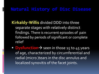
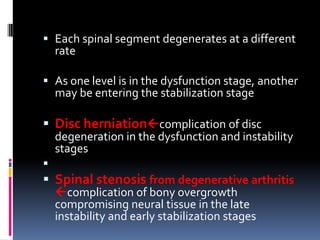
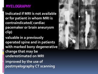
Degenerative disc disease is a condition characterized by changes in the discs between vertebrae. As discs degenerate they lose water content and height. Fissures can form in the annulus fibrosus and the nucleus pulposus loses structure. This can lead to bulging of the disc and potentially protrusion or extrusion of disc material. Kirkaldy-Willis divided the process into three stages: dysfunction, instability, and stabilization. Symptoms include back pain and pain that may radiate into the legs. Diagnostic imaging includes x-rays, CT, MRI, and discography which can help identify problematic discs.
Treatment modality of non union fracture neck of femurAvik Sarkar
The document discusses treatment modalities for non-union of femoral neck fractures. It describes causes of non-union and investigatory imaging. For the elderly, replacement arthroplasty is recommended, while for young adults a classification system is used to determine treatment. Type I involves bone grafting and fixation, Type II an osteotomy to change shear to compressive forces, and Type III drilling and fixation. Rehabilitation includes restricted weight bearing and physiotherapy. Osteotomies can correct alignment and reduce shearing forces at the non-union site.
Osteoarthritis of the knee is a degenerative joint disease affecting the articular cartilage and subchondral bone. It is more common in those over age 60 and females after menopause. Risk factors include obesity, previous knee injury, heredity, and muscle weakness. The pathophysiology involves biomechanical stress causing wear and tear of cartilage and bone. Patients experience pain, stiffness, swelling, and decreased range of motion. Treatment includes non-pharmacological measures, medications, injections, and surgery for advanced cases.
This document provides an overview of rotator cuff injuries, including anatomy, causes, symptoms, diagnosis, and treatment. It describes how the rotator cuff is composed of four tendons that stabilize the shoulder joint. Rotator cuff tears occur when one or more of these tendons becomes damaged and can range from partial to full thickness. Symptoms may include shoulder pain that is worsened with movement. Diagnosis involves physical examination along with imaging tests like x-rays, MRI, or ultrasound. Treatment options include non-operative measures like medication and physical therapy or surgical repair if conservative treatment fails.
The document discusses injuries to the acromioclavicular (AC) joint. It provides details on the anatomy and biomechanics of the AC joint and surrounding ligaments. Common mechanisms of injury include falling on an outstretched arm or direct force to the lateral shoulder. Injuries are classified using the Rockwood system from Type I to VI based on the degree of ligament disruption and bone displacement. Treatment options include nonoperative measures for lower grades and surgery for higher grades or failed nonoperative treatment. Surgical techniques and associated conditions are also reviewed.
The document discusses the anatomy, types, mechanisms of injury, evaluation, and management of proximal tibial fractures. Key points include:
- Proximal tibial fractures can be articular or nonarticular, with the lateral plateau most commonly involved. Injuries occur from shear, compressive, or axial forces on the knee.
- Evaluation includes physical exam of neurovascular status and knee stability as well as imaging like x-rays and CT to classify the fracture.
- Treatment depends on the fracture classification system used (Schatzker or AO/OTA) and involves non-operative management with immobilization for non-displaced fractures or operative management with open reduction and internal fixation for displaced or
Spondylolisthesis is the forward or backward slipping of one vertebra over another. This document discusses various types and classifications of spondylolisthesis. The key classifications discussed are the Wiltse, Newman, and Macnab classification (which categorizes spondylolisthesis based on its location and cause), the Meyerding classification (which grades the severity of slip based on percentage of vertebral translation), and the Marchetti-Bartolozzi classification (which categorizes spondylolisthesis as developmental or acquired based on etiology). Risk factors for progression include young age at presentation, female gender, high slip angle, and high grade slip.
Dr. Mahak Jain presented on spondylolisthesis. Key points include:
1) Spondylolisthesis is the forward translation of one vertebra on another, commonly caused by defects in the pars interarticularis known as spondylolysis.
2) It is classified based on etiology, with dysplastic, isthmic, degenerative, traumatic, and pathological types.
3) Treatment depends on factors like grade, symptoms, and etiology, ranging from conservative care to surgical options like decompression, fusion, and instrumentation.
4) Studies show surgery with fusion has better outcomes for pain and function than nonsurgical treatment or decompression alone for degenerative
The document discusses slipped capital femoral epiphysis (SCFE), a condition where the femoral head slides out of position in the hip. It begins by defining SCFE and describing the typical displacements seen, which are usually upward and anterior. It then covers the pathoanatomy of SCFE, focusing on the growth plate and zones of the physis. Finally, it discusses the incidence, risk factors, clinical presentation, investigations, and treatment options for SCFE.
This document discusses fractures around the shoulder joint, including proximal humerus fractures, shoulder dislocations, scapular fractures, and clavicular fractures. It provides details on the anatomy, classifications, clinical presentations, imaging, and treatment options for each type of injury. Treatment may involve closed reduction, open reduction with various surgical techniques like plating or nailing depending on the fracture pattern and bone quality. Post-operative rehabilitation is important for optimal outcomes.
This document defines and describes cavus foot, including its causes, clinical features, diagnosis, and treatment options. A cavus foot has an abnormally high arch and accompanying toe deformities. Causes include neuromuscular conditions like Charcot-Marie-Tooth disease and polio. Clinical features include a high arch and clawing of the toes. Diagnosis involves physical exam and x-rays. Treatment depends on flexibility and severity but may include tendon lengthening, osteotomies, and joint fusions to correct deformities in the forefoot, midfoot, and hindfoot. The goal is to create a plantigrade foot.
Dr. Ankur Mittal presented on diagnostic tests and imaging for Achilles tendon injuries. [1] Ultrasound is often used to determine tendon thickness and gap size for complete ruptures and is inexpensive and fast. [2] MRI is better for detecting incomplete tears and planning surgery for chronic tears but is more expensive. [3] Imaging is rarely needed for acute cases but can help with chronic cases for diagnosis and surgical planning.
Carpal instability can result from injuries to ligaments like the scapholunate ligament. Examination may reveal tenderness over injured ligaments or pain with wrist motion. X-rays can detect instability patterns and MRI is sensitive for detecting ligament tears. Arthroscopy is the gold standard for diagnosis. Treatment depends on injury chronicity and severity, and may include ligament repair, reconstruction, capsuldesis, limited fusions, or total wrist fusion.
SPONDYLOLISTHESIS: DIAGNOSIS, CLASSIFICATION, EVALUATION AND MANAGEMENTBenthungo Tungoe
Spondylolisthesis is the forward slippage of one vertebra on another. There are several classifications including developmental, isthmic, degenerative, traumatic, and postsurgical types. Developmental spondylolisthesis is usually asymptomatic and rarely progresses after adulthood. Isthmic spondylolisthesis has a risk of progression over 25% slippage or in symptomatic cases. Degenerative spondylolisthesis results from sagittal facet orientation or disc degeneration and increases in older females. The natural history depends on factors like age, gender, slip severity and progression.
This document summarizes a case of cubital tunnel syndrome in a 40-year-old Thai Buddhist monk. He experienced numbness and tingling in his right ring and little fingers for 1 month after injuring his right elbow. Examination found signs of ulnar nerve dysfunction including clawing of the fingers. Imaging showed no abnormalities. He was diagnosed with cubital tunnel syndrome and scheduled for ulnar nerve decompression surgery.
Adhesive capsulitis is a condition characterized by a painful and progressive loss of shoulder range of motion. It typically progresses through painful, freezing, and thawing phases over 1-2 years. Treatment involves medications to manage pain, physical therapy to restore range of motion, and in refractory cases, procedures like corticosteroid injections or surgery. While pain is usually transient, some patients may develop permanent loss of range of motion.
Here are the key steps in the ORIF procedure:
1. Patient is placed in lateral decubitus position and a right-angled lateral incision is made to minimize soft tissue damage.
2. The fracture line at the angle of Gissane is identified.
3. Fragments are temporarily held in place with K-wires under fluoroscopy while the reconstruction restores the 3D shape of the calcaneus.
4. The "constant" sustentacular fragment is used to begin the reconstruction, working anteriorly and medially.
5. Traction may be needed to restore the posterior facet.
6. The lateral wall fragment is closed like a door last to complete the
This document discusses shoulder instability, including definitions, anatomy, evaluation, and treatment. It defines instability as the inability to maintain the humeral head in the glenoid fossa, ranging from dislocation to laxity. Static stabilizers include the labrum and ligaments, while dynamic stabilizers are the rotator cuff and scapulothoracic muscles. Evaluation involves history, exam, and imaging to classify instability by direction, degree, and etiology. Treatment depends on classification but may include immobilization, rehabilitation, or surgical repair of labral tears or bone defects.
This document provides an overview of thoracolumbar fractures, including epidemiology, clinical evaluation, classification systems, radiographic evaluation, treatment approaches, and specific surgical techniques. It discusses the anatomy of the thoracolumbar region, mechanisms of injury, neurological assessment tools, radiographic indicators of instability, and non-operative and operative treatment options depending on the fracture classification.
This document discusses several types of hand injuries including Bennett's fracture, Rolando's fracture, and tendon injuries. Bennett's fracture is a fracture of the base of the first metacarpal bone that extends into the carpometacarpal joint, often accompanied by subluxation or dislocation. Rolando's fracture is an intra-articular fracture of the base of the first metacarpal bone that extends into the carpometacarpal joint. Tendon injuries can involve the flexor or extensor tendons and are classified based on the zone of injury. Treatment depends on the specific injury but may involve closed or open reduction, internal fixation, splinting, or surgery.
Cervical myelopathy is caused by compression of the cervical spinal cord, most commonly from cervical spondylosis. Cervical spondylosis involves degenerative changes to the spine that decrease space for the spinal cord. This can lead to static or dynamic compression of the cord, impairing circulation. Patients experience symptoms like neck pain, weakness, spasticity and sensory changes. Diagnosis involves assessing severity using scales and investigating spinal changes through imaging like CT which shows stenosis and compression more clearly than x-rays.
Common peroneal nerve lesions often present with foot drop and loss of sensation in the lower leg and foot. The common peroneal nerve is susceptible to injury where it passes between muscles in the leg and around the fibula bone. Injuries can occur from trauma, fractures, compression, ischemia or tight footwear and result in weakness of ankle dorsiflexion. Physical exams may show reduced foot movement and sensation loss while nerve conduction studies can identify injuries. Treatment focuses on bracing, stimulation, positioning and protective devices to prevent foot drop and sprains.
Spinal cord injuries can cause paralysis, sensory loss, and autonomic dysfunction below the level of injury. There are two main types - complete lesions with no preserved function below the injury, and incomplete lesions with some preserved sensation or motor function. Complications include neurogenic bladder, spasticity, autonomic dysreflexia, pressure ulcers, and orthostatic hypotension. Early rehabilitation is important to prevent further issues and maximize recovery of function.
Pp for lumbarization and sacralization by Dr Dhruv Taneja Assistant ProfessorDhruv Taneja
Lumbarization is a condition where the first sacral vertebra appears like a lumbar vertebra rather than being fused with the sacrum. This occurs when the first and second sacral segments fail to fuse during development. A lumbarized S1 vertebra may have its own disc or an underdeveloped disc space, making it difficult to accommodate and more prone to injury with age. Sacralization is a related condition where the fifth lumbar vertebra fuses with the sacrum, reducing mobility and increasing stress on the L4 vertebra. Both conditions can potentially lead to back pain and disc problems.
Degenerative spine disease involves three main areas: the intervertebral disc, vertebral bodies/end plates, and posterior elements. Changes to the intervertebral disc include decreased water/proteoglycan content leading to distorted collagen fibers and tears in the annulus fibrosis. Vertebral endplates can show three stages of degeneration. Posterior element changes include facet joint osteoarthritis with osteophytes/hypertrophy, ligamentum flavum hypertrophy/cysts, and spinal canal/foraminal stenosis. Imaging plays an important role in evaluating these degenerative changes and their effects.
Degenerative disc disease (DDD) is a condition caused by the deterioration of discs between the vertebrae in the spine. It commonly affects the cervical and lumbar regions and causes chronic back pain that may radiate into the legs. As people age, the nucleus of discs loses fluid and deteriorates, resulting in the narrowing of discs and pressure on spinal nerves. Common symptoms include back pain, sciatica, and numbness or weakness in the arms or legs. Diagnosis involves physical exams, x-rays, CT scans, and MRIs to evaluate the spine and rule out other causes. Treatment focuses on modifying activities, physical therapy, medications, injections, and sometimes surgery. While DDD cannot be prevented due to
INTERVERTEBRAL DISC ANATOMY AND PIVD OF LUMBAR SPINE AND ITS MANAGEMENTBenthungo Tungoe
1) The intervertebral disc consists of the nucleus pulposus surrounded by the annulus fibrosus and endplates. The nucleus contains water and proteoglycans to absorb pressure, while the annulus contains collagen fibers for strength and flexibility.
2) Degeneration of discs occurs over time as the nucleus loses water content and the annulus becomes weaker, altering load distribution and potentially leading to herniation.
3) Herniated discs occur when part of the nucleus extrudes from the annulus, most commonly posterolaterally, and can impinge nerves causing radicular pain. Classification is based on location and involvement of surrounding tissues.
This document discusses intervertebral disc prolapse. It begins by describing disc anatomy, development, and location in the spine. It then explains the pathology of prolapse, which involves nuclear degeneration, displacement, and fibrosis. Risk factors for prolapse include heavy lifting, smoking, obesity, and improper posture. Clinical features include low back pain radiating to the buttocks, aggravated by certain movements. Investigations include CT, MRI, and myelography. Treatment options range from conservative measures like rest and physical therapy to operative procedures like fenestration, hemi-laminectomy, and endoscopic discectomy.
The document provides an overview of spinal anatomy including:
1) It describes the coronal, sagittal, and axial planes used to view the spine on imaging and their anatomical divisions.
2) The basic structures and functions of vertebrae are outlined including protection of the spinal cord, flexibility, and load distribution.
3) Ligaments, joints, vasculature and innervation of the spine are summarized at different regions from cervical to lumbar.
This document discusses degenerative disc disease, which is a common cause of back and nerve pain. It begins by explaining the importance of correlating imaging findings with clinical symptoms to determine the appropriate treatment. Magnetic resonance imaging is often the preferred method for evaluating lumbar disc disease. The document then describes the normal anatomy of cervical, thoracic, and lumbar discs. It identifies the C5-C6, C6-C7, L4-L5, and L5-S1 levels as being most commonly affected. The remainder of the document discusses the pathophysiology and phases of disc degeneration, including changes that can be seen on imaging studies.
The document discusses intervertebral discs, disc prolapse, and degenerative changes in the spine. It provides the following key points:
- Intervertebral discs act as shock absorbers between vertebrae and allow some spinal motion. Discs are made up of an inner gelatinous nucleus pulposus surrounded by the annulus fibrosus.
- Disc prolapse occurs when the nucleus pulposus extrudes through tears in the annulus fibrosus. Imaging like MRI and CT can detect disc prolapses and herniations.
- Degenerative changes in the spine include osteophytosis, ligament calcification and ossification, and a reduction in the size of the spinal
This document discusses research on running and degenerative disc disease. It summarizes several studies that found short term effects of running like temporary loss of disc height and volume but recovery within weeks. One study found no increase in degenerative changes on MRI in long term runners. Another study found that running in rats for 3 weeks stimulated production of extracellular matrix proteins in discs. The document also discusses potential benefits of activities like climbing, tai chi, yoga and mindfulness for chronic low back pain.
When do we operate the degenerative disease ?
Pain not responding to conservative treatment, lasting more than 3 months
Non improving neurologic deficit
Persistence or deterioration of symptoms of intermitent claudication
Significant restriction of the common daily working and social activities
1) Degenerative disc disease involves the degeneration of intervertebral discs most commonly in the lower cervical and lumbar regions. The degenerated discs may herniate and press on nerves, causing pain and neurological deficits.
2) MRI is useful for evaluating degenerative disc disease as it can identify disc bulges and herniations, facet joint changes, and compression of nerves or the spinal cord.
3) Common sites for lumbar disc herniations are the L4-L5 and L5-S1 levels. Over 1/3 of herniated discs are asymptomatic, so clear evidence of nerve root compression is needed for surgery.
A 6-year-old girl presented with a 6-7 month history of dull, aching pain in her right groin that was aggravated by exertion and squatting. On examination, she had tenderness over the anterior hip joint line and limited internal rotation. X-rays confirmed Perthe's disease. At 3-month follow up, her pain and limp had increased with further limitation of internal rotation.
This is a short educational presentation on osteoporosis and its causes. This presentation also gives brief idea of management and prevention of osteoporosis.
This is a short patient education presention on Arthroscopy or Key hole surgery. It gives brief outline of the procedure.
Arhroscopy or key hole surgery is surgical procedure in which joints like knee, shoulder , elbow, ankle, hip joints could be examined for diagnosis and some times as used to provide definitive treatment. This surgical pocedure can be carried out as outpatient or inpatient basis. The main advantage of this procedure is it is minimally invasive, faster recovery, small scars.
This document discusses anti-inflammatory drugs and their mechanisms of action. It summarizes that steroids inhibit phospholipase A2 while nonsteroidal anti-inflammatory drugs (NSAIDs) inhibit cyclooxygenase (COX) enzymes, decreasing prostaglandin levels. NSAIDs are analgesic, antipyretic, and anti-inflammatory. The document further discusses the inflammatory process and mediators, different COX enzymes, prostaglandin functions, classes of NSAIDs including mechanisms and examples, and individual drug profiles of aspirin, indomethacin, ibuprofen, and paracetamol.
This presentation gives idea of managing osteoporotic fractures. This presentation also describes different surgical options in treating osteoporotic fractures.
Dr.A.Mohan krishna,
M.S.Ortho., MCh Ortho(U.K),
Consultant orthopedic surgeon
Apollo Hospitals,
V Care hospitals,
Fehmi care hospital,
Apollo clinic SR nagar
This is a patient education presentation. It gives a brief a description of causes of back pain during pregnancy. It also give tips for avoiding and managing back pain during pregnancy.
Search terms for the article:
Back pain in pregnancy, Lumbar pain, labour pain, back bone problems, pregnancy and back pain
Dr.A.Mohan krishna
M.s.Ortho., MCh Ortho(U.K)
Consultant Orthopedic surgeon,
Apollo Hospitals.
Vcare Multispeciality Hospital,
Apollo Clinic SR nagar,
This is a short patient education and awarness presentation on tail bone pain (coccydynia). This presentation delivers a brief information on causes, diagnosis, investigations and treatment of tail bone pain,
Disclaimer:
This presentation is solely for educational purpose.
This document provides an overview of approaches to musculoskeletal neoplasms (tumors). It discusses the classification and staging of bone tumors, the evaluation and workup of patients, and treatment approaches including biopsy, surgery, radiation therapy, and chemotherapy. Specifically, it summarizes a case of a 51-year-old woman who presented with knee pain and was found to have a low-grade chondrosarcoma in her fibula that was successfully treated with wide excision without preoperative biopsy.
This is a brief presentation of most common fracture around the wrist i.e Colles fracture. This presentation gives a brief information on anatomy, mechanism of injury, causes and treatment methods of colles fracture.
1) Spondylolisthesis is the slipping of one vertebra over another, usually caused by a defect in the pars interarticularis. It is classified based on its etiology and degree of slip.
2) Symptoms depend on the severity and include back pain, hamstring tightness, and sciatica. Examination may reveal a step in the back, tenderness over the pars defect, and limited back movement.
3) Imaging shows the degree of slip. Treatment focuses on pain relief through non-operative measures like physiotherapy. Surgery is considered if conservative treatment fails or neurological symptoms are present.
The document provides a history of lumbar disc disease and intervertebral disc discoveries from the 5th century to modern times. It then summarizes lumbar spine anatomy, the pathophysiology of disc degeneration and herniation, associated signs and symptoms, diagnostic tests including imaging, and conservative and surgical treatment options. Key events include Vesalius describing the intervertebral disc in 1543, Mixter and Barr identifying disc herniation as a cause of sciatica in 1934, and Kambin describing percutaneous lumbar discectomy in 1983.
This document discusses biological treatment options for avascular necrosis (AVN) of the femoral head. It provides details on the anatomy and blood supply of the femoral head. AVN occurs when there is interruption of blood flow to the femoral head, leading to bone cell death. Imaging plays an important role in diagnosis and staging of AVN. Conservative options include restricted weight bearing, medications, and physical therapies. Surgical options become necessary with more advanced stages to prevent femoral head collapse. The document covers various classification and staging systems used to determine the appropriate treatment based on the individual case.
The document discusses various chronic pain syndromes including low back pain, sciatica, complex regional pain syndrome, trigeminal neuralgia, and cancer pain. It provides details on the definition, causes, symptoms, diagnostic tools and treatment options for low back pain and sciatica, which are the most commonly discussed chronic pain conditions. The treatment sections cover medications, physical therapy, injections including epidural steroid injections, radiofrequency ablation, and other minimally invasive procedures.
This document discusses low back pain, sciatica, and lumbar disc prolapse. It provides details on the anatomy of the lumbar discs and describes how disc prolapses typically occur at the L4/L5 and L5/S1 levels. Clinical features of disc prolapses are outlined for different levels. Conservative and surgical treatment options are discussed. Cervical disc prolapse is also covered, including typical levels of involvement, clinical presentations, and management approaches.
This document discusses various degenerative spine diseases. It begins with the anatomy of the spine, including details on intervertebral discs, facet joints, and other structures. Common imaging tests are then outlined, such as plain x-rays, MRI, CT, and myelography. Common causes of back pain like muscle strains, herniated discs, and spinal stenosis are reviewed. The document also discusses Waddell signs which are used to evaluate non-organic causes of back pain. Finally, conditions such as degenerative disc disease, spondylosis, and spondylolisthesis are introduced.
This document provides an overview of cervical spine anatomy, cervical spondylosis, grading systems for cervical spine injuries, and treatment approaches. It defines cervical spondylosis as a widespread age-related degenerative condition affecting the cervical spine. Symptoms can include neck pain and neurological deficits. Treatment involves conservative approaches like physical therapy or surgical options like laminectomy, laminoplasty, or anterior cervical discectomy and fusion. Whiplash injuries and spinal cord injuries without radiographic abnormality (SCIWORA) are also discussed.
Cervical disc disorders include cervical spondylosis, radiculopathy, and myelopathy. Cervical spondylosis is a general term referring to degenerative changes in the cervical spine. It commonly causes neck pain but can also cause radiculopathy or myelopathy. Cervical radiculopathy involves compression of a cervical nerve root, causing pain and weakness along the nerve distribution. Cervical myelopathy refers to compression of the spinal cord, which can cause gait abnormalities, leg weakness, and hand/arm symptoms. Management involves conservative treatments like physical therapy initially, with surgery considered for worsening or persistent symptoms.
Lubar Arthritis : Lumbar Stenosis by Pablo Pazmino, MDPablo Pazmino
This video explains Lumbar Stenosis. When arthritis begins to encroach around the spinal cord and neural elements this is called Lumbar Stenosis. This video highlights the history, epidemiology, and treatment options both conservative and surgical. If you or someone you know needs to be seen in regards to Lumbar Stenosis/Arthritis feel free to look us up online www.beverlyspine.com or www.santamonicaspine.com OR call toll free 1-8SPINECAL-1
The document discusses avascular necrosis (also known as osteonecrosis), which is the death of bone tissue due to impaired blood supply. It affects over 20,000 new patients per year in the US, most commonly in the ages of 30-60. Common causes include steroid use, alcoholism, trauma, and blood clotting disorders. Diagnosis involves imaging like x-rays, CT, MRI, and bone scans to detect bone changes. Treatment aims to delay disease progression and joint breakdown through nonsurgical methods or sometimes surgery like joint replacement.
This document discusses avascular necrosis (also known as osteonecrosis), which is the death of bone tissue due to impaired blood supply. It affects over 20,000 new patients per year in the US, most commonly males ages 30-60. Risk factors include steroid use, alcoholism, blood clotting disorders, and autoimmune diseases like SLE. Symptoms may include joint pain. Diagnosis involves imaging modalities like x-ray, CT, MRI, and bone scans. Treatment aims to delay disease progression and prevent joint breakdown, and may include nonsurgical options or eventually joint replacement if collapse occurs.
Cervical spondylosis is a degenerative condition affecting the bones and joints in the neck. It causes pain, stiffness, and weakness and can compress nerves leading to sensory and motor problems. Symptoms range from mild neck pain to major dysfunction. While it mainly affects older adults, injuries or occupations involving heavy lifting or straining of the neck can also trigger it. Treatment focuses on relieving pain and addressing weakness, sensory loss, and other symptoms through analgesics, cervical collars, physiotherapy, surgery if needed, and encouraging patients to seek medical help. Healthcare assistants should explain cervical spondylosis to patients and ensure any problems are referred to doctors.
This document discusses the investigation and management of discitis/diskitis. Key points include:
- Elevated ESR, CRP, and procalcitonin levels can indicate discitis, along with abnormal findings on imaging like MRI and X-rays. Blood and sputum cultures help identify the infection source.
- MRI is the most sensitive test for discitis, showing abnormalities in the disc space and vertebrae on T1 and T2 weighted images. CT also shows bone destruction.
- Treatment involves intravenous antibiotics for 6-8 weeks, immobilization, and pain management. Surgery may be needed for neurological complications, non-response to treatment, or deformity correction.
-
Cervical radiculopathy is caused by spinal nerve root dysfunction resulting in dermatomal pain, parasthesias, myotomal weakness, and impaired deep tendon reflexes. It is commonly caused by herniated discs or bony spurs compressing nerve roots. Diagnosis involves history, physical exam testing dermatomes and myotomes, and imaging such as MRI. Treatment includes immobilization, traction, medications, injections, and sometimes surgery for severe or progressive cases.
A 66-year-old male presented with non-specific lower back pain. MRI revealed disc degeneration including annular bulging and Schmorl's nodes in the upper lumbar spine. The diagnosis was lumbar spondylosis. Disc degeneration results from mechanical stress and age-related changes and can lead to annular tears, nuclear material changes, and instability. Later stages involve further disc resorption and replacement by fibrocartilage. Nerve fibers in the outer annulus can contribute to discogenic pain.
This document discusses spondylolisthesis, defined as the anterior or posterior displacement of one vertebra on another. It describes classifications based on anatomy and etiology, radiological grading scales, clinical presentation, diagnostic imaging including X-rays, CT, MRI, and myelography. Treatment options are discussed including non-operative care with bracing and exercise, as well as surgical techniques such as decompression with or without fusion, interbody fusion approaches, reduction methods, instrumentation, and complications. Surgical treatment aims to prevent slip progression, stabilize the segment, correct deformity, relieve pain and reverse neurological deficits.
The document discusses various causes of neck and back pain including degenerative changes to the spine like thinning of the annulus and bulging discs which can press on nerves. It describes cervical radiculopathy causing arm pain and cervical myelopathy with neck stiffness and finger tingling. Diagnosis involves x-rays and MRI to view the spine and rule out other causes. Treatment ranges from conservative measures to surgery to relieve pressure on nerves or decompress the spinal cord.
Prolapsed lumbar intervertebral disc occurs when the soft central portion of an intervertebral disc bulges out beyond the damaged outer rings of the disc. It commonly affects the lower back and can cause lower back pain and leg pain. Diagnosis involves physical examination, imaging studies like MRI, and ruling out other potential causes of back pain. Treatment options include conservative measures like physical therapy and epidural steroid injections or surgery like discectomy if conservative options fail. Surgery aims to relieve nerve compression and associated back pain.
This document discusses failed back syndrome, specifically defining it as any condition where there is failure to improve satisfactorily following back surgery. It then classifies failures into those with no immediate improvement and temporary relief but recurrence of pain within weeks, months, or years. Causes of failures include wrong diagnosis, technical errors during surgery, infection, arachnoiditis, and recurrent stenosis. The document also discusses juxtafacet cysts near facet joints.
This document discusses intervertebral disc herniation. It begins by describing the anatomy of the lumbar spine motion segment and intervertebral disc. It then discusses the causes, risk factors, symptoms, diagnosis and management of intervertebral disc herniation. Common locations for herniation are the L4-L5 and L5-S1 discs. Symptoms can include back pain and radicular leg pain. Diagnosis involves physical exam, imaging like MRI, and ruling out other conditions. Management includes conservative options like physical therapy and injections initially. Surgery is considered if conservative options fail or if neurological deficits are present. Microdiscectomy is a common surgical procedure for disc herniation.
This document discusses diabetes and its effects on the orthopedic system, with a focus on the diabetic foot. It covers the pathophysiology of diabetes and how it can lead to complications like neuropathy, vasculopathy, and immunopathy. These complications increase the risk of issues like diabetic foot ulcers and Charcot arthropathy. The document outlines treatments for various conditions like total contact casting, antibiotics, and surgery. It also describes classifications of diabetic foot ulcers and Charcot arthropathy to help guide treatment decisions.
Diabetes can cause many complications affecting the feet including neuropathy, vasculopathy, and immunopathy which increase risks for foot ulcers and infections. Neuropathy damages nerves through multiple pathways like reactive oxygen species, impairing sensation and motor function. Vasculopathy damages blood vessels increasing risks of ischemia. Immunopathy impairs immune response to infection. Charcot arthropathy is a condition where loss of autonomic control and sensation leads to repetitive micro-trauma and bone destruction in the foot. Treatment involves wound care, offloading pressure through casting, surgery to correct deformities or remove infected tissue, and managing underlying complications of diabetes.
The document describes various surgical approaches to the elbow joint. The posterior approach is described in detail, including indications such as ORIF of distal humerus fractures. Key steps involve identifying the ulnar nerve, protecting it, and exposing the distal fourth of the humerus through a longitudinal incision over the posterior olecranon. The medial, lateral, anterior cubital fossa, and posterolateral radial head approaches are also outlined, identifying structures at risk and ways to optimize exposure for various procedures.
This document describes various surgical approaches to the elbow, including the posterior, medial, anterolateral, anterior of cubital fossa, and posterolateral approaches. For each approach, it provides the indications, patient positioning, landmarks, incision details, internervous planes, superficial and deep dissections, and structures at risk. The posterior approach is used for fractures of the distal humerus. The medial approach provides access to the medial condyle and coronoid process. The anterolateral approach is used for fractures of the radial head and capitellum.
The document provides an overview of plates and screws used in orthopedic surgery. It discusses the different parts and types of screws, including cortical screws, cancellous screws, and locking screws. It also describes the mechanical functions of plates, including neutralization plates, compression plates, and buttress plates. The document outlines the features and uses of various plate systems, such as the dynamic compression plate (DCP), limited contact-DCP (LC-DCP), reconstruction plates, and one-third tubular plates. It also introduces locking compression plates (LCP), which provide angular stability through the locking head of the screw instead of friction between the plate and bone.
The document discusses hip joint anatomy and biomechanics from the perspective of total hip arthroplasty. It describes key terms like kinematics and kinetics. It provides details on normal ranges of motion for the hip. It discusses femoral head anatomy and the forces acting on the hip during single leg stance, which can be up to 4 times body weight. Factors like leg length, weight, and abductor lever arm influence joint loading.
This document provides information on spondylolisthesis, including its definition, classification systems, etiology, natural history, clinical evaluation, radiographic findings, and management. Spondylolisthesis is defined as the slipping of one vertebra over another, most commonly of L5 over S1. It is classified based on etiology, including dysplastic, isthmic, degenerative, traumatic, and pathological types. Causes include developmental defects, stress fractures of the pars interarticularis, degeneration of the disc and facets, acute fractures, and bone diseases. Progression risks include young age, female sex, slip angle over 10 degrees, and sacral morphology. Evaluation involves history, exam looking for signs
The scaphoid bone forms the radial part of the wrist and is susceptible to fractures due to its oblique orientation crossing two rows of carpus bones. It receives its blood supply from three main arterial groups. Common mechanisms of injury involve falls onto outstretched hands and forced wrist motions. Clinical evaluation for scaphoid fractures includes wrist pain, swelling over the anatomical snuffbox, and tenderness over the scaphoid tubercle. While initial x-rays may not detect up to 25% of fractures, other imaging like bone scans, MRI, CT, and ultrasound provide higher sensitivity. Fractures are classified based on location and displacement, with displaced fractures or those in the proximal pole requiring operative treatment
2. DEFINITION
Degenerative disc disease (DDD) has been
used to describe a wide variety of
morphologic and radiographic changes in the
adult spine
3. DEFINITION: DISC DEGENERATION
The North American Spine Society Consensus
Committee on Nomenclature :
Changes in a disc characterized by desiccation,
fibrosis, or cleft formation in the nucleus;
fissuring or mucinous degeneration of the
annulus; defects and sclerosis of the endplates;
and/or osteophytes at the vertebral apophysis.
4. DEFINITION: DDD
Degenerative disc disease:
as a clinical syndrome characterized by
manifestations of disc degeneration and
symptoms thought to be related to those
changes
5. Intervertebral disc
Total 23
Hydrostatic, load bearing ,
shock absorbing structure
between the vertebral
bodies
Each disc unit has
strong outer ring of fibers
called the annulus fibrosus
a soft , jelly like center
called the nucleus
pulposus
2 endplates (Campbell)
6. Intervertebral Disc
Annulus Fibrosus
Outer portion of the disc
Annulus
Fibrosus
– Made up of lamellae fibrocartilage
– Layers of collagen fibers Type I
Arranged obliquely 30°
Some radial fibers
Thicker anteriorly >posteriolry
Attached to end plates
Great tensile, torsional
& radial strength
Lamellae
7. Intervertebral Disc
Nucleus Pulposus
– Inner structure
– Notochord remnant
– Type II collagen
+Gelatinous GAG,H2O
– High water content (7090%)
– Resists axial forces
Nucleus
Pulposus
9. The intervertebral disc in the adult
is avascular.
blood vessels in the annulus until
the age of 20 years and within the
cartilage endplates until the age
of 7 years.
The cells within the disc are
sustained by diffusion of nutrients
into the disc through the porous
central concavity of the vertebral
endplate
The discs vary in size and shape
with their position in the spine.
Discs also decrease in volume,
resulting in a 16% to 21% loss in
disc height after 6 hours of
standing or sitting.
11. Anatomical House with windows
Window of opportunity to the disc space, interlaminar and intertransverse window
12. Spine Motion Segment
The FUNCTIONAL UNIT of
the spine
Comprises of:
1.Two adjacent vertebrae
2.Intervertebral disc
3.Connecting ligaments:
including the ligamentum
flavum, interspinous,
supraspinous, intertransverse
ligament
4.Two facet joints and
capsules
13. Natural History of Disc Disease
Kirkaldy-Willis divided DDD into three
separate stages with relatively distinct
findings. There is recurrent episodes of pain
followed by periods of significant or complete
relief
Dysfunction seen in those 15 to 45 years
of age, characterized by circumferential and
radial (micro )tears in the disc annulus and
localized synovitis of the facet joints.
14. Instability found in 35- to 70-year-old
patients, characterized by internal disruption of
the disc, progressive disc resorption,
degeneration of the facet joints with capsular
laxity, subluxation, and joint erosion
Stabilization present in patients older than
60 years, the progressive development of
hypertrophic bone about the disc and facet
joints leads to segmental stiffening or frank
ankylosis
15. Each spinal segment degenerates at a different
rate
As one level is in the dysfunction stage, another
may be entering the stabilization stage
Disc herniationcomplication of disc
degeneration in the dysfunction and instability
stages
Spinal stenosis from degenerative arthritis
complication of bony overgrowth
compromising neural tissue in the late
instability and early stabilization stages
16. Pathoanatomy & pathogenesis
Kirkaldy-Willis Three
DYSFUNCTION
phases of
Degenerative process
INSTABILITY
STABILIZATION
17. Mechanism of DYSFUNCTION
Episode of rotational or compressive trauma
( uncoordinated muscle contraction)
Posterior joint strain
( also annular strain)
Splinting
Posterior joint
Subluxation
maintained
Minor facet subluxation
Synovitis( pain)
Sustained segmental
Hypertonicity of muscle
Ischemia ( pain)
Altered muscle metabolism
18. Symptom sign & radiological changes in
dysfunction
Symptom:
Low back pain
Often localised axial
Sometimes referred /radicular
Movement painful
Sign:
Local tenderness
Muscle contracted: PSM spasm
Hypomobility
Extension painful
Neurologically usually normal
Radiograph:
Loss of physiological curvatures
Spinous process malalignment
Irregular facet
Early disc changes
19. Mechanism of unstable phase
Severe dysfunction
Continuing stress
Trauma
Increased dysfunction
disc
facets
Degeneration of cartilage
Coalescence of tears
Loss of nucleus, internal disruption
Attenuation of capsule
Bulging of annulus
Laxity of capsule
Increased abnormal movement
Unstable phase
20. INSTABILITY
Symptom: Those of dysfunction
Giving away of back, “catch” in back( on movement)
Pain on coming to standing position after flexion
Sign: Detection of abnormal movement( LOOK/FEEL)
Observation of “catch” sway or shift when coming erect after
flexion
Radiograph: AP: Lateral shift
Rotation
Abnormal tilt
Malaligned spinous process
OBLIQUE: Opening facets
LATERAL: Spondylolisthesis( in flexion)
Retrolisthesis ( in extension)
Narrowing foramen( in extension).Abnormal opening of disc
Abrupt change in pedicle height.Traction spurs
21. Mechanism of stabilization
Disc
Facets
Destruction of cartilage
Loss of nucleus
Fibrosis in joints
Approximation of bodies
Enlargement of facets
Destruction of plates
Locking facets
Fibrosis in disc
osteophytes
Fibrosis arund joints
Increased stiffness
stabilization
22. STABILIZATION
Symptom: Low back pain of decreasing severity
Sign:
Muscle tenderness
Stiffness
Reduced movement
Scoliosis
Radiograph:
Enlarged facet
Loss of disc height
Osteophytes
Small foramen
Reduced movement
Scoliosis
23. Diagnostic Studies
ROENTGENOGRAPHY
1. AP and Lateral
2. Oblique views:
useful in defining spondylolisthesis
and spondylolysis
3. Lateral flexion and extension:
X-ray may reveal segmental
instability
4. Ferguson view (20-degree
caudocephalic anteroposterior ):
value in the diagnosis of the "far out
syndrome," that is, L5 compression
produced by a large transverse
process of the fifth lumbar vertebra
against the ala of the sacrum
24. MYELOGRAPHY
Indicated if MRI is not available
or for patient in whom MRI is
contraindicated( cardiac
pacemaker or brain aneurysm
clip)
valuable in a previously
operated spine and in patients
with marked bony degenerative
change that may be
underestimated on MRI
improved by the use of
postmyelography CT scanning
25. COMPUTED TOMOGRAPHY
extremely useful diagnostic tool
noninvasive, painless, outpatient procedure can supply
more information about spinal disease
Unfortunately, CT does not demonstrate intraspinal tumors
or arachnoiditis and is unable to differentiate scar from
recurrent disc herniation.
26. MAGNETIC RESONANCE IMAGING
newest technological
advance in spinal imaging
The advantages : ability to demonstrate
intraspinal tumors, examine
the entire spine, and identify
degenerative discs based on
decreased H2O content
costly and requires specially
constructed facilities.
27. Modic Change
Type I
Signal intensity on
low T1-weighted
High T2-weighted
replacement of the
end-plate marrow with
vascular fibrous tissue
in response to chronic
“injury.”
Clinical: annular tear,
fissure
28. MODIC CHANGE
Type II
signal intensity
high T1-weighted and on
FSE T2-weighted
Low T2
represents
replacement of the
end-plate marrow with
fatty tissue.
Chronic marrow disuse
Type II changes tend to
remain stable with
time.
29. Modic Change
Type III
signal intensity
lowT1-weighted
lowT2-weighted
severely degenerated end
plates
only end plate change visible
on CT scans or radiographssclerosis
Part of the normal aging
process and must not be
confused with other
pathologic processes, such as
tumor and infection
30. Other diagnostic tests
PET / SPECT- experimental & few centers
have this facilities
Electromyography/ NCVadvantage of
electromyography is in the identification of
peripheral neuropathy and diffuse
neurological involvement indicative of higher
or lower lesions.
The SSEP is an extremely sensitive
monitoring technique.
31. Bone scans positive findings usually are not
indicative of intervertebral disc disease, but they
can confirm neoplastic, traumatic, and arthritic
problems in the spine.
complete blood count, differential white cell count,
biochemical profile, urinalysis, and sedimentation
rate good screening procedures
Rheumatoid screening studies such as rheumatoid
arthritis latex, antinuclear antibody, lupus
erythematosus cell preparation, and HLA-B27 also
are useful when indicated by the clinical picture.
32. ZYGAPOPHYSEAL (FACET) JOINT
INJECTIONS
Cause of facet joint pain: Meniscoid entrapment and extrapment, synovial
impingement, chondromalacia facetae, capsular and synovial
inflammation, and mechanical injury to the joint capsule.
Osteoarthritis
No noninvasive pathognomonic findings distinguish
facet joint–mediated pain from other sources of spine
pain.
Fluoroscopically guided facet joint injections therefore
are commonly considered the gold standard for
isolating or excluding the facet joint as a source of
spine or extremity pain.
33. DISCOGRAPHY
provocative testing for
concordant pain to
provide information regarding
the clinical significance of
the disc abnormality.
Indications :surgical planning of spinal fusion, testing of the structural
integrity of an adjacent disc to a known abnormality such as
spondylolisthesis or fusion, identifying a painful disc among
multiple degenerative discs, ruling out secondary internal disc
disruption or suspected lateral or recurrent disc herniation, and
determining the primary symptom-producing level when
chemonucleolysis is being considered.
34. Lumbar spine in
an oblique
position with
superior articular
process (arrow)
dividing disc
space (d) in half
Disc entry point is
just anterior
(arrow) to base of
superior articular
process (s) and
just above superior
endplate of
vertebral body
Curved procedure
needle (c) passing
through straight
introducer needle (n
35. Lumbar Disc Disease
Symptomatic LDH occurs during the lifetime of
approx. 2% of the general population
Factors associated with LDH:
Male gender
Age 30 -50 yrs
Job requiring heavy lifting
Lifting in a twisted or asymmetric posture
Stressful occupation
Lower income
Cigarette smoking
Exposure to prolonged vibration in the range of 4 to 5
Hz
36. Degenerative Disc Disease
Pathophysiology:
1.
Disc gradually dries out, loses height and
volume.
2. NP changes from a turgid gelatinous
bulb to brownish dessicated structure.
3.
4.
5.
6.
7.
8.
9.
AF develops fissures parrallel to the vertebral
end plates.
Compressive loads transfer away from nucleus
to margins
Sclerosis of endplate reduces disc nutrition.
Facet joints wear away cartilage, begin to
override
Motion segment becomes hypermobile
Osteophytes develop to attempt to stabilize
motion segment
Osteophytes may encroach on neural
structures.
37. Prolapse intervertebral disc
Pathophysiology:
1. Acute disc prolapse is due to flexion
+compression.
2. More at L4/5,L5/S1 (stress is more severe).
3. Disc rupture = stress + disturbances in the
hydrophilic properties of the NP.
4. Disc rupture = fibrocartilaginous material
extruded posteriorly and annulus bulges to one
side.
5. Part of the nucleus may sequestrated freely.
6. Large central rupture may cause pressure of the
cauda equina.
38. Boos et al.
decrease in
nutritional transport
water content
absolute number of viable cells
proteoglycans
pH
increase in
an increase keratin sulfate to chondroitin sulfate ratio
lactate
degradative enzyme activity
39. Pathophysiology:
Pain= arises due to disruption of outermost layer
of the annulus fibrosus, stretching or tearing of
the posterior longitudanal ligament and pressure
on the dura. symptoms worsened by
coughing, valsalva, sneezing
Sciatica= if disc protudes to oneside it may irritate
the dural covering of the adjacent nerve root
causing pain in the buttock, posterior thigh and
calf.
Pressure on the nerve root itself causes
paraesthesia and/or numbness in the
corresponding dermatome, as well as weakness
and decreased reflexes in the muscles suppllied
by that nerve root.
40. STAGES OF DDD
Dessication
Loss of fluid in nucleus pulposus.
Disc bulge:- diffuse symmetrical outpouching of the annulus fibrosus
caused by early disc degeneration& collapse
Protrusion:- base wider than any diameter of the material displaced
beyond disc space
Extrusion:- displaced portion has a greater diameter than its connection
with the parent disc at its base
Sequestration:- when disc extrusion has lost all connection with the
parent disc
Migration:- an extruded disc, whether sequestrated or not, that has been
displaced above or below the edge of the disc space
41. CLASSIFICATION DD
Depending upon whether the
displaced portion is
completely enveloped by
intact outer annulus or
combination of annulus and
PLL( s/t called capsule): Contained: Un contained: Subligamentous:- disc material
contained beneath the PLL
Transligamentous
Submembranous:- disc material
contained only by peridural
menbrane
42. CLASSIFICATION
Depending upon the relationship of the herniated
material to the posterior annulus and PLL:
Central( midline):- herniation along the posterior annulus
Posterolateral:- along the weaker lateral expansion of PLL
Foraminal( lateral )
Extraforaminal ( far lateral)
In relation to nerve root:
Shoulder herniation
Axillary herniation
According to the level of LDH: High LDH:- L1-L2, L2-L3, L3-L4
Low LDH:- L4-L5, L5-S1
43. SIGNS AND SYMPTOMS LDD
Age:- 3rd or 4th decade in healthy adult
Mostly relate to traumatic incident but
Intermittent back pain of months or year
Back or leg pain, radiating
Aggravating :-heavy exertion, repetitive
bending, twisting, or heavy lifting, relieved
with rest in semi-Fowler position
weakness and paresthesias, localized to the
neurological level of involvement
44. Clinical examination
Standing pt who declines to sit, with loss of
normal lumbar lordosis & PVM spasm
suggestive of PIVD
List
Limited spine ROM
Point tenderness may be present over the
spinous process at the level of the disc
involved
Atrophy of muscles – chronic cases
45. LSR testing
During SLR maneuver , the L5 and S1 nerve root
either moves or passively deforms approx. 2 to 6
mm at the level of foramen
Maximum tension is realized in the sciatic nerve at
30* to 70* of elevation from the supine
Crossed SLR is more specific of a disc herniation(
pathognomonic of micromotion in affected side
nerve roots while raising normal side leg)
Large cenrtal or lateral recess herniation
Free disc fragment
Lasegue sign
Bowstring sign
46.
47.
48.
49. Management
Goal: Prompt return to normal function and pain relief through the
efficient and effective use of diagnostic tests and efficacious
treatments
Non operative: Reassurance, medications, and activity modification
Bed rest in a semi Fowlers position for 1 to 2 days in acute
cases
Aerobic conditioning including abdominal and back
strengthening exercises
Application of heat, ice, TENS, USG massage, Traction
Manipulative therapy
Back school programme
50.
EPIDURAL STEROIDS:-
offer relatively prolonged pain relief without
excessive narcotic intake if conservative care is
elected.
Methylprednisolone is the usual steroid injected.
The dosage may vary from 80 to 120 mg.
The anesthetics used may include lidocaine,
bupivacaine, or procaine.
current protocol is to inject the patient three times.
These injections are made at 7- to 10-day intervals.
51. Indication of surgery
Emergent/ absolute: Presence of cauda equina syndrome
Progressive neurologic deficit
Relative: Persistent radiculopathy despite an adequate trial of non surgical
treatment( min of 6 wks)
Recurrent episodes of incapacitating sciatica
Significant motor deficit with persistent tension signs and pain
Pseudoclaudication( activity related leg pain) caused by canal stenosis
resulting from a disc herniation
Goal of surgery: Alleviate the neural compression without further injury to the affected
nerve root
Minimal disruption of surrounding normal tissues and maintenance of
spinal stability
52. Waddell’s Non-organic sign
(DOReST)
Finding
Description
1. Tenderness
a. superficial - pain with light touch
to skin
b. deep - nonanatomic widespread
deep pain
2. Simulation
a. pain with light axial compression
on skull
b. pain with light twisting of pelvis
3. Distraction
No pain with distracted SLR
4. Regional
a.nonanatomic or inconsistent
motor findings during entire exam
b. nonanatomic or inconsistent
sensory findings during entire exam
5. Overreaction
Overreaction noted at any time
during exam
53. Surgical procedure
Standard open lumbar disectomy
Microlumbar disc excision
Endoscopic disc excision
Additional exposure
Hemilaminectomy usually is required when identifying
the root is a problem. This may occur with a conjoined
root.
Total laminectomy usually is reserved for patients with
spinal stenoses that are central in nature, which occurs
typically in cauda equina syndrome.
Facetectomy usually is reserved for foraminal stenosis or
severe lateral recess stenosis.
degeneration involves: 1) declining disc nutrition, 2) loss of proteoglycan organization and concentration, 3) decrease in water content, 4) a decline in cell numerical density and synthetic activity, 5) increased degradative enzyme activity relative to matrix synthesis. An increased keratin sulfate to chondroitin sulfate ratio annulus fibrosis outer structure that encases the nucleus pulposuscomposed of type I collagen that is obliquely oriented, water, and proteoglycanscharacterized by high tensile strength and its ability to prevent intervertebral distractionremains flexible enough to allow for motionhigh collagen / low proteoglycan ratio (low % dry weight of proteoglycans) fibroblast-like cells responsible for producing type I collagen and proteoglycans
nucleus pulposus central portion of the intervertebral disc that is surrounded by the annulus fibrosiscomposed of type II collagen, water, and proteoglycansapproximately 88% waterhydrophilic matrix is responsible for height of the intervertebral disccharacterized by compressibility a hydrated gel due to high polysacharide content and high water content (88%) proteoglycans interact with water and resist compressionviscoelastic matrix distributes the forces smoothly to the annulus and the end plates low collagen / high proteoglycan ratio (high % dry weight of proteoglycans) chondrocyte-like cells responsible for producing type II collagen and proteoglycanssurvive in hypoxic conditions
With aging of the intervertebral disc there is an increase in the keratin sulfate to chondroitin sulfate ratioAt birth, the disc surface area is 50% nucleus pulposus (NP) and 50% annulus. The notochordal cells of the NP are gradually replaced by chondrocytes throughout the early teenage years. The demarcation betweenthe annulus and the nucleus becomes less distinct. The older NP has a higher collagen content with more structured fibers. In these fibers, the ratio of type II to type I collagen increases
Stresses annulus fibrosushighest tensile stressesnucleus pulposushighest compressive stressintradiscal pressure is position dependent pressure is lowest when lying supinepressure is intermediate when standingpressure is highest when sitting and flexed forward with weights in the handswhen carrying weight, the closer the object is to the body the lower the pressure
Epidemiology 95% involve L4/5 or L5/S1 levels L5/S1 most common levelpeak incidence is 4th and 5th decadesonly ~5% become symptomatic3:1 male:female ratio
Disc Herniation herniated disks are associated with a spontaneous increase in the production of osteoprotegrin (OPG)interleukin-1 betareceptor activator of nuclear factor-kBligand (RANKL)parathyroid hormone (PTH)Disc aging leads to an overall loss of water content and conversion to fibrocartilage. Specifically there is a decrease in nutritional transport water contentabsolute number of viable cellsproteoglycanspHincrease inan increase keratin sulfate to chondroitin sulfate ratio lactatedegradative enzyme activityno change inabsolute quantity of collagen
Containedbulge:circumferential symmetric disc extension upto the vertebral border within the annulus fibrosusContained protrusion: focal or asymmetrical extension of disc beyond the vertebral borderContained extrusion: extruded thru annulus but not thru PLLUncontained sequestration: disc material thru both annulus and PLL , not continuous with the parent disc materialMigration: Disc material displaced from the site of extrusion (either sequestrated or not)
motor exam ankle dorsiflexion (L4 or L5) test by having patient walk on heelsEHL weakness (L5) manual testinghip abduction weakness (L5)have patient lie on side on exam table and abduct leg against resistanceankle plantar flexion (S1) have patient do 10 single leg toes stands
Presence of 3 out of 5 of these sign correlated poor outcome with surgery even in the presence of true structural abnormalities.Waddell et al described and standardized these non-organic signs of low-back pain in 350 North American and British patients. They divided them into 5 categories (tenderness tests, simulation tests, distraction tests, regional disturbances, and overreaction), and found that when three or more categories were positive, the finding was considered clinically significant. This was also correlated with high scores for depression, hysteria and hypochondriasis on the Minnesota Multiphasic Personality Inventory (MMPI)