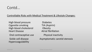
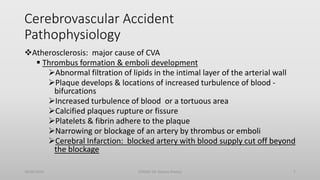
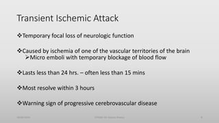
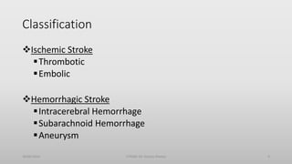
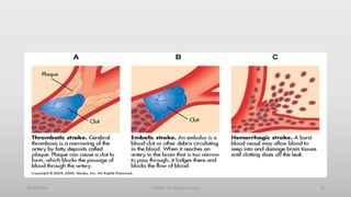
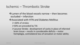
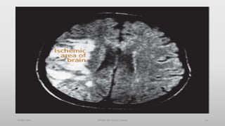
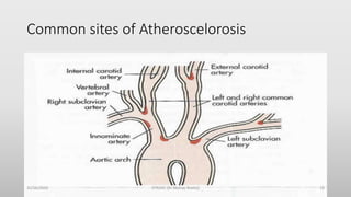
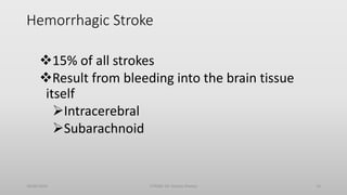
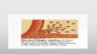
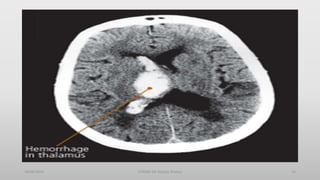
This document discusses stroke, including defining it as resulting from ischemia or hemorrhage in the brain, listing common risk factors like age, hypertension and diabetes, classifying strokes as ischemic or hemorrhagic, and describing diagnostic tools like CT scans and MRI. Ischemic strokes are further divided into thrombotic and embolic types while hemorrhagic strokes are intracerebral or subarachnoid. Clinical effects and sites of pathology are outlined for different stroke types.
Is characterized by the sudden loss of blood circulation to an area of the brain, resulting in a corresponding loss of neurologic function. Acute ischemic stroke is caused by thrombotic or embolic occlusion of a cerebral artery and is more common than hemorrhagic stroke.
It can occur
in the carotid
artery of the
neck as well as
other arteries.
When an artery is acutely occluded by thrombus or embolus, the area of the CNS supplied by it will undergo infarction if there is no adequate collateral blood supply.
Surrounding a central necrotic zone, an ‘ischemic penumbra’ remains viable for a time, i.e. it may recover function if blood flow is restored.
CNS ischemia may be accompanied by swelling for two reasons:
● cytotoxic oedema – accumulation of water in damaged glial cells and neurones,
● vasogenic oedema – extracellular fluid accumulation as a result of breakdown of the blood–brain barrier.
In the brain, this swelling may be sufficient to produce clinical deterioration in the days following a major stroke, as a result of a rise in intracranial pressure and compression of adjacent structures.
Hemiparesis is unilateral paresis, that is, weakness of the entire left or right side of the body (hemi- means "half"). Hemiplegia is, in its most severe form, complete paralysis of half of the body. Hemiparesis and hemiplegia can be caused by different medical conditions, including congenital causes, trauma, tumors, or stroke
Hypenension: Commonest cause of intracerebral haemorrhage.
Rupture of an intracranial aneurysm, angioma or A-V malformation: commonest cause of subarachnoid haemorrhage.
Haemorrhagic blood diseases: purpura, haemophilia.
Anticoagulants.
Trauma to the head: commonest of subdural haematoma.
II. Infective: ;
Encephalitis
Meningitis – Brain abscess.
III. Neoplastic: e.g. Meningioma.
IV. Demyelination: multiple sclerosis may present with hemiplegia.
V. Traumatic: e.g. Cerebral laceration and subdural haematoma.
VI. Hysterical: patient suffering from paralysis in the absence of organic lesion.
A stroke occurs when the blood supply to part of your brain is interrupted or reduced, depriving brain tissue of oxygen and nutrients. Within minutes, brain cells begin to die.
Keunikan anatomi small vessel of the brain dan neurovascular unit, kontroversi peran stganasi vena dalam patofisiologi, klasifikasi small vessel disease, variasi kriteria diagnostik, pitfall dalam neuroimaging, pilihan antiplatelet untuk prevensi sekundar, dampaknya bagi outcome pasien, hubungannya dengan gangguan fungsi kognitif.
Hmm, apa lagi nih yang baru?
I am a Neurosurgeon with advanced training in Interventional vascular Neurosurgery(FINR) from Zurich, Switzerland, and FMINS-Fellowship in minimally invasive and Endoscopic Neurosurgery from Germany.
I am presently working in Columbia asia hospitals, Bangalore.
My areas of interest are Vascular Neurosurgery, Stroke specialist, interventional neuroradiology, Endoscopic and minimally invasive Neurosurgery, Endoscopic spine surgery.
Stroke : Introduction, types and treatment.Obed Adams
Stroke is defined as the rapid loss of brain function due to disturbance in blood flow and supply to the brain.
OR
An acute episode of focal dysfunction of the brain, retina, or spinal cord.
It is clinically defined as the rapid onset of cerebral deficit lasting more than 24hours and is caused by acute vascular injury to parts of the brain. Presented by Obed Adams.
Title: Sense of Smell
Presenter: Dr. Faiza, Assistant Professor of Physiology
Qualifications:
MBBS (Best Graduate, AIMC Lahore)
FCPS Physiology
ICMT, CHPE, DHPE (STMU)
MPH (GC University, Faisalabad)
MBA (Virtual University of Pakistan)
Learning Objectives:
Describe the primary categories of smells and the concept of odor blindness.
Explain the structure and location of the olfactory membrane and mucosa, including the types and roles of cells involved in olfaction.
Describe the pathway and mechanisms of olfactory signal transmission from the olfactory receptors to the brain.
Illustrate the biochemical cascade triggered by odorant binding to olfactory receptors, including the role of G-proteins and second messengers in generating an action potential.
Identify different types of olfactory disorders such as anosmia, hyposmia, hyperosmia, and dysosmia, including their potential causes.
Key Topics:
Olfactory Genes:
3% of the human genome accounts for olfactory genes.
400 genes for odorant receptors.
Olfactory Membrane:
Located in the superior part of the nasal cavity.
Medially: Folds downward along the superior septum.
Laterally: Folds over the superior turbinate and upper surface of the middle turbinate.
Total surface area: 5-10 square centimeters.
Olfactory Mucosa:
Olfactory Cells: Bipolar nerve cells derived from the CNS (100 million), with 4-25 olfactory cilia per cell.
Sustentacular Cells: Produce mucus and maintain ionic and molecular environment.
Basal Cells: Replace worn-out olfactory cells with an average lifespan of 1-2 months.
Bowman’s Gland: Secretes mucus.
Stimulation of Olfactory Cells:
Odorant dissolves in mucus and attaches to receptors on olfactory cilia.
Involves a cascade effect through G-proteins and second messengers, leading to depolarization and action potential generation in the olfactory nerve.
Quality of a Good Odorant:
Small (3-20 Carbon atoms), volatile, water-soluble, and lipid-soluble.
Facilitated by odorant-binding proteins in mucus.
Membrane Potential and Action Potential:
Resting membrane potential: -55mV.
Action potential frequency in the olfactory nerve increases with odorant strength.
Adaptation Towards the Sense of Smell:
Rapid adaptation within the first second, with further slow adaptation.
Psychological adaptation greater than receptor adaptation, involving feedback inhibition from the central nervous system.
Primary Sensations of Smell:
Camphoraceous, Musky, Floral, Pepperminty, Ethereal, Pungent, Putrid.
Odor Detection Threshold:
Examples: Hydrogen sulfide (0.0005 ppm), Methyl-mercaptan (0.002 ppm).
Some toxic substances are odorless at lethal concentrations.
Characteristics of Smell:
Odor blindness for single substances due to lack of appropriate receptor protein.
Behavioral and emotional influences of smell.
Transmission of Olfactory Signals:
From olfactory cells to glomeruli in the olfactory bulb, involving lateral inhibition.
Primitive, less old, and new olfactory systems with different path
NVBDCP.pptx Nation vector borne disease control programSapna Thakur
NVBDCP was launched in 2003-2004 . Vector-Borne Disease: Disease that results from an infection transmitted to humans and other animals by blood-feeding arthropods, such as mosquitoes, ticks, and fleas. Examples of vector-borne diseases include Dengue fever, West Nile Virus, Lyme disease, and malaria.
Rasamanikya is a excellent preparation in the field of Rasashastra, it is used in various Kushtha Roga, Shwasa, Vicharchika, Bhagandara, Vatarakta, and Phiranga Roga. In this article Preparation& Comparative analytical profile for both Formulationon i.e Rasamanikya prepared by Kushmanda swarasa & Churnodhaka Shodita Haratala. The study aims to provide insights into the comparative efficacy and analytical aspects of these formulations for enhanced therapeutic outcomes.
Adv. biopharm. APPLICATION OF PHARMACOKINETICS : TARGETED DRUG DELIVERY SYSTEMSAkankshaAshtankar
MIP 201T & MPH 202T
ADVANCED BIOPHARMACEUTICS & PHARMACOKINETICS : UNIT 5
APPLICATION OF PHARMACOKINETICS : TARGETED DRUG DELIVERY SYSTEMS By - AKANKSHA ASHTANKAR
Basavarajeeyam is an important text for ayurvedic physician belonging to andhra pradehs. It is a popular compendium in various parts of our country as well as in andhra pradesh. The content of the text was presented in sanskrit and telugu language (Bilingual). One of the most famous book in ayurvedic pharmaceutics and therapeutics. This book contains 25 chapters called as prakaranas. Many rasaoushadis were explained, pioneer of dhatu druti, nadi pareeksha, mutra pareeksha etc. Belongs to the period of 15-16 century. New diseases like upadamsha, phiranga rogas are explained.
263778731218 Abortion Clinic /Pills In Harare ,sisternakatoto
263778731218 Abortion Clinic /Pills In Harare ,ABORTION WOMEN’S CLINIC +27730423979 IN women clinic we believe that every woman should be able to make choices in her pregnancy. Our job is to provide compassionate care, safety,affordable and confidential services. That’s why we have won the trust from all generations of women all over the world. we use non surgical method(Abortion pills) to terminate…Dr.LISA +27730423979women Clinic is committed to providing the highest quality of obstetrical and gynecological care to women of all ages. Our dedicated staff aim to treat each patient and her health concerns with compassion and respect.Our dedicated group ABORTION WOMEN’S CLINIC +27730423979 IN women clinic we believe that every woman should be able to make choices in her pregnancy. Our job is to provide compassionate care, safety,affordable and confidential services. That’s why we have won the trust from all generations of women all over the world. we use non surgical method(Abortion pills) to terminate…Dr.LISA +27730423979women Clinic is committed to providing the highest quality of obstetrical and gynecological care to women of all ages. Our dedicated staff aim to treat each patient and her health concerns with compassion and respect.Our dedicated group of receptionists, nurses, and physicians have worked together as a teamof receptionists, nurses, and physicians have worked together as a team wwww.lisywomensclinic.co.za/
ABDOMINAL TRAUMA in pediatrics part one.drhasanrajab
Abdominal trauma in pediatrics refers to injuries or damage to the abdominal organs in children. It can occur due to various causes such as falls, motor vehicle accidents, sports-related injuries, and physical abuse. Children are more vulnerable to abdominal trauma due to their unique anatomical and physiological characteristics. Signs and symptoms include abdominal pain, tenderness, distension, vomiting, and signs of shock. Diagnosis involves physical examination, imaging studies, and laboratory tests. Management depends on the severity and may involve conservative treatment or surgical intervention. Prevention is crucial in reducing the incidence of abdominal trauma in children.
These lecture slides, by Dr Sidra Arshad, offer a quick overview of the physiological basis of a normal electrocardiogram.
Learning objectives:
1. Define an electrocardiogram (ECG) and electrocardiography
2. Describe how dipoles generated by the heart produce the waveforms of the ECG
3. Describe the components of a normal electrocardiogram of a typical bipolar lead (limb II)
4. Differentiate between intervals and segments
5. Enlist some common indications for obtaining an ECG
6. Describe the flow of current around the heart during the cardiac cycle
7. Discuss the placement and polarity of the leads of electrocardiograph
8. Describe the normal electrocardiograms recorded from the limb leads and explain the physiological basis of the different records that are obtained
9. Define mean electrical vector (axis) of the heart and give the normal range
10. Define the mean QRS vector
11. Describe the axes of leads (hexagonal reference system)
12. Comprehend the vectorial analysis of the normal ECG
13. Determine the mean electrical axis of the ventricular QRS and appreciate the mean axis deviation
14. Explain the concepts of current of injury, J point, and their significance
Study Resources:
1. Chapter 11, Guyton and Hall Textbook of Medical Physiology, 14th edition
2. Chapter 9, Human Physiology - From Cells to Systems, Lauralee Sherwood, 9th edition
3. Chapter 29, Ganong’s Review of Medical Physiology, 26th edition
4. Electrocardiogram, StatPearls - https://www.ncbi.nlm.nih.gov/books/NBK549803/
5. ECG in Medical Practice by ABM Abdullah, 4th edition
6. Chapter 3, Cardiology Explained, https://www.ncbi.nlm.nih.gov/books/NBK2214/
7. ECG Basics, http://www.nataliescasebook.com/tag/e-c-g-basics
2. Contents
• Objectives
• Definition of Stroke
• Risk factors of Stroke
• Pathophysiology of Stroke
• Classification of stroke
• Ischemic stroke
• Haemorrhagic stroke
• Diagnosis
• Summary
• References
30/06/2020 STROKE (Dr Akshay Shetty) 2
3. Objectives
By the end of the presentation the students must be able to
1. Define Stroke
2. List Risk factors of Stroke
3. Explain Pathophysiology of Stroke
4. Classify stroke
5. Discuss Ischemic stroke
6. Discuss Haemorrhagic stroke
7. State Diagnosis
30/06/2020 STROKE (Dr Akshay Shetty) 3
4. Definition
• Stroke or CVA results from ischemia to a part of the brain or
hemorrhage into the brain that results in death of brain cells.
30/06/2020 STROKE (Dr Akshay Shetty) 4
5. Cerebrovascular Accident
Risk Factors
Nonmodifiable:
Age – Occurrence doubles each decade >55 years
Gender – Equal for men & women; women die more frequently than men
Race – African Americans, Hispanics, Native Americans, Asian Americans --
higher incidence
Heredity – family history, prior transient ischemic attack, or prior stroke
increases risk
30/06/2020 STROKE (Dr Akshay Shetty) 5
6. Contd…
Controllable Risks with Medical Treatment & Lifestyle Changes:
High blood pressure Diabetes
Cigarette smoking TIA (Aspirin)
High blood cholesterol Obesity
Heart Disease Atrial fibrillation
Oral contraceptive use Physical inactivity
Sickle cell disease Asymptomatic carotid stenosis
Hypercoagulability
30/06/2020 STROKE (Dr Akshay Shetty) 6
7. Cerebrovascular Accident
Pathophysiology
Atherosclerosis: major cause of CVA
Thrombus formation & emboli development
Abnormal filtration of lipids in the intimal layer of the arterial wall
Plaque develops & locations of increased turbulence of blood -
bifurcations
Increased turbulence of blood or a tortuous area
Calcified plaques rupture or fissure
Platelets & fibrin adhere to the plaque
Narrowing or blockage of an artery by thrombus or emboli
Cerebral Infarction: blocked artery with blood supply cut off beyond
the blockage
30/06/2020 STROKE (Dr Akshay Shetty) 7
8. Transient Ischemic Attack
Temporary focal loss of neurologic function
Caused by ischemia of one of the vascular territories of the brain
Micro emboli with temporary blockage of blood flow
Lasts less than 24 hrs. – often less than 15 mins
Most resolve within 3 hours
Warning sign of progressive cerebrovascular disease
30/06/2020 STROKE (Dr Akshay Shetty) 8
11. Ischemic – Thrombotic Stroke
Lumen of the blood vessels narrow – then becomes
occluded – infarction
Associated with HTN and Diabetes Mellitus
>60% of strokes
50% are preceded by TIA
Lacunar Stroke: development of cavity in place of infarcted
brain tissue – results in considerable deficits – motor
hemiplegia, contralateral loss of sensation or motor ability
30/06/2020 STROKE (Dr Akshay Shetty) 11
13. Common sites of Atheroscelorosis
30/06/2020 STROKE (Dr Akshay Shetty) 13
14. Hemorrhagic Stroke
15% of all strokes
Result from bleeding into the brain tissue
itself
Intracerebral
Subarachnoid
30/06/2020 STROKE (Dr Akshay Shetty) 14
17. Hemorrhagic-Subarachnoid
Commonly caused by rupture of cerebral aneurysm
(congenital or acquired)
Saccular or berry – few to 20-30 mm in size
Majority occur in the Circle of Willis
Other causes: Arteriovenous malformation (AVM),
trauma, illicit drug abuse
Incidence: 6-16/100,000
Increases with age and more common in women
30/06/2020 STROKE (Dr Akshay Shetty) 17
18. Clinical manifestations
• Motor activity
• Elimination
• Intellectual function
• Spatial-perceptual alterations
• Personality
• Affect
• Sensation
• Communication
30/06/2020 STROKE (Dr Akshay Shetty) 18
20. Characteristic motor deficits (contra-lateral)
•Loss of skilled voluntary movement
•Impairment of integration of movements
•Alterations in muscle tone (flaccid → spastic)
•Alterations in reflexes (hypo → hyper)
30/06/2020 STROKE (Dr Akshay Shetty) 20
21. Communication
• Patient may experience aphasia when stroke damages the
dominant hemisphere of the brain
Aphasia: total loss of comprehension and use of language
Dysphasia: difficulty with comprehension and use of
language
• Dysarthria
Disturbance in the muscular control of speech
Impairments in pronunciation, articulation, and
phonation; NOT meaning or comprehension
30/06/2020 STROKE (Dr Akshay Shetty) 21
22. Affect
• May have difficulty controlling their emotions
• Emotional responses may be exaggerated or unpredictable
• Depression , impaired body image and loss of function can
make this worse
• May be frustrated by mobility and communication problems
30/06/2020 STROKE (Dr Akshay Shetty) 22
23. Intellectual function
•Memory and judgment may be impaired
•Left-brain stroke: more likely to result in memory
problems related to language
30/06/2020 STROKE (Dr Akshay Shetty) 23
25. Spatial-Perceptual Alterations
• Stroke on the right side of the brain is more likely to cause problems
in spatial-perceptual orientation
• However, this may occur with left-brain stroke
30/06/2020 STROKE (Dr Akshay Shetty) 25
26. Spatial-perceptual problems may be divided into four
categories
1. Incorrect perception of self and illness (may deny
illness or body parts)
2. Erroneous perception of self in space (e.g., neglect all
input from affected side; distance judgement)
3. Inability to recognize an object by sight, touch, or
hearing
4. Inability to carry out learned sequential movements
on command
30/06/2020 STROKE (Dr Akshay Shetty) 26
27. Elimination
• Most problems with elimination occur initially and are
temporary
• Prognosis for normal bladder function is excellent
when only one hemisphere of brain is affected
30/06/2020 STROKE (Dr Akshay Shetty) 27
29. Summary
Define stroke
1.List Risk factors of Stroke
1.Explain Pathophysiology of Stroke
Classify stroke
Discuss Ischemic stroke
Discuss Haemorrhagic stroke
State Diagnosis
30/06/2020 STROKE (Dr Akshay Shetty) 29
30. References
• Davidson Principles Of Medicine & Practice Of Medicine .20th Ed.
• Harrison’s PRINCIPLES OF INTERNAL MEDICINE Seventeenth Edition
• https://www.cdc.gov/stroke/types_of_stroke.htm
• https://www.nhs.uk/conditions/stroke/
30/06/2020 STROKE (Dr Akshay Shetty) 30