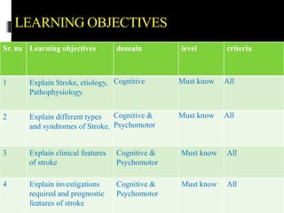
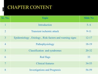
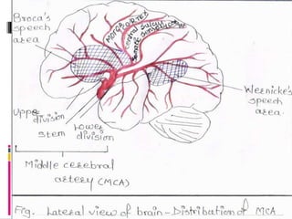
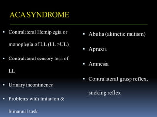
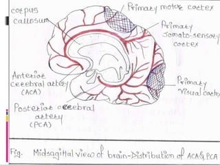
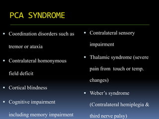
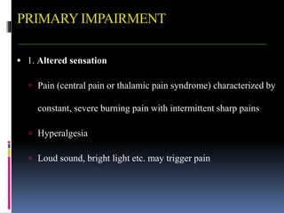
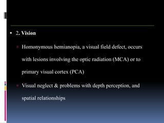
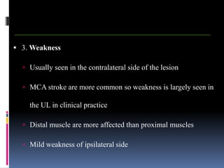
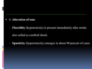
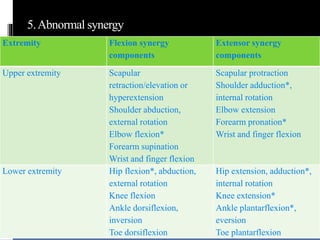
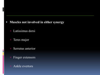
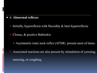
DEPARTMENT OF
NEUROSCIENCES.
DATTA MEGHE COLLEGE OF
PHYSIOTHERAPY. NAGPUR.
STROKE
(INTRODUCTION, CLASSIFICATION AND
CLINICAL FEATURES)
DR. NEHA INGALE CHAUDHARY
MPT (Neuro)
PROFESSOR & HEAD
DR. SACHIN CHAUDHARY
MPT (Cardio-respi. Physiotherapy)
PROFESSOR & HEAD
DATTA MEGHE COLLEGE OF PHYSIOTHERAPY.
Nagpur.
PREFACE
 This PPT is intended primarily for Bachelor of
Physiotherapy (BPTh) Final year students those are under
preparation for their University Examination.
 I have attempted to cover different areas of stroke.
 Despite my best efforts there might have some errors.
 I like to thank all those who have helped me.
Dr. Neha Ingale Chaudhary
Dr. Sachin Chaudhary
CONTENT
Sr. No. Topic Slide No
1 Objectives 5- 6
2 Introduction of stroke and Transient Ischemic Stroke. 9-11
3 Epidemiology , Etiology , Risk factors and warning signs 12-17
4 Pathophysiology 18-19
5 Classification and syndromes 20-33
6 Red flags 33
7 Clinical features 34-55
8 Investigations and Prognosis 56-59
9 Summary 60
10 References 61
11 Questions 62
GENERALOBJECTIVES OF THE
SYLLABUS
At the end of the session student should be able to
understand –
 The definition, epidemiology, Pathophysiology, and
classification of stroke.
 The clinical features and the investigations done for
stroke.
 The investigations and prognosis outcome of stroke.
LEARNING OBJECTIVES
Sr. no Learning objectives domain level criteria
1 Explain Stroke, etiology,
Pathophysiology.
Cognitive Must know All
2 Explain different types
and syndromes of Stroke.
Cognitive &
Psychomotor
Must know All
3 Explain clinical features
of stroke
Cognitive &
Psychomotor
Must know All
4 Explain investigations
required and prognostic
features of stroke
Cognitive &
Psychomotor
Must know All
CHAPTER CONTENT
Sr. No. Topic Slide No
1 Introduction 5- 6
2 Transient ischemic attack 9-11
3 Epidemiology , Etiology , Risk factors and warning signs 12-17
4 Pathophysiology 18-19
5 Classification and syndromes 20-32
6 Red flags 33
7 Clinical features 34-55
8 Investigations and Prognosis 56-59
INTRODUCTION AND CLASSIFICATION
STROKE
INTRODUCTION
 Stroke is an acute onset of neurological dysfunction
due to an abnormality in cerebral circulation with
resultant signs & symptoms which corresponds to
involvement of focal areas of the brain
ACC. TO WHO
 It is defined as the sudden onset of neurological
deficits due to an abnormality in cerebral
circulation with the signs and symptoms lasting for
more than 24 hours or longer
TRANSIENT ISCHEMIC ATTACK
 It is defined as the sudden onset of neurological
deficits due to an abnormality in cerebral
circulation with the signs and symptoms lasting
for less than 24 hours
EPIDEMIOLOGY
 Third leading cause of death
 The incidence of stroke is about 1.25 times greater
for males than females
 Most common cause of disability among adults
ETIOLOGY
 Atherosclerosis.
 Cerebral Thrombus.
 Cerebral embolus.
 Embolism from the heart (cardiac origin)
 Intracranial hemorrhage.
 Subarachnoid hemorrhage.
 Intracranial small vessel disease.
 Arterial aneurysms and Arterio-venous malformation.
 Haematological disorders (haemoglobinopathies, leukemia)
Atherothromboembolism
MISCELLANEOUS RARE CAUSES OF
STROKE
 Infective endocarditis & HIV infection
 Tumour, Hypoglycemia, Chronic meningitis
 Perioperative stroke (hypotension and boundary zone infarction,
trauma, dissection of neck arteries, paradoxical embolism, fat
embolism, infective endocarditis)
 Migraine, Snake bite
 Inflammatory bowel disease (Ulcerative and Crohn's colitis)
RISK FACTORS
 Smoking
 Obesity
 Lack of physical exercise or
sedentary life style
 Diet & excess alcohol
consumption
 Oral contraceptives
 Infection (meningeal
infection)
 Psychological factors
 Vasectomy
 Ageing & gender
 Positive family history
 Circadian and seasonal factors
 Heart disease
 Diabetes mellitus
 Hypertension
 Peripheral arterial disease
 Blood pathology
 Hyperlipidemia , TIA
Non Modifiable Modifiable
STROKE EARLYWARNING SIGN
 F- Face Drooping
 A- Arm Weakness
 S- Speech Difficulty
 T- Time To Call Hospital
OTHER THAN FAST SIGN-
 Sudden Weakness, Numbness Of Leg
 Sudden Confusion Or Trouble In Understanding
 Sudden Trouble In Seeing or Walking
 Sudden Severe Headache With No Known Cause
 Other important but less common stroke symptoms include:
 Sudden nausea, fever, & vomiting distinguished from a viral
illness by speed of onset
 Brief loss of consciousness (fainting, confusion, convulsions)
PATHOPHYSIOLOGY
 Ischemia results in irreversible cellular damage with a
core area of focal infarction within minutes
 Transitional area surrounding core is termed ischemic
penumbra & consists of viable but metabolically
lethargic cells
 Ischemia produce cerebral edema, that begins within
minutes of insult & reaches a maximum by 3 to 4 days.
 Swelling gradually subsides & generally disappears by 2 - 3 weeks.
 Edema elevates ICP, leading to intracranial HT & neurological
deterioration associated with contra lateral & caudal shifts of brain
structures.
 Cerebral edema is the most frequent cause of death in acute stroke
& is characteristic of large infarcts involving MCA & ICA.
CLASSIFICATION
 Depending on the cause
 Hemorrhagic stroke
 Intracranial hemorrhage
 Subarachnoid hemorrhage
 Signs of raised ICP will be evident with a history
of a traumatic accident
Contd…
 Ischemic stroke
 Thrombotic: more common. Usually occurs in the sleeping
hours. Characterized by gradual onset of symptoms
 Embolic: Occurs in the waking hours of the day. Sudden
onset of symptoms preceded by giddiness in most
conditions
 Depending on the severity
 Mild stroke: symptoms subside with no deficit in a week
period.
 Moderate stroke: symptoms recover in a period of 3 - 6
months with minimal neurological deficit.
 Severe stroke: no complete recovery of the symptoms, even
after 1 year. Always ends up with severe neurological deficit.
 Depending on the duration
 Acute stroke: to a period of one week or until spasticity develops
 Sub acute stroke: after the development of spasticity & last for a
period of 3-12 months
 Chronic stroke: more than 12 months
Depending on the artery
involved-
Depending on the artery
involved-
 MCA Syndrome
 ACA Syndrome
 PCA syndrome
 Vertebro basilar artery
syndrome
 Vertebral, basilar artery
 Internal carotid artery
 Lacunar syndrome
MCA SYNDROME
 Contralateral hemiplegia (UL &
face >LL)
 Contralateral hemi sensory loss
(UL & face more affected than
LL)
 Ideomotor apraxia
 Ataxia of contra lateral limb
 Contralateral Homonymous
hemianopia
 Left hemisphere infarction
 Contralateral neglect
 Possible Contralateral visual
field deficit
 Aphasia: Broca’s (expressive)
or Wernicke’s (receptive)
ACASYNDROME
 Contralateral Hemiplegia or
monoplegia of LL (LL >UL)
 Contralateral sensory loss of
LL
 Urinary incontinence
 Problems with imitation &
bimanual task
 Abulia (akinetic mutism)
 Apraxia
 Amnesia
 Contralateral grasp reflex,
sucking reflex
PCA SYNDROME
 Coordination disorders such as
tremor or ataxia
 Contralateral homonymous
field deficit
 Cortical blindness
 Cognitive impairment
including memory impairment
 Contralateral sensory
impairment
 Thalamic syndrome (severe
pain from touch or temp.
changes)
 Weber’s syndrome
(Contralateral hemiplegia &
third nerve palsy)
VERTIBRO-BASILARARTERY
SYNDROMES
 Medial medullary syndrome
(VA)
 Lateral medullary (Wallenberg's)
syndrome (PICA)
 Complete basilar artery
syndrome (locked-in syndrome)
 Med inferior pontine syndrome
 Lateral inferior pontine syndrome
(AICA)
 Medial midpontine syndrome
 Lateral midpontine syndrome
 Medial superior pontine syndrome
 Lateral superior pontine syndrome
Locked-in syndrome (LIS)
 Acute hemiparesis rapidly progressing to tetraplegia & lower bulbar
paralysis (CN V through XII are involved)
 Initially patient is dysarthric & dysphonic & progresses to mutism
 There is preserved consciousness & sensation
 Horizontal eye movements are impaired but vertical eye movements
& blinking remain intact.
 Communication can be established via these eye movements.
LACUNAR SYNDROME
 Caused by small vessel disease
of deep white mater
 Pure motor Lacunar stroke
 Pure sensory Lacunar stroke
 Ataxic hemiparesis
 Dysarthria
 Clumsy hand syndrome
 Sensory/motor stroke
 Dystonia/involuntary
movements
RED FLAG
Changes in neurological status
Symptoms Possible causes Management
 Decreased level of
arousal
 enlargement of pupil
on the side of stoke
 sudden change in
muscle tone and /or
deep tendon reflexes
 Cerebral edema
 Another attack of
stroke
Cease treatment and
seek immediate
medical attention
CLINICALFEATURES AND
INVESTIGATIONS OF STROKE.
PRIMARYIMPAIRMENT
 1. Altered sensation
 Pain (central pain or thalamic pain syndrome) characterized by
constant, severe burning pain with intermittent sharp pains
 Hyperalgesia
 Loud sound, bright light etc. may trigger pain
 2. Vision
 Homonymous hemianopia, a visual field defect, occurs
with lesions involving the optic radiation (MCA) or to
primary visual cortex (PCA)
 Visual neglect & problems with depth perception, and
spatial relationships
 3. Weakness
 Usually seen in the contralateral side of the lesion
 MCA stroke are more common so weakness is largely seen in
the UL in clinical practice
 Distal muscle are more affected than proximal muscles
 Mild weakness of ipsilateral side
 4. Alteration of tone
 Flaccidity (hypotonicity) is present immediately after stroke
also called as cerebral shock.
 Spasticity (hypertonicity) emerges in about 90 percent of cases
5.Abnormal synergy
Extremity Flexion synergy
components
Extensor synergy
components
Upper extremity Scapular
retraction/elevation or
hyperextension
Shoulder abduction,
external rotation
Elbow flexion*
Forearm supination
Wrist and finger flexion
Scapular protraction
Shoulder adduction*,
internal rotation
Elbow extension
Forearm pronation*
Wrist and finger flexion
Lower extremity Hip flexion*, abduction,
external rotation
Knee flexion
Ankle dorsiflexion,
inversion
Toe dorsiflexion
Hip extension, adduction*,
internal rotation
Knee extension*
Ankle plantarflexion*,
eversion
Toe plantarflexion
 Muscles not involved in either synergy
 Latissimus dorsi
 Teres major
 Serratus anterior
 Finger extensors
 Ankle evertors
 6. Abnormal reflexes
 Initially, hyporeflexia with flaccidity & later hyperreflexia
 Clonus, & positive Babinskie
▪ Asymmetric tonic neck reflex (ATNR) present most of times.
 Associated reactions are also present by stimulation of yawning,
sneezing, or coughing.
 7. Altered co ordination
 Proprioceptive loss can result in sensory ataxia.
 Strokes affecting cerebellum typically produce cerebellar ataxia
(e.g. Basilar Artery Syndrome, Pontine Syndromes) & motor
weakness.
 Basal ganglia involvement (PCA syndrome) may lead to
bradykinesia or involuntary movements.
 8. Altered motor programming
 Lesions of premotor frontal cortex of either hemisphere, left
inferior parietal lobe, & corpus callosum can produce Apraxia.
 Apraxia is more evident with left hemisphere damage than right
and is commonly seen with aphasia.
▪ Ideational apraxia
▪ Ideomotor apraxia
 9. Postural Control & Balance
 Impairments in steadiness, symmetry, & dynamic stability.
 Reactive postural control and Anticipatory postural control
affected.
 Pusher syndrome: Active pushing with stronger extremities
toward affected side, leading to lateral postural imbalance.
 10. Speech, Language, and Swallowing
 Lesions of dominant hemisphere.
 Aphasia: impairment of language comprehension, formulation,
and use.
 Dysarthria: Motor speech disorders -lesions of CNS or PNS.
 Dysphagia: Lesions affecting medullary brainstem (CN IX and
X), large vessel pontine lesions, acute MCA and PCA lesion
 11. Perception and Cognition
 Disorders of body scheme/body image, spatial relations, and
agnosias.
 Result of lesions in right parietal cortex & seen more with left
hemiplegia than right.
 12. Emotional Status
 Pseudobulbar affect (PBA), also known as emotional liability
or emotional dysregulation syndrome.
▪ emotional outbursts or exaggerated laughing or crying.
 Lesions of brain affecting frontal lobe, hypothalamus, & limbic
system
 Depression is extremely common
 13. Bladder and Bowel Function
 Common during acute phase.
 Urinary incontinence- bladder hyperreflexia or hyporeflexia,
loss of sphincter control, or sensory loss.
 Bowel function affection: Incontinence & Diarrhea or
Constipation
HEMISPHERIC BEHAVIORAL
DIFFERENCES
INDIRECT IMPAIRMENTS
 1. Musculoskeletal changes
 Loss of ROM & Contractures.
 Disuse atrophy & muscle weakness.
 Osteoporosis, results from decreased physical activity, changes
in protein nutrition, hormonal deficiency, & calcium deficiency.
 2. Neurological signs
 Seizures occur in a small % of patients - more common in
occlusive carotid disease than in MCA disease
 Hydrocephalus - rare but can occur with subarachnoid or
intracerebral hemorrhage.
 3. Thrombophlebitis & deep venous thrombosis (DVT)
 Commonest complications for all immobilized patients.
 4. Cardiac Function
 Stroke as a result of underlying coronary artery disease (CAD) -
impaired CO, cardiac decompensation, & rhythm disorders.
 If problems persist, can alter cerebral perfusion & produce
additional focal signs (e.g., mental confusion).
 Cardiac limitations in exercise tolerance
 5. Pulmonary Function
 Decreased lung volume, decreased pulmonary perfusion & vital
capacity & altered chest wall excursion
 Aspiration- due to Dysphagia.
 6. Integumentary
 Pressure sore/ decubitus ulcer.
 Fragile skin- reduced blood supply to epidermis.
 The skin breaks down over bony prominences from pressure,
friction, shearing, and/or maceration
TESTSAND MEASURES
 Urine analysis
 CBC count
 Blood sugar level
 Blood cholesterol & lipid profile
 Cardiac evaluation
 Lumbar puncture
IMAGING
 CT Scan.
 Magnetic Resonance Imaging (MRI).
 Cerebral Angiography.
RECOVERYAND PROGNOSIS
 Fastest in first weeks after onset
 Measurable neurological & functional recovery –
in first month after stroke.
 Continue to make measurable functional gains for months or years
after insult.
 Late recovery of function- seen in patients with chronic stroke
undergoing extensive functional training
VARIATION OF RECOVERY
 Recovery depends on severity of stroke .
 Depends on type of stroke – hemorrhagic or ischemic.
 Varies from individual to individual.
 Depends on intensity of therapy.
 Depends on age of the patient.
SUMMARY
 Introduction
 Etiology, epidemiology
 Risk factors, warning signs
 Pathophysiology, syndromes
 Clinical features
 Investigations and prognosis
REFERENCES
 O’ Sullivan SB, Schmitz TJ. Stroke. Physical rehabilitation.
7th ed., New Delhi: Jaypee Brothers, 2007, chapter 15, P(592-
661)
 Darcy A. Umphred. Neurological Rehabilitation, 6th ed.,
Mosby Elsevier, Missouri.Chapter 23, P(711-752).
QUESTIONS
 Define stroke and explain causes.
 Describe various syndromes occurring in stroke.
 Illustrate various clinical features and prognosis of stroke
STROKE INTRODUCTION, CLASSIFICATION AND CLINICAL FEATURES.pptx

STROKE INTRODUCTION, CLASSIFICATION AND CLINICAL FEATURES.pptx

  • 1.
    DEPARTMENT OF NEUROSCIENCES. DATTA MEGHECOLLEGE OF PHYSIOTHERAPY. NAGPUR.
  • 2.
    STROKE (INTRODUCTION, CLASSIFICATION AND CLINICALFEATURES) DR. NEHA INGALE CHAUDHARY MPT (Neuro) PROFESSOR & HEAD DR. SACHIN CHAUDHARY MPT (Cardio-respi. Physiotherapy) PROFESSOR & HEAD DATTA MEGHE COLLEGE OF PHYSIOTHERAPY. Nagpur.
  • 3.
    PREFACE  This PPTis intended primarily for Bachelor of Physiotherapy (BPTh) Final year students those are under preparation for their University Examination.  I have attempted to cover different areas of stroke.  Despite my best efforts there might have some errors.  I like to thank all those who have helped me. Dr. Neha Ingale Chaudhary Dr. Sachin Chaudhary
  • 4.
    CONTENT Sr. No. TopicSlide No 1 Objectives 5- 6 2 Introduction of stroke and Transient Ischemic Stroke. 9-11 3 Epidemiology , Etiology , Risk factors and warning signs 12-17 4 Pathophysiology 18-19 5 Classification and syndromes 20-33 6 Red flags 33 7 Clinical features 34-55 8 Investigations and Prognosis 56-59 9 Summary 60 10 References 61 11 Questions 62
  • 5.
    GENERALOBJECTIVES OF THE SYLLABUS Atthe end of the session student should be able to understand –  The definition, epidemiology, Pathophysiology, and classification of stroke.  The clinical features and the investigations done for stroke.  The investigations and prognosis outcome of stroke.
  • 6.
    LEARNING OBJECTIVES Sr. noLearning objectives domain level criteria 1 Explain Stroke, etiology, Pathophysiology. Cognitive Must know All 2 Explain different types and syndromes of Stroke. Cognitive & Psychomotor Must know All 3 Explain clinical features of stroke Cognitive & Psychomotor Must know All 4 Explain investigations required and prognostic features of stroke Cognitive & Psychomotor Must know All
  • 7.
    CHAPTER CONTENT Sr. No.Topic Slide No 1 Introduction 5- 6 2 Transient ischemic attack 9-11 3 Epidemiology , Etiology , Risk factors and warning signs 12-17 4 Pathophysiology 18-19 5 Classification and syndromes 20-32 6 Red flags 33 7 Clinical features 34-55 8 Investigations and Prognosis 56-59
  • 8.
  • 9.
    INTRODUCTION  Stroke isan acute onset of neurological dysfunction due to an abnormality in cerebral circulation with resultant signs & symptoms which corresponds to involvement of focal areas of the brain
  • 10.
    ACC. TO WHO It is defined as the sudden onset of neurological deficits due to an abnormality in cerebral circulation with the signs and symptoms lasting for more than 24 hours or longer
  • 11.
    TRANSIENT ISCHEMIC ATTACK It is defined as the sudden onset of neurological deficits due to an abnormality in cerebral circulation with the signs and symptoms lasting for less than 24 hours
  • 12.
    EPIDEMIOLOGY  Third leadingcause of death  The incidence of stroke is about 1.25 times greater for males than females  Most common cause of disability among adults
  • 13.
    ETIOLOGY  Atherosclerosis.  CerebralThrombus.  Cerebral embolus.  Embolism from the heart (cardiac origin)  Intracranial hemorrhage.  Subarachnoid hemorrhage.  Intracranial small vessel disease.  Arterial aneurysms and Arterio-venous malformation.  Haematological disorders (haemoglobinopathies, leukemia) Atherothromboembolism
  • 14.
    MISCELLANEOUS RARE CAUSESOF STROKE  Infective endocarditis & HIV infection  Tumour, Hypoglycemia, Chronic meningitis  Perioperative stroke (hypotension and boundary zone infarction, trauma, dissection of neck arteries, paradoxical embolism, fat embolism, infective endocarditis)  Migraine, Snake bite  Inflammatory bowel disease (Ulcerative and Crohn's colitis)
  • 15.
    RISK FACTORS  Smoking Obesity  Lack of physical exercise or sedentary life style  Diet & excess alcohol consumption  Oral contraceptives  Infection (meningeal infection)  Psychological factors  Vasectomy  Ageing & gender  Positive family history  Circadian and seasonal factors  Heart disease  Diabetes mellitus  Hypertension  Peripheral arterial disease  Blood pathology  Hyperlipidemia , TIA Non Modifiable Modifiable
  • 16.
    STROKE EARLYWARNING SIGN F- Face Drooping  A- Arm Weakness  S- Speech Difficulty  T- Time To Call Hospital
  • 17.
    OTHER THAN FASTSIGN-  Sudden Weakness, Numbness Of Leg  Sudden Confusion Or Trouble In Understanding  Sudden Trouble In Seeing or Walking  Sudden Severe Headache With No Known Cause  Other important but less common stroke symptoms include:  Sudden nausea, fever, & vomiting distinguished from a viral illness by speed of onset  Brief loss of consciousness (fainting, confusion, convulsions)
  • 18.
    PATHOPHYSIOLOGY  Ischemia resultsin irreversible cellular damage with a core area of focal infarction within minutes  Transitional area surrounding core is termed ischemic penumbra & consists of viable but metabolically lethargic cells  Ischemia produce cerebral edema, that begins within minutes of insult & reaches a maximum by 3 to 4 days.
  • 19.
     Swelling graduallysubsides & generally disappears by 2 - 3 weeks.  Edema elevates ICP, leading to intracranial HT & neurological deterioration associated with contra lateral & caudal shifts of brain structures.  Cerebral edema is the most frequent cause of death in acute stroke & is characteristic of large infarcts involving MCA & ICA.
  • 20.
    CLASSIFICATION  Depending onthe cause  Hemorrhagic stroke  Intracranial hemorrhage  Subarachnoid hemorrhage  Signs of raised ICP will be evident with a history of a traumatic accident
  • 21.
    Contd…  Ischemic stroke Thrombotic: more common. Usually occurs in the sleeping hours. Characterized by gradual onset of symptoms  Embolic: Occurs in the waking hours of the day. Sudden onset of symptoms preceded by giddiness in most conditions
  • 22.
     Depending onthe severity  Mild stroke: symptoms subside with no deficit in a week period.  Moderate stroke: symptoms recover in a period of 3 - 6 months with minimal neurological deficit.  Severe stroke: no complete recovery of the symptoms, even after 1 year. Always ends up with severe neurological deficit.
  • 23.
     Depending onthe duration  Acute stroke: to a period of one week or until spasticity develops  Sub acute stroke: after the development of spasticity & last for a period of 3-12 months  Chronic stroke: more than 12 months
  • 24.
    Depending on theartery involved- Depending on the artery involved-  MCA Syndrome  ACA Syndrome  PCA syndrome  Vertebro basilar artery syndrome  Vertebral, basilar artery  Internal carotid artery  Lacunar syndrome
  • 25.
    MCA SYNDROME  Contralateralhemiplegia (UL & face >LL)  Contralateral hemi sensory loss (UL & face more affected than LL)  Ideomotor apraxia  Ataxia of contra lateral limb  Contralateral Homonymous hemianopia  Left hemisphere infarction  Contralateral neglect  Possible Contralateral visual field deficit  Aphasia: Broca’s (expressive) or Wernicke’s (receptive)
  • 27.
    ACASYNDROME  Contralateral Hemiplegiaor monoplegia of LL (LL >UL)  Contralateral sensory loss of LL  Urinary incontinence  Problems with imitation & bimanual task  Abulia (akinetic mutism)  Apraxia  Amnesia  Contralateral grasp reflex, sucking reflex
  • 29.
    PCA SYNDROME  Coordinationdisorders such as tremor or ataxia  Contralateral homonymous field deficit  Cortical blindness  Cognitive impairment including memory impairment  Contralateral sensory impairment  Thalamic syndrome (severe pain from touch or temp. changes)  Weber’s syndrome (Contralateral hemiplegia & third nerve palsy)
  • 30.
    VERTIBRO-BASILARARTERY SYNDROMES  Medial medullarysyndrome (VA)  Lateral medullary (Wallenberg's) syndrome (PICA)  Complete basilar artery syndrome (locked-in syndrome)  Med inferior pontine syndrome  Lateral inferior pontine syndrome (AICA)  Medial midpontine syndrome  Lateral midpontine syndrome  Medial superior pontine syndrome  Lateral superior pontine syndrome
  • 31.
    Locked-in syndrome (LIS) Acute hemiparesis rapidly progressing to tetraplegia & lower bulbar paralysis (CN V through XII are involved)  Initially patient is dysarthric & dysphonic & progresses to mutism  There is preserved consciousness & sensation  Horizontal eye movements are impaired but vertical eye movements & blinking remain intact.  Communication can be established via these eye movements.
  • 32.
    LACUNAR SYNDROME  Causedby small vessel disease of deep white mater  Pure motor Lacunar stroke  Pure sensory Lacunar stroke  Ataxic hemiparesis  Dysarthria  Clumsy hand syndrome  Sensory/motor stroke  Dystonia/involuntary movements
  • 33.
    RED FLAG Changes inneurological status Symptoms Possible causes Management  Decreased level of arousal  enlargement of pupil on the side of stoke  sudden change in muscle tone and /or deep tendon reflexes  Cerebral edema  Another attack of stroke Cease treatment and seek immediate medical attention
  • 34.
  • 35.
    PRIMARYIMPAIRMENT  1. Alteredsensation  Pain (central pain or thalamic pain syndrome) characterized by constant, severe burning pain with intermittent sharp pains  Hyperalgesia  Loud sound, bright light etc. may trigger pain
  • 36.
     2. Vision Homonymous hemianopia, a visual field defect, occurs with lesions involving the optic radiation (MCA) or to primary visual cortex (PCA)  Visual neglect & problems with depth perception, and spatial relationships
  • 37.
     3. Weakness Usually seen in the contralateral side of the lesion  MCA stroke are more common so weakness is largely seen in the UL in clinical practice  Distal muscle are more affected than proximal muscles  Mild weakness of ipsilateral side
  • 38.
     4. Alterationof tone  Flaccidity (hypotonicity) is present immediately after stroke also called as cerebral shock.  Spasticity (hypertonicity) emerges in about 90 percent of cases
  • 39.
    5.Abnormal synergy Extremity Flexionsynergy components Extensor synergy components Upper extremity Scapular retraction/elevation or hyperextension Shoulder abduction, external rotation Elbow flexion* Forearm supination Wrist and finger flexion Scapular protraction Shoulder adduction*, internal rotation Elbow extension Forearm pronation* Wrist and finger flexion Lower extremity Hip flexion*, abduction, external rotation Knee flexion Ankle dorsiflexion, inversion Toe dorsiflexion Hip extension, adduction*, internal rotation Knee extension* Ankle plantarflexion*, eversion Toe plantarflexion
  • 40.
     Muscles notinvolved in either synergy  Latissimus dorsi  Teres major  Serratus anterior  Finger extensors  Ankle evertors
  • 41.
     6. Abnormalreflexes  Initially, hyporeflexia with flaccidity & later hyperreflexia  Clonus, & positive Babinskie ▪ Asymmetric tonic neck reflex (ATNR) present most of times.  Associated reactions are also present by stimulation of yawning, sneezing, or coughing.
  • 42.
     7. Alteredco ordination  Proprioceptive loss can result in sensory ataxia.  Strokes affecting cerebellum typically produce cerebellar ataxia (e.g. Basilar Artery Syndrome, Pontine Syndromes) & motor weakness.  Basal ganglia involvement (PCA syndrome) may lead to bradykinesia or involuntary movements.
  • 43.
     8. Alteredmotor programming  Lesions of premotor frontal cortex of either hemisphere, left inferior parietal lobe, & corpus callosum can produce Apraxia.  Apraxia is more evident with left hemisphere damage than right and is commonly seen with aphasia. ▪ Ideational apraxia ▪ Ideomotor apraxia
  • 44.
     9. PosturalControl & Balance  Impairments in steadiness, symmetry, & dynamic stability.  Reactive postural control and Anticipatory postural control affected.  Pusher syndrome: Active pushing with stronger extremities toward affected side, leading to lateral postural imbalance.
  • 45.
     10. Speech,Language, and Swallowing  Lesions of dominant hemisphere.  Aphasia: impairment of language comprehension, formulation, and use.  Dysarthria: Motor speech disorders -lesions of CNS or PNS.  Dysphagia: Lesions affecting medullary brainstem (CN IX and X), large vessel pontine lesions, acute MCA and PCA lesion
  • 46.
     11. Perceptionand Cognition  Disorders of body scheme/body image, spatial relations, and agnosias.  Result of lesions in right parietal cortex & seen more with left hemiplegia than right.
  • 47.
     12. EmotionalStatus  Pseudobulbar affect (PBA), also known as emotional liability or emotional dysregulation syndrome. ▪ emotional outbursts or exaggerated laughing or crying.  Lesions of brain affecting frontal lobe, hypothalamus, & limbic system  Depression is extremely common
  • 48.
     13. Bladderand Bowel Function  Common during acute phase.  Urinary incontinence- bladder hyperreflexia or hyporeflexia, loss of sphincter control, or sensory loss.  Bowel function affection: Incontinence & Diarrhea or Constipation
  • 49.
  • 50.
    INDIRECT IMPAIRMENTS  1.Musculoskeletal changes  Loss of ROM & Contractures.  Disuse atrophy & muscle weakness.  Osteoporosis, results from decreased physical activity, changes in protein nutrition, hormonal deficiency, & calcium deficiency.
  • 51.
     2. Neurologicalsigns  Seizures occur in a small % of patients - more common in occlusive carotid disease than in MCA disease  Hydrocephalus - rare but can occur with subarachnoid or intracerebral hemorrhage.
  • 52.
     3. Thrombophlebitis& deep venous thrombosis (DVT)  Commonest complications for all immobilized patients.
  • 53.
     4. CardiacFunction  Stroke as a result of underlying coronary artery disease (CAD) - impaired CO, cardiac decompensation, & rhythm disorders.  If problems persist, can alter cerebral perfusion & produce additional focal signs (e.g., mental confusion).  Cardiac limitations in exercise tolerance
  • 54.
     5. PulmonaryFunction  Decreased lung volume, decreased pulmonary perfusion & vital capacity & altered chest wall excursion  Aspiration- due to Dysphagia.
  • 55.
     6. Integumentary Pressure sore/ decubitus ulcer.  Fragile skin- reduced blood supply to epidermis.  The skin breaks down over bony prominences from pressure, friction, shearing, and/or maceration
  • 56.
    TESTSAND MEASURES  Urineanalysis  CBC count  Blood sugar level  Blood cholesterol & lipid profile  Cardiac evaluation  Lumbar puncture
  • 57.
    IMAGING  CT Scan. Magnetic Resonance Imaging (MRI).  Cerebral Angiography.
  • 58.
    RECOVERYAND PROGNOSIS  Fastestin first weeks after onset  Measurable neurological & functional recovery – in first month after stroke.  Continue to make measurable functional gains for months or years after insult.  Late recovery of function- seen in patients with chronic stroke undergoing extensive functional training
  • 59.
    VARIATION OF RECOVERY Recovery depends on severity of stroke .  Depends on type of stroke – hemorrhagic or ischemic.  Varies from individual to individual.  Depends on intensity of therapy.  Depends on age of the patient.
  • 60.
    SUMMARY  Introduction  Etiology,epidemiology  Risk factors, warning signs  Pathophysiology, syndromes  Clinical features  Investigations and prognosis
  • 61.
    REFERENCES  O’ SullivanSB, Schmitz TJ. Stroke. Physical rehabilitation. 7th ed., New Delhi: Jaypee Brothers, 2007, chapter 15, P(592- 661)  Darcy A. Umphred. Neurological Rehabilitation, 6th ed., Mosby Elsevier, Missouri.Chapter 23, P(711-752).
  • 62.
    QUESTIONS  Define strokeand explain causes.  Describe various syndromes occurring in stroke.  Illustrate various clinical features and prognosis of stroke