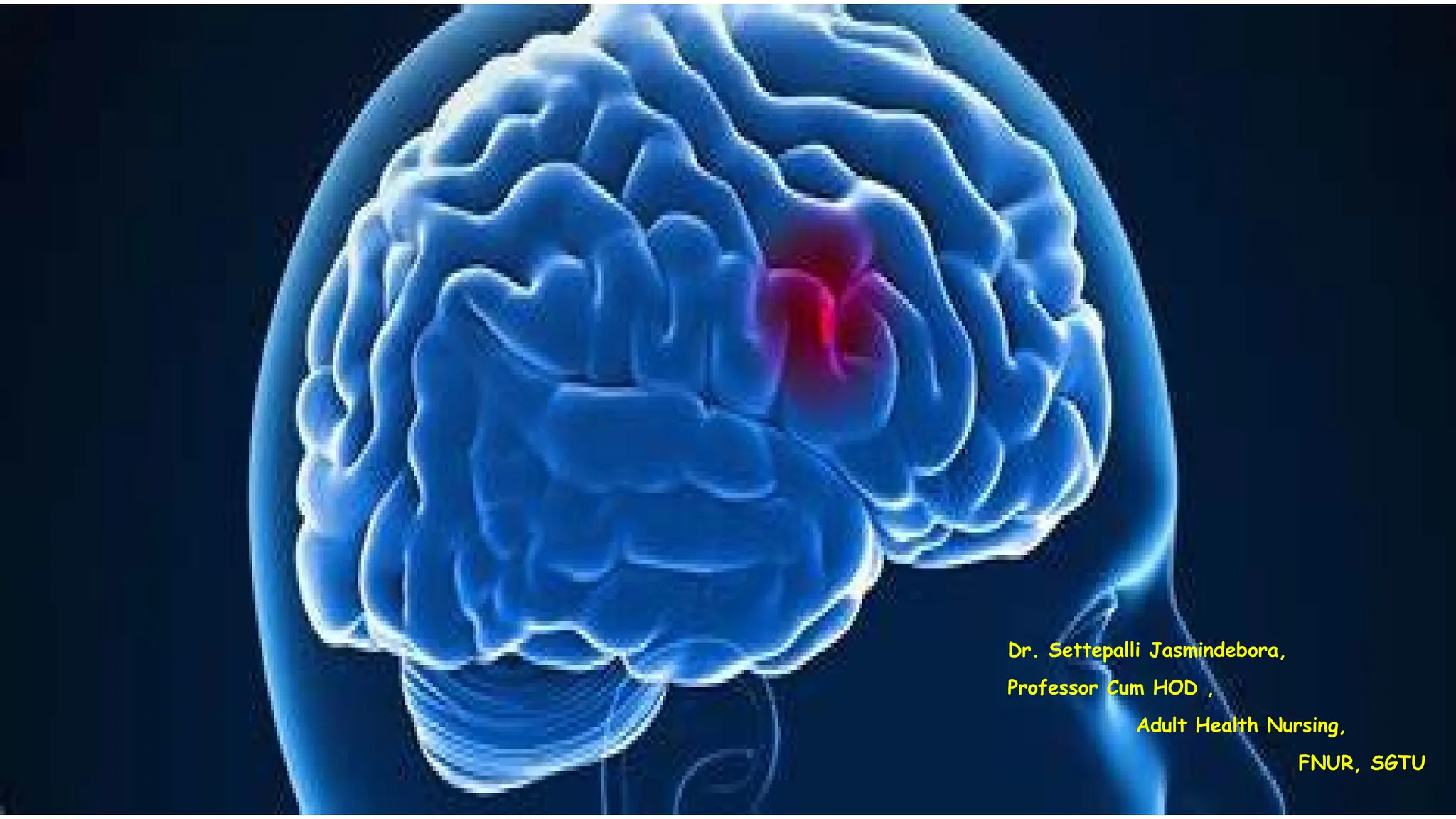
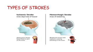
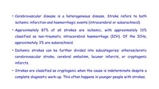
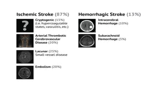
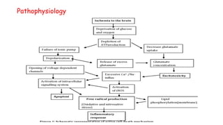
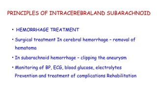
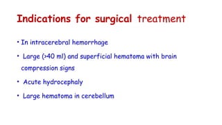
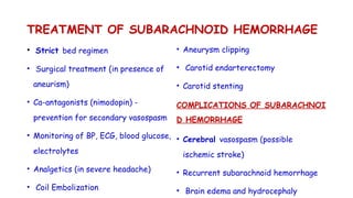
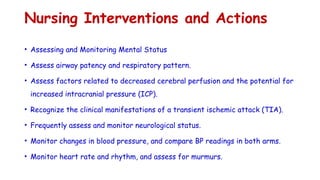
Dr. Settepalli Jasmindebora,
Professor Cum HOD ,
Adult Health Nursing,
FNUR, SGTU
cerebrovascular accident (CVA)
• A cerebrovascular accident (CVA), an ischemic stroke or
“brain attack,” is a sudden loss of brain function resulting
from a disruption of the blood supply to a part of the brain.
Epidemiology
• Stroke is an abrupt and serious vascular event that affects approximately 795,000 Americans each year,
with approximately 610,000 being first attacks and 185,000 being recurrent. In the United States,
someone has a stroke about every 40 seconds, and every four minutes someone dies from a stroke.
• Approximately 129,000 deaths per year result from stroke, making it the fifth leading cause of death in
the nation (behind heart disease, cancer, chronic lower respiratory diseases, and unintentional
accidents).
• Stroke is the most common acute neurological illness and cause for hospital admission and is the leading
cause of serious long-term disability in our nation.
 The total direct and indirect costs of stroke, as of 2010, are estimated to be $73.7 billion.
 Strokes extoll a huge cost in human suffering. Among survivors, approximately 31% require assistance
with activities of daily living, 20% require assistance with ambulation, and 16% require institutional
care.
TYPES OF STROKES
• Cerebrovascular disease is a heterogeneous disease. Stroke refers to both
ischemic infarction and haemorrhagic events (intracerebral or subarachnoid).
• Approximately 87% of all strokes are ischemic, with approximately 13%
classified as non-traumatic intracerebral haemorrhage (ICH). Of the ICHs,
approximately 3% are subarachnoid.
• Ischemic strokes can be further divided into subcategories: atherosclerotic
cerebrovascular stroke, cerebral embolism, lacunar infarcts, or cryptogenic
infarcts.
• Strokes are classified as cryptogenic when the cause is indeterminate despite a
complete diagnostic work-up. This often happens in younger people with strokes.
• Cerebral venous thrombosis represents approximately 0.5-1% of all
strokes.
ISCHEMIC STROKE
Mechanisms of ischemia
An ischemic stroke results from an interruption in the flow of
blood to the brain. The mechanisms of ischemia can be divided
into five main categories: thrombosis, embolism, systemic
hypoperfusion, arterial luminal obliteration, and venous
congestion.
Thrombosis A thrombus is the formation of a clot in an
artery. If the clot persists long enough, it can cause
ischemic damage to the cerebral tissue supplied by the
affected artery. A thrombosis is often triggered by
pathology at the endothelium. Thrombotic occlusions are
formed when the artery is damaged or diseased due to
atherosclerosis, platelet adherence, or arterial stenosis.
• Symptoms of thrombotic strokes
evolve slowly over a period of
minutes to days.
• A less common cause of
thrombotic stroke is migraine
headache. In severe cases, a
migraine headache can cause the
artery to spasm for a long time,
which allows a blood clot to form.
• Embolism
An embolus is most often a piece of a thrombus that has
broken free and is carried toward the brain by the
bloodstream. Bits of plaque, fat, air bubbles, and other
material also qualify as emboli.
Embolic occlusions are often blood clots that originate in the
heart due to atrial fibrillation, myocardial infarction, or
valvular disease. The blood clots travel through the
bloodstream until they become lodged and cannot travel any
further, blocking cerebral arteries. Stasis in the posterior left
atrium and appendage, associated with atrial fibrillation or
flutter,
• creates an environment for thrombus formation.
• In infectious endocarditis, vegetations composed of a mixture
of platelets, fibrin, and bacteria can fragment, sending emboli
into the cerebral circulation.
• Atheromatous plaques in the aorta and carotid arteries can
ulcerate or be mechanically disrupted, such as during
intravascular procedures, leading to embolization. The result is
almost immediate physical and neurological deficits.
Systemic hypoperfusion due to a loss of arterial
pressure can also cause an ischemic stroke. Several
processes can lead to hypoperfusion, but the most
recognized is cardiac arrest. When this occurs, the
most distal areas of the brain between the larger
main arteries are usually affected. This area is
called the watershed area.
Arterial luminal obliteration
A fourth mechanism of ischemic stroke is narrowing
of the arterial lumen. Causes of narrowing can be
non-inflammatory, such as in sickle cell or with
migraines, or due to inflammatory or infectious
vasculitis, vasospasm, or compression by an external
source, such as a mass or herniation.
Venous congestion
• Cerebral venous thrombosis is linked to disorder of coagulation, blood
vessel wall damage and/or decreased blood flow. It can cause ischemic
and/or haemorrhagic damage to the brain.
RISK FACTORS
Several factors are known to increase the risk of stroke. Some of these factors are modifiable; others are
not. The most common risk factors are:
Modifiable
• Hypertension
• Heart disease
• Atrial fibrillation
• Diabetes mellitus
• Hyperlipidaemia
• History of stroke/transient ischemic attack (TIA)
• Family history
• Tobacco use
Non-modifiable risk factors
• Non-modifiable risk factors are variables that cannot be changed. These risk factors include:
• Age - stroke risk doubles for each decade of life after age 55. Increasing age is the strongest non-
modifiable risk factor, as the incidence of stroke increases dramatically with increasing age.
• Heredity - stroke risk increases if a family member has had a stroke.
• Sex - men have greater risk of stroke than women until the age of 75, and then the risk levels out.
• Race/ethnicity - African Americans have a greater risk of stroke than Caucasians, probably due to a
higher incidence of hypertension and diabetes.
• History of stroke or TIA - people who have had a stroke or TIA are ten times more likely to have a
second stroke. The prevalence of stroke, which began leveling off around 2006 after a three-decade
drop, may soon be on the rise as a result of an aging population and a growing number of Hispanics and
African Americans.
Pathophysiology
WARNING SIGNS
It's important to know the warning signs for a stroke. Early
intervention when symptoms first appear can positively impact
outcomes. The signs of a stroke include:
1. Sudden numbness or weakness of the face, arm, or leg,
especially on one side of the body
2. Sudden confusion, trouble speaking, or understanding
3. Sudden trouble seeing in one or both eyes
4. Sudden trouble walking, dizziness, or lightheadedness
(vertigo), loss of balance or coordination
5. Sudden, severe headache with no known cause
6. Mental status changes
7. Seizure (relatively rare)
8. Sudden decrease in the level of consciousness’
Diagnostic tests commonly used during stroke
evaluation
 Non-contrast Computed Tomography (CT scan)
o Emergently at presentation
o 24 hours post alteplase
o Sensitivity increases 24 hours after the
ischemic event; approximately 6-12 hours
after the event, oedema occurs in the stroke
area to produce a hypodense area on CT scan.
o If hypodense area is seen within the first
three hours of symptom onset, re-evaluate
the time of onset.
 CT Perfusion
o Uses IV contrast
o Useful in identifying early ischemia
o Able to determine areas of viability (ischemic
penumbra)
 CT Angiography
o Identifies filling defects or vessel occlusion
o Higher sensitivity in identifying small vessel
strokes
 Higher sensitivity in identifying subarachnoid
haemorrhage
 Magnetic Resonance Imaging (MRI)
o Improved sensitivity in detecting acute
ischemic and haemorrhagic strokes
o Diffusion-weighted images (DWI) can detect
infarcts earlier
o Can detect small areas of ischemia and in
poorly visualized areas such as the cerebellum
and brain stem
o Perfusion-weighted images (PWI) measures
perfused areas of the brain after contrast
has been given
 Magnetic Resonance Angiography (MRA)
o Identifies blocked vessels early
 Ultrasonography (carotid and transcranial
doppler)
o Assessment for carotid artery stenosis
or occlusion
o Assessment of intracranial vascular
anatomy (helpful in evaluating the
proximal anatomy)
 Cerebral Angiography – useful in
characterizing the cerebrovascular
anatomy to guide treatment strategies.
It is also important in evaluating carotid
artery disease prior to surgery.
 Transoesophageal (TEE) and Transthoracic
Echocardiography (TTE) – useful in
detecting thoracic aortic dissection, aortic
arch atherosclerosis, thrombi in the atrial
appendage and patent foramen ovale.
 Chest Radiograph (CXR) – not
recommended as routine work-up during
evaluation in ED to prevent delay in
alteplase administration. Should be done
during hospitalization.
 Electrocardiogram (ECG) – acute stroke
has been associated with cardiac
dysfunction and arrhythmias. Continuous
monitoring is also recommended.
 Electroencephalogram (EEG) – used to rule
out seizures post-stroke
TRANSIENT ISCHEMIC ATTACK
• Definition
• A TIA is a prognostic indicator of ischemic stroke. The definition of TIA is controversial, and a
formal definition has not yet been accepted by authoritative organizations. The 2009 American
Heart Association (AHA)/American Stroke Association (ASA) scientific paper “Definition and
Evaluation of Transient Ischemic Attack” recommends the following definition:
• “TIAs are brief episodes of neurological dysfunction resulting from focal cerebral ischemia not
associated with permanent cerebral infarction.”
• (The old method of defining TIAs as any cerebral ischemic event with symptoms lasting < 24 hours
brought scrutiny because worldwide studies showed the arbitrary time threshold to be too broad.
Some cases that were classified as TIAs based on how long the symptoms lasted were strokes.
When reviewing the MRIs of these cases, 30-50% showed strokes on diffusion-weighted MRI.)
Management
• Emergency IV medicine. An IV medicine that
can break up a clot has to be given within 4.5
hours from when symptoms began. The sooner
the medicine is given, the better. Quick
treatment improves your chances of survival
and may reduce complications.
• An IV injection of recombinant tissue
plasminogen activator (TPA) is the gold
standard treatment for ischemic stroke. The
two types of TPA are alteplase (Activase) and
tenecteplase (TNKase). An injection of TPA is
usually given through a vein in the arm within
the first three hours. Sometimes, TPA can be
given up to 4.5 hours after stroke symptoms
started.
• This medicine restores blood flow by dissolving
the blood clot causing the stroke. By quickly
removing the cause of the stroke, it may help
people recover more fully from a stroke. Your
healthcare professional considers certain risks,
such as potential bleeding in the brain, to
determine whether TPA is appropriate for you.
• Emergency endovascular procedures. Healthcare professionals sometimes treat ischemic strokes
directly inside the blocked blood vessel. Endovascular therapy has been shown to improve outcomes and
reduce long-term disability after ischemic stroke. These procedures must be performed as soon as
possible:
• Medicines delivered directly to the brain. During this procedure, a long, thin tube called a catheter is
inserted through an artery in the groin. The catheter is moved through the arteries to the brain to
deliver TPA directly where the stroke is happening. The time window for this treatment is somewhat
longer than for injected TPA but is still limited.
• Removing the clot with a stent retriever. A device attached to a catheter can directly remove the clot
from the blocked blood vessel in the brain. This procedure is especially helpful for people with large clots
that can't be completely dissolved with TPA. This procedure often is performed in combination with
injected TPA.
• The time window when these procedures can be considered has been expanding due to newer imaging
technology. Perfusion imaging tests done with CT or MRI help determine if that someone may benefit
from endovascular therapy.
Other procedures
• Carotid endarterectomy. Carotid arteries are the blood vessels that run along
each side of the neck, supplying the brain with blood. This surgery removes the
plaque blocking a carotid artery and may reduce the risk of ischemic stroke. A
carotid endarterectomy also involves risks, especially for people with heart
disease or other medical conditions.
• Angioplasty and stents. In an angioplasty, a surgeon threads a catheter to the
carotid arteries through an artery in the groin. A balloon is then inflated to
expand the narrowed artery. Then a stent can be inserted to support the
opened artery.
HAEMORRHAGIC STROKES
Intracerebral haemorrhage
• ICH occurs when a blood vessel directly within the brain bursts. Blood leaking into the brain
causes damage to surrounding tissues from increased pressure due to the hematoma’s mass
effect. The expanding hematoma may damage neighbouring arteries, resulting in further
bleeding. Expansion may move into the subarachnoid and intraventricular space.
• Eventually the growth stops from a combination of haemostasis and increased pressure.
Hematoma growth (approximately 30 to 40%) is common within the first three to four hours
after the onset of symptoms. The amount of blood and increased pressure can lead to
seizures, unconsciousness, and/or death. Mortality is increased when blood is present in the
ventricles.
Pathophysiology
Acute hydrocephaly
Increased intracranial pressure
Brain edema
Dislocation of brain structures
Brainstem compression
Haematoma resolution occurs in 4-8 weeks, leaving a cystic cavity
LOCATION OF BLEEDING IN HEMORRHAGIC STROKES
Intracerebral Haemorrhage
Within the Meninges
or the Associated
Spaces
Intraventricular Haemorrhage (IVH)
Parenchymal Haemorrhage
(IPH)
Subarachnoid Haemorrhage
(SAH)
Epidural Hematoma
(EDH)
Subdural Hematoma (SDH)
Intracerebral Haemorrhage
• Parenchymal, intraventricular, and subarachnoid haemorrhages are usually stroke-related.
Subdural and epidural hematomas are usually caused by trauma.
INTRACEREBERAL HAEMORRHAGE
• ICH typically occurs in the basal ganglia, cerebral cortex, cerebellum, or
brain stem. Most hemorrhages occur in the basal ganglia and cerebral
cortex.
• Basal ganglia (40-50%)
• Cerebral cortex (20-50%)
• Thalamus (10-15%)
• Brain stem (5-12%)
• Cerebellum (5-10%)
• ICH accounts for 10-15% of all strokes. It is more likely to result in death
or major disability than ischemic stroke or subarachnoid haemorrhage.
Gonzalez-Perez found 42% mortality in ICH compared with 28.7% in SAH.
• Haemorrhages can be classified as non-traumatic or traumatic.
This course focuses on non-traumatic causes of ICH.
• Non-traumatic ICH
• Non-traumatic ICH is most often due to hypertension, which
damages the blood vessel walls.
• The most common areas of hypertensive bleeds are basal
ganglia, thalamus, brainstem and cerebellum.
• Less common causes of ICH, which are often seen in the parietal or
occipital lobes, include:
• High vessel pressure - eclampsia, drug abuse
• Excessive cerebral blood flow - reperfusion injury, hemorrhagic
transformation associated with ischemic infarct
• Rupture of an aneurysm or arteriovenous malformation (AVM)
• Arteriopathy - cerebral amyloid angiopathy, moyamoya, vasculitis
• Altered hemostasis - thrombolysis, anticoagulation
• Hemorrhagic necrosis - tumor, infection
• Venous outflow obstruction - cerebral venous thrombosis
Symptoms
Hemorrhagic strokes often cause a sudden, intense headache. They may be associated with
nausea and vomiting. ICH can occur at any age and are typically seen in younger people due
to the lack of knowledge of hypertension.
Symptoms of hemorrhagic strokes include:
•Headache
•Numbness, tingling or decreased sensation
•Weakness or paralysis
•Change in mental status
•Change in level of consciousness
•Nausea and vomiting
•Dizziness and/or loss of coordination
•Visual impairments
•Speech difficulties
•Seizures
Risk factors
• Haemorrhagic risk factors are listed below:
 Hypertension
 Diabetes
 Drug abuse - cocaine and amphetamines
 Alcohol abuse - > 2 drinks a day
 Use of anticoagulation
 Age - risk doubles every 10 years after age 45
 Gender - occurs more frequently in men
 Race - occurs more frequently in African Americans
 Menopause - for those being treated with hormone replacement therapy
 Caffeine - possible association seen with increased caffeine in pharmaceuticals
SUBARACHNOID HAEMORRHAGE
• Subarachnoid haemorrhage (SAH) occurs when a blood vessel outside the brain ruptures in
the subarachnoid space. The epidemiology of SAH is described below:
 80% of SAHs are due to ruptured aneurysms (primarily Berry aneurysms).
 AVM contribute to the other 20% of cases.
 The mean age of a patient experiencing an SAH is 50.
 SAHs are more common in women than men.
 SAHs are more common in African Americans than Caucasians.
 SAHs are responsible for the death and/or disability of 18,000 people per year.
 60% of the patients experiencing an SAH die within the first 30 days of the event.
 10% of patients die immediately without any warning.
 Hospitalized patients have an average mortality rate of 40% in the first month. Re-bleeding
carries a mortality rate of 51-80%.
Aetiology
• SAHs are usually caused by an aneurysm that
causes the blood vessel wall to weaken and rupture.
A normal artery wall consists of three layers: the
intima, which is the innermost endothelial layer; the
media, which consists of smooth muscle; and the
adventitia, or the outermost layer, which consists
of connective tissue. The aneurysmal sac is usually
composed of only intima and adventitia, with the
internal membrane thin or absent.
Saccular aneurysms are rounded berrylike outpouchings that arise at arterial bifurcation points,
most commonly in the Circle of Willis.
• Approximately 90% of aneurysms occur in the anterior circulation. The most common site is
the anterior cerebral artery and the anterior communicating artery, followed by the middle
cerebral artery at the M1/M2 junction.
• Most intracranial aneurysms probably result from hemodynamically induced vascular injury.
The rupture of aneurysms can be explained by abnormal hemodynamic stresses on the walls of
large cerebral arteries, particularly at bifurcation points. Less common causes of saccular
aneurysms include trauma, infection, tumour, drug abuse (cocaine), and high-flow states
associated with AVMs or fistulae.
• In a 25-year autopsy study of 125 patients with ruptured or unruptured aneurysms conducted
at Johns Hopkins, hypertension, cerebral atherosclerosis, vascular asymmetry in the Circle of
Willis, persistent headache, pregnancy-induced hypertension, long-term analgesic use, and
family history of stroke all were correlated positively with the formation of saccular
aneurysms.
• SAH can be spontaneous or traumatic.
Spontaneous SAHs usually occur in the setting
of a ruptured aneurysm or arteriovenous
malformations (AVM). Saccular or berry
aneurysms are specific to the intracranial
arteries and are the most common.
• The following risk factors are associated with
aneurysmal rupture and SAH:
 Hypertension
 Smoking
 Alcohol
 Multiple aneurysms
 Increasing aneurysm size
 Fatty metamorphosis of the liver
 Long-term analgesic use
 Oral contraceptives
 Family history
Symptoms
• Typical presentation includes:
 Headache - described as the “worst
headache of my life”
 Neck stiffness
 Photophobia
 Nausea and vomiting
 Seizure
 Often accompanied by loss of
consciousness
Risk factors
• Subarachnoid haemorrhage risk factors are listed below:
 Aneurysm
 Vascular malformations (AVM)
 Age - increases with age and peaks between the ages of 40 and 60
 Gender - more common in women than men
 Race - African Americans are twice as likely as Caucasians
 Cigarette use - increases risk of aneurysm rupture
 Alcohol abuse - increases risk of aneurysm rupture
 Family history - certain types of aneurysms are hereditary
Diagnostic Tests
• CT scan
• MRI scan
• MRA with contrast to identify Aneurysms & AV malformations
• Lumbar Puncture
PRINCIPLES OF INTRACEREBRALAND SUBARACHNOID
• HEMORRHAGE TREATMENT
• Surgical treatment In cerebral hemorrhage – removal of
hematoma
• In subarachnoid hemorrhage – clipping the aneurysm
• Monitoring of BP, ECG, blood glucose, electrolytes
Prevention and treatment of complications Rehabilitation
Indications for surgical treatment
• In intracerebral hemorrhage
• Large (>40 ml) and superficial hematoma with brain
compression signs
• Acute hydrocephaly
• Large hematoma in cerebellum
TREATMENT OF SUBARACHNOID HEMORRHAGE
• Strict bed regimen
• Surgical treatment (in presence of
aneurism)
• Ca-antagonists (nimodopin) -
prevention for secondary vasospasm
• Monitoring of BP, ECG, blood glucose,
electrolytes
• Analgetics (in severe headache)
• Coil Embolization
• Aneurysm clipping
• Carotid endarterectomy
• Carotid stenting
COMPLICATIONS OF SUBARACHNOI
D HEMORRHAGE
• Cerebral vasospasm (possible
ischemic stroke)
• Recurrent subarachnoid hemorrhage
• Brain edema and hydrocephaly
Nursing Interventions and Actions
• Assessing and Monitoring Mental Status
• Assess airway patency and respiratory pattern.
• Assess factors related to decreased cerebral perfusion and the potential for
increased intracranial pressure (ICP).
• Recognize the clinical manifestations of a transient ischemic attack (TIA).
• Frequently assess and monitor neurological status.
• Monitor changes in blood pressure, and compare BP readings in both arms.
• Monitor heart rate and rhythm, and assess for murmurs.
• Monitor respirations, noting patterns and rhythm, Cheyne-Stokes respiration.
• Evaluate pupils, noting size, shape, equality, and light reactivity.
• Document changes in vision: reports of blurred vision, alterations in the visual field,
and depth perception.
• Assess higher functions, including speech, if the client is alert.
• Assess for nuchal rigidity, twitching, increased restlessness, irritability, and the
onset of seizure activity.
• Use the National Institutes of Health Stroke Scale (NIHSS) for assessing
neurologic impairment.
• Screen the client for stroke risk.
• Monitor blood glucose levels.
• Position with head slightly elevated and in a neutral position.
• Maintain bedrest, provide a quiet and relaxing environment, and restrict visitors and
activities. Cluster nursing interventions and provide rest periods between care
activities. Limit the duration of procedures.
• Prevent straining at stool, holding breath, and physical exertion.
• Stress smoking cessation.
• Administer supplemental oxygen as indicated.
• Administer medications and insulin as indicated.
• Monitor laboratory studies as indicated: prothrombin time (PT), activated partial
thromboplastin time (aPTT), and Dilantin level.
• Prepare for surgery, as appropriate.
• Improving Physical Mobility and Preventing Contractures
• Managing Aphasia and Promoting Effective Communication
• Managing Post-Stroke Pain
• Promoting Effective Coping Strategies and Providing Emotional Support
• Promoting Independence Through Self-Care
• Preventing Dysphagia and Promoting Effective Swallowing
• Managing Fatigue and Tolerance to Activity
• Assessing and Monitoring for Unilateral Neglect
• Assessing and Monitoring for Disuse Syndrome
Reference
• 14 Stroke (CVA) Nursing Diagnosis and Nursing Care Plans – Nurseslabs
• Stroke,Pathophysiology, Diagnosis, and Management,7th Edition - February 6,
2021,Editors: James C. Grotta, Gregory W Albers, Joseph P Broderick, Scott E Kasner, Eng H.
Lo, Ralph L Sacco, Lawrence KS Wong, Arthur L. Day,Hardback ISBN: 9780323694247,eBook
ISBN: 9780323694254.
• Norrving, Bo (ed.), Oxford Textbook of Stroke and Cerebrovascular Disease, Oxford
Textbooks in Clinical Neurology (Oxford, 2014; online edn, Oxford Academic, 1 Mar. 2014),
https://doi.org/10.1093/med/9780199641208.001.0001, accessed 3 Apr. 2024.

Stroke Management for Nurse Practitioners

  • 1.
    Dr. Settepalli Jasmindebora, ProfessorCum HOD , Adult Health Nursing, FNUR, SGTU
  • 3.
    cerebrovascular accident (CVA) •A cerebrovascular accident (CVA), an ischemic stroke or “brain attack,” is a sudden loss of brain function resulting from a disruption of the blood supply to a part of the brain.
  • 4.
    Epidemiology • Stroke isan abrupt and serious vascular event that affects approximately 795,000 Americans each year, with approximately 610,000 being first attacks and 185,000 being recurrent. In the United States, someone has a stroke about every 40 seconds, and every four minutes someone dies from a stroke. • Approximately 129,000 deaths per year result from stroke, making it the fifth leading cause of death in the nation (behind heart disease, cancer, chronic lower respiratory diseases, and unintentional accidents). • Stroke is the most common acute neurological illness and cause for hospital admission and is the leading cause of serious long-term disability in our nation.  The total direct and indirect costs of stroke, as of 2010, are estimated to be $73.7 billion.  Strokes extoll a huge cost in human suffering. Among survivors, approximately 31% require assistance with activities of daily living, 20% require assistance with ambulation, and 16% require institutional care.
  • 5.
  • 6.
    • Cerebrovascular diseaseis a heterogeneous disease. Stroke refers to both ischemic infarction and haemorrhagic events (intracerebral or subarachnoid). • Approximately 87% of all strokes are ischemic, with approximately 13% classified as non-traumatic intracerebral haemorrhage (ICH). Of the ICHs, approximately 3% are subarachnoid. • Ischemic strokes can be further divided into subcategories: atherosclerotic cerebrovascular stroke, cerebral embolism, lacunar infarcts, or cryptogenic infarcts. • Strokes are classified as cryptogenic when the cause is indeterminate despite a complete diagnostic work-up. This often happens in younger people with strokes.
  • 8.
    • Cerebral venousthrombosis represents approximately 0.5-1% of all strokes.
  • 9.
    ISCHEMIC STROKE Mechanisms ofischemia An ischemic stroke results from an interruption in the flow of blood to the brain. The mechanisms of ischemia can be divided into five main categories: thrombosis, embolism, systemic hypoperfusion, arterial luminal obliteration, and venous congestion. Thrombosis A thrombus is the formation of a clot in an artery. If the clot persists long enough, it can cause ischemic damage to the cerebral tissue supplied by the affected artery. A thrombosis is often triggered by pathology at the endothelium. Thrombotic occlusions are formed when the artery is damaged or diseased due to atherosclerosis, platelet adherence, or arterial stenosis.
  • 10.
    • Symptoms ofthrombotic strokes evolve slowly over a period of minutes to days. • A less common cause of thrombotic stroke is migraine headache. In severe cases, a migraine headache can cause the artery to spasm for a long time, which allows a blood clot to form.
  • 11.
    • Embolism An embolusis most often a piece of a thrombus that has broken free and is carried toward the brain by the bloodstream. Bits of plaque, fat, air bubbles, and other material also qualify as emboli. Embolic occlusions are often blood clots that originate in the heart due to atrial fibrillation, myocardial infarction, or valvular disease. The blood clots travel through the bloodstream until they become lodged and cannot travel any further, blocking cerebral arteries. Stasis in the posterior left atrium and appendage, associated with atrial fibrillation or flutter,
  • 12.
    • creates anenvironment for thrombus formation. • In infectious endocarditis, vegetations composed of a mixture of platelets, fibrin, and bacteria can fragment, sending emboli into the cerebral circulation. • Atheromatous plaques in the aorta and carotid arteries can ulcerate or be mechanically disrupted, such as during intravascular procedures, leading to embolization. The result is almost immediate physical and neurological deficits.
  • 13.
    Systemic hypoperfusion dueto a loss of arterial pressure can also cause an ischemic stroke. Several processes can lead to hypoperfusion, but the most recognized is cardiac arrest. When this occurs, the most distal areas of the brain between the larger main arteries are usually affected. This area is called the watershed area.
  • 14.
    Arterial luminal obliteration Afourth mechanism of ischemic stroke is narrowing of the arterial lumen. Causes of narrowing can be non-inflammatory, such as in sickle cell or with migraines, or due to inflammatory or infectious vasculitis, vasospasm, or compression by an external source, such as a mass or herniation.
  • 15.
    Venous congestion • Cerebralvenous thrombosis is linked to disorder of coagulation, blood vessel wall damage and/or decreased blood flow. It can cause ischemic and/or haemorrhagic damage to the brain.
  • 16.
    RISK FACTORS Several factorsare known to increase the risk of stroke. Some of these factors are modifiable; others are not. The most common risk factors are: Modifiable • Hypertension • Heart disease • Atrial fibrillation • Diabetes mellitus • Hyperlipidaemia • History of stroke/transient ischemic attack (TIA) • Family history • Tobacco use
  • 17.
    Non-modifiable risk factors •Non-modifiable risk factors are variables that cannot be changed. These risk factors include: • Age - stroke risk doubles for each decade of life after age 55. Increasing age is the strongest non- modifiable risk factor, as the incidence of stroke increases dramatically with increasing age. • Heredity - stroke risk increases if a family member has had a stroke. • Sex - men have greater risk of stroke than women until the age of 75, and then the risk levels out. • Race/ethnicity - African Americans have a greater risk of stroke than Caucasians, probably due to a higher incidence of hypertension and diabetes. • History of stroke or TIA - people who have had a stroke or TIA are ten times more likely to have a second stroke. The prevalence of stroke, which began leveling off around 2006 after a three-decade drop, may soon be on the rise as a result of an aging population and a growing number of Hispanics and African Americans.
  • 18.
  • 19.
  • 20.
    It's important toknow the warning signs for a stroke. Early intervention when symptoms first appear can positively impact outcomes. The signs of a stroke include: 1. Sudden numbness or weakness of the face, arm, or leg, especially on one side of the body 2. Sudden confusion, trouble speaking, or understanding 3. Sudden trouble seeing in one or both eyes 4. Sudden trouble walking, dizziness, or lightheadedness (vertigo), loss of balance or coordination 5. Sudden, severe headache with no known cause 6. Mental status changes 7. Seizure (relatively rare) 8. Sudden decrease in the level of consciousness’
  • 21.
    Diagnostic tests commonlyused during stroke evaluation  Non-contrast Computed Tomography (CT scan) o Emergently at presentation o 24 hours post alteplase o Sensitivity increases 24 hours after the ischemic event; approximately 6-12 hours after the event, oedema occurs in the stroke area to produce a hypodense area on CT scan. o If hypodense area is seen within the first three hours of symptom onset, re-evaluate the time of onset.  CT Perfusion o Uses IV contrast o Useful in identifying early ischemia o Able to determine areas of viability (ischemic penumbra)  CT Angiography o Identifies filling defects or vessel occlusion o Higher sensitivity in identifying small vessel strokes  Higher sensitivity in identifying subarachnoid haemorrhage  Magnetic Resonance Imaging (MRI) o Improved sensitivity in detecting acute ischemic and haemorrhagic strokes o Diffusion-weighted images (DWI) can detect infarcts earlier o Can detect small areas of ischemia and in poorly visualized areas such as the cerebellum and brain stem o Perfusion-weighted images (PWI) measures perfused areas of the brain after contrast has been given
  • 22.
     Magnetic ResonanceAngiography (MRA) o Identifies blocked vessels early  Ultrasonography (carotid and transcranial doppler) o Assessment for carotid artery stenosis or occlusion o Assessment of intracranial vascular anatomy (helpful in evaluating the proximal anatomy)  Cerebral Angiography – useful in characterizing the cerebrovascular anatomy to guide treatment strategies. It is also important in evaluating carotid artery disease prior to surgery.  Transoesophageal (TEE) and Transthoracic Echocardiography (TTE) – useful in detecting thoracic aortic dissection, aortic arch atherosclerosis, thrombi in the atrial appendage and patent foramen ovale.  Chest Radiograph (CXR) – not recommended as routine work-up during evaluation in ED to prevent delay in alteplase administration. Should be done during hospitalization.  Electrocardiogram (ECG) – acute stroke has been associated with cardiac dysfunction and arrhythmias. Continuous monitoring is also recommended.  Electroencephalogram (EEG) – used to rule out seizures post-stroke
  • 23.
    TRANSIENT ISCHEMIC ATTACK •Definition • A TIA is a prognostic indicator of ischemic stroke. The definition of TIA is controversial, and a formal definition has not yet been accepted by authoritative organizations. The 2009 American Heart Association (AHA)/American Stroke Association (ASA) scientific paper “Definition and Evaluation of Transient Ischemic Attack” recommends the following definition: • “TIAs are brief episodes of neurological dysfunction resulting from focal cerebral ischemia not associated with permanent cerebral infarction.” • (The old method of defining TIAs as any cerebral ischemic event with symptoms lasting < 24 hours brought scrutiny because worldwide studies showed the arbitrary time threshold to be too broad. Some cases that were classified as TIAs based on how long the symptoms lasted were strokes. When reviewing the MRIs of these cases, 30-50% showed strokes on diffusion-weighted MRI.)
  • 24.
    Management • Emergency IVmedicine. An IV medicine that can break up a clot has to be given within 4.5 hours from when symptoms began. The sooner the medicine is given, the better. Quick treatment improves your chances of survival and may reduce complications. • An IV injection of recombinant tissue plasminogen activator (TPA) is the gold standard treatment for ischemic stroke. The two types of TPA are alteplase (Activase) and tenecteplase (TNKase). An injection of TPA is usually given through a vein in the arm within the first three hours. Sometimes, TPA can be given up to 4.5 hours after stroke symptoms started. • This medicine restores blood flow by dissolving the blood clot causing the stroke. By quickly removing the cause of the stroke, it may help people recover more fully from a stroke. Your healthcare professional considers certain risks, such as potential bleeding in the brain, to determine whether TPA is appropriate for you.
  • 25.
    • Emergency endovascularprocedures. Healthcare professionals sometimes treat ischemic strokes directly inside the blocked blood vessel. Endovascular therapy has been shown to improve outcomes and reduce long-term disability after ischemic stroke. These procedures must be performed as soon as possible: • Medicines delivered directly to the brain. During this procedure, a long, thin tube called a catheter is inserted through an artery in the groin. The catheter is moved through the arteries to the brain to deliver TPA directly where the stroke is happening. The time window for this treatment is somewhat longer than for injected TPA but is still limited. • Removing the clot with a stent retriever. A device attached to a catheter can directly remove the clot from the blocked blood vessel in the brain. This procedure is especially helpful for people with large clots that can't be completely dissolved with TPA. This procedure often is performed in combination with injected TPA. • The time window when these procedures can be considered has been expanding due to newer imaging technology. Perfusion imaging tests done with CT or MRI help determine if that someone may benefit from endovascular therapy.
  • 26.
    Other procedures • Carotidendarterectomy. Carotid arteries are the blood vessels that run along each side of the neck, supplying the brain with blood. This surgery removes the plaque blocking a carotid artery and may reduce the risk of ischemic stroke. A carotid endarterectomy also involves risks, especially for people with heart disease or other medical conditions. • Angioplasty and stents. In an angioplasty, a surgeon threads a catheter to the carotid arteries through an artery in the groin. A balloon is then inflated to expand the narrowed artery. Then a stent can be inserted to support the opened artery.
  • 27.
    HAEMORRHAGIC STROKES Intracerebral haemorrhage •ICH occurs when a blood vessel directly within the brain bursts. Blood leaking into the brain causes damage to surrounding tissues from increased pressure due to the hematoma’s mass effect. The expanding hematoma may damage neighbouring arteries, resulting in further bleeding. Expansion may move into the subarachnoid and intraventricular space. • Eventually the growth stops from a combination of haemostasis and increased pressure. Hematoma growth (approximately 30 to 40%) is common within the first three to four hours after the onset of symptoms. The amount of blood and increased pressure can lead to seizures, unconsciousness, and/or death. Mortality is increased when blood is present in the ventricles.
  • 28.
    Pathophysiology Acute hydrocephaly Increased intracranialpressure Brain edema Dislocation of brain structures Brainstem compression Haematoma resolution occurs in 4-8 weeks, leaving a cystic cavity
  • 29.
    LOCATION OF BLEEDINGIN HEMORRHAGIC STROKES Intracerebral Haemorrhage Within the Meninges or the Associated Spaces Intraventricular Haemorrhage (IVH) Parenchymal Haemorrhage (IPH)
  • 30.
  • 31.
    • Parenchymal, intraventricular,and subarachnoid haemorrhages are usually stroke-related. Subdural and epidural hematomas are usually caused by trauma. INTRACEREBERAL HAEMORRHAGE
  • 32.
    • ICH typicallyoccurs in the basal ganglia, cerebral cortex, cerebellum, or brain stem. Most hemorrhages occur in the basal ganglia and cerebral cortex. • Basal ganglia (40-50%) • Cerebral cortex (20-50%) • Thalamus (10-15%) • Brain stem (5-12%) • Cerebellum (5-10%) • ICH accounts for 10-15% of all strokes. It is more likely to result in death or major disability than ischemic stroke or subarachnoid haemorrhage. Gonzalez-Perez found 42% mortality in ICH compared with 28.7% in SAH.
  • 33.
    • Haemorrhages canbe classified as non-traumatic or traumatic. This course focuses on non-traumatic causes of ICH. • Non-traumatic ICH • Non-traumatic ICH is most often due to hypertension, which damages the blood vessel walls. • The most common areas of hypertensive bleeds are basal ganglia, thalamus, brainstem and cerebellum.
  • 34.
    • Less commoncauses of ICH, which are often seen in the parietal or occipital lobes, include: • High vessel pressure - eclampsia, drug abuse • Excessive cerebral blood flow - reperfusion injury, hemorrhagic transformation associated with ischemic infarct • Rupture of an aneurysm or arteriovenous malformation (AVM) • Arteriopathy - cerebral amyloid angiopathy, moyamoya, vasculitis • Altered hemostasis - thrombolysis, anticoagulation • Hemorrhagic necrosis - tumor, infection • Venous outflow obstruction - cerebral venous thrombosis
  • 35.
    Symptoms Hemorrhagic strokes oftencause a sudden, intense headache. They may be associated with nausea and vomiting. ICH can occur at any age and are typically seen in younger people due to the lack of knowledge of hypertension. Symptoms of hemorrhagic strokes include: •Headache •Numbness, tingling or decreased sensation •Weakness or paralysis •Change in mental status •Change in level of consciousness •Nausea and vomiting •Dizziness and/or loss of coordination •Visual impairments •Speech difficulties •Seizures
  • 36.
    Risk factors • Haemorrhagicrisk factors are listed below:  Hypertension  Diabetes  Drug abuse - cocaine and amphetamines  Alcohol abuse - > 2 drinks a day  Use of anticoagulation  Age - risk doubles every 10 years after age 45  Gender - occurs more frequently in men  Race - occurs more frequently in African Americans  Menopause - for those being treated with hormone replacement therapy  Caffeine - possible association seen with increased caffeine in pharmaceuticals
  • 37.
  • 38.
    • Subarachnoid haemorrhage(SAH) occurs when a blood vessel outside the brain ruptures in the subarachnoid space. The epidemiology of SAH is described below:  80% of SAHs are due to ruptured aneurysms (primarily Berry aneurysms).  AVM contribute to the other 20% of cases.  The mean age of a patient experiencing an SAH is 50.  SAHs are more common in women than men.  SAHs are more common in African Americans than Caucasians.  SAHs are responsible for the death and/or disability of 18,000 people per year.  60% of the patients experiencing an SAH die within the first 30 days of the event.  10% of patients die immediately without any warning.  Hospitalized patients have an average mortality rate of 40% in the first month. Re-bleeding carries a mortality rate of 51-80%.
  • 39.
    Aetiology • SAHs areusually caused by an aneurysm that causes the blood vessel wall to weaken and rupture. A normal artery wall consists of three layers: the intima, which is the innermost endothelial layer; the media, which consists of smooth muscle; and the adventitia, or the outermost layer, which consists of connective tissue. The aneurysmal sac is usually composed of only intima and adventitia, with the internal membrane thin or absent.
  • 40.
    Saccular aneurysms arerounded berrylike outpouchings that arise at arterial bifurcation points, most commonly in the Circle of Willis.
  • 41.
    • Approximately 90%of aneurysms occur in the anterior circulation. The most common site is the anterior cerebral artery and the anterior communicating artery, followed by the middle cerebral artery at the M1/M2 junction. • Most intracranial aneurysms probably result from hemodynamically induced vascular injury. The rupture of aneurysms can be explained by abnormal hemodynamic stresses on the walls of large cerebral arteries, particularly at bifurcation points. Less common causes of saccular aneurysms include trauma, infection, tumour, drug abuse (cocaine), and high-flow states associated with AVMs or fistulae. • In a 25-year autopsy study of 125 patients with ruptured or unruptured aneurysms conducted at Johns Hopkins, hypertension, cerebral atherosclerosis, vascular asymmetry in the Circle of Willis, persistent headache, pregnancy-induced hypertension, long-term analgesic use, and family history of stroke all were correlated positively with the formation of saccular aneurysms.
  • 42.
    • SAH canbe spontaneous or traumatic. Spontaneous SAHs usually occur in the setting of a ruptured aneurysm or arteriovenous malformations (AVM). Saccular or berry aneurysms are specific to the intracranial arteries and are the most common. • The following risk factors are associated with aneurysmal rupture and SAH:  Hypertension  Smoking  Alcohol  Multiple aneurysms  Increasing aneurysm size  Fatty metamorphosis of the liver  Long-term analgesic use  Oral contraceptives  Family history
  • 43.
    Symptoms • Typical presentationincludes:  Headache - described as the “worst headache of my life”  Neck stiffness  Photophobia  Nausea and vomiting  Seizure  Often accompanied by loss of consciousness
  • 44.
    Risk factors • Subarachnoidhaemorrhage risk factors are listed below:  Aneurysm  Vascular malformations (AVM)  Age - increases with age and peaks between the ages of 40 and 60  Gender - more common in women than men  Race - African Americans are twice as likely as Caucasians  Cigarette use - increases risk of aneurysm rupture  Alcohol abuse - increases risk of aneurysm rupture  Family history - certain types of aneurysms are hereditary
  • 45.
    Diagnostic Tests • CTscan • MRI scan • MRA with contrast to identify Aneurysms & AV malformations • Lumbar Puncture
  • 46.
    PRINCIPLES OF INTRACEREBRALANDSUBARACHNOID • HEMORRHAGE TREATMENT • Surgical treatment In cerebral hemorrhage – removal of hematoma • In subarachnoid hemorrhage – clipping the aneurysm • Monitoring of BP, ECG, blood glucose, electrolytes Prevention and treatment of complications Rehabilitation
  • 47.
    Indications for surgicaltreatment • In intracerebral hemorrhage • Large (>40 ml) and superficial hematoma with brain compression signs • Acute hydrocephaly • Large hematoma in cerebellum
  • 48.
    TREATMENT OF SUBARACHNOIDHEMORRHAGE • Strict bed regimen • Surgical treatment (in presence of aneurism) • Ca-antagonists (nimodopin) - prevention for secondary vasospasm • Monitoring of BP, ECG, blood glucose, electrolytes • Analgetics (in severe headache) • Coil Embolization • Aneurysm clipping • Carotid endarterectomy • Carotid stenting COMPLICATIONS OF SUBARACHNOI D HEMORRHAGE • Cerebral vasospasm (possible ischemic stroke) • Recurrent subarachnoid hemorrhage • Brain edema and hydrocephaly
  • 49.
    Nursing Interventions andActions • Assessing and Monitoring Mental Status • Assess airway patency and respiratory pattern. • Assess factors related to decreased cerebral perfusion and the potential for increased intracranial pressure (ICP). • Recognize the clinical manifestations of a transient ischemic attack (TIA). • Frequently assess and monitor neurological status. • Monitor changes in blood pressure, and compare BP readings in both arms. • Monitor heart rate and rhythm, and assess for murmurs.
  • 50.
    • Monitor respirations,noting patterns and rhythm, Cheyne-Stokes respiration. • Evaluate pupils, noting size, shape, equality, and light reactivity. • Document changes in vision: reports of blurred vision, alterations in the visual field, and depth perception. • Assess higher functions, including speech, if the client is alert. • Assess for nuchal rigidity, twitching, increased restlessness, irritability, and the onset of seizure activity. • Use the National Institutes of Health Stroke Scale (NIHSS) for assessing neurologic impairment. • Screen the client for stroke risk. • Monitor blood glucose levels.
  • 51.
    • Position withhead slightly elevated and in a neutral position. • Maintain bedrest, provide a quiet and relaxing environment, and restrict visitors and activities. Cluster nursing interventions and provide rest periods between care activities. Limit the duration of procedures. • Prevent straining at stool, holding breath, and physical exertion. • Stress smoking cessation. • Administer supplemental oxygen as indicated. • Administer medications and insulin as indicated. • Monitor laboratory studies as indicated: prothrombin time (PT), activated partial thromboplastin time (aPTT), and Dilantin level. • Prepare for surgery, as appropriate.
  • 52.
    • Improving PhysicalMobility and Preventing Contractures • Managing Aphasia and Promoting Effective Communication • Managing Post-Stroke Pain • Promoting Effective Coping Strategies and Providing Emotional Support • Promoting Independence Through Self-Care • Preventing Dysphagia and Promoting Effective Swallowing • Managing Fatigue and Tolerance to Activity • Assessing and Monitoring for Unilateral Neglect • Assessing and Monitoring for Disuse Syndrome
  • 53.
    Reference • 14 Stroke(CVA) Nursing Diagnosis and Nursing Care Plans – Nurseslabs • Stroke,Pathophysiology, Diagnosis, and Management,7th Edition - February 6, 2021,Editors: James C. Grotta, Gregory W Albers, Joseph P Broderick, Scott E Kasner, Eng H. Lo, Ralph L Sacco, Lawrence KS Wong, Arthur L. Day,Hardback ISBN: 9780323694247,eBook ISBN: 9780323694254. • Norrving, Bo (ed.), Oxford Textbook of Stroke and Cerebrovascular Disease, Oxford Textbooks in Clinical Neurology (Oxford, 2014; online edn, Oxford Academic, 1 Mar. 2014), https://doi.org/10.1093/med/9780199641208.001.0001, accessed 3 Apr. 2024.