BRAIN ABSCESS
introduction
• A brain abscess, also known as a cerebral
abscess or CNS abscess, is an abscess within
the brain tissue caused by inflammation and
collection of immune cells and infected
material originating from local or remote
infection sources.
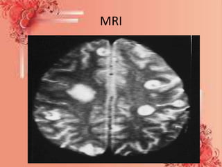
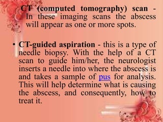
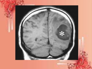
• A brain abscess is a rare, life-threatening
infection of the brain.
• Local sources may include ear infections, a
dental abscess, infection of the paranasal
sinuses, or epidural abscess, while remote
sources may include infections in the lung,
heart or kidney.
definition
It is free or encapsulated collection
of infectious material of brain
parenchyma between dura and
archnoid lining.
Types
1. intracranial subdural abscess
2.intracranial epidural abscess
3.spinal epidural infection/ abscess
4.intermedullary abscess
5.cerebritis
6.fungal brain abscess
causes
• Streptococcus, candida
• Chronic mastoiditis, sinusitis
• Head trauma, craniotomy
• Spinal injury, meningitis
• Immunosuppressed (HIV)
• M. tuberculosis
pathophysiology
Due to etilogical factor
That cross the blood brain barrier reaches to brain
Inflammation of parenchymal layer of brain
Cerebritis necrotic lesions(1)
Organism through the meningis or middle ear infection
Cause inflammation and purulant drainage between dura and
archnoid layer
Subdural abscess(2)
Any head trauma or sinusitis
Epidural abscess(3)
Causative agent may be from spinal injury
Cause spinal epidural abscess(4)
If bacteria reaches to lumbosacral region
Intermedullary abscess(5)
Clinical feature
• Headache with purulent material collect in brain,
increase ICP
• Nausea , vomiting, otitis media
• Nucchal rigidity
• Kerning sign-inability to extend knees more than
135 degress
• , brudzinski sign-severe neck stiffness causes a
person hips and knees to flex when neck is flexed
• Behaviour changes
• Decrease sensory and motor functioning
• Aphasia , ataxia- impaired coordination
Diagnostic test
• Blood test - to determine whether there is an
infection. If results show elevated levels of white-
blood cells, there most probably is an infection.
• Chest X-ray - to determine whether the patient has a
lung infection.
Imaging scans:
• MRI (magnetic resonance imaging) scan - an MRI
machine uses a magnetic field and radio waves to
create detailed images of the inside of the body.
Unlike CT scanning or general x-ray studies, no
ionizing radiation is involved with an MRI. It is a
painless procedure
MRI
CT (computed tomography) scan -
In these imaging scans the abscess
will appear as one or more spots.
• CT-guided aspiration - this is a type of
needle biopsy. With the help of a CT
scan to guide him/her, the neurologist
inserts a needle into where the abscess is
and takes a sample of pus for analysis.
This will help determine what is causing
the abscess, and consequently, how to
treat it.
Management
• Antibiotics
• Laminectomy of surgical drainage
• needle biopsy, under CT guidance may used
for drainage
• Antimicrobial therapy for streptococcus.
• Anti tuberculosis THERAPY if cause is tuberculi
• Corticosteroids for cerebral edema
• Anticonvulsants to manage seizures
Craniotomy - this is a surgical operation in
which an opening is made in the skull.
• The surgeon shaves a small area of scalp hair.
• A small piece of bone is removed in order to
gain access to the brain.
• Often with the a CT scanner for guidance, the
abscess is either drained of pus (aspiration), or
removed (excision).
• The bone is replaced.
• The skin is stitched
Complications
• Brain abscess rupture into ventriclar space
causing increase severity of headache.
• Brain stem herniation
• Permanent neurologic deficit,
• Flaccid paralysis with sensory loss
• Intracranial temporal complications
Nursing DIAGNOSIS
• Headache related to brain abscess.
• Altered mental activity related to pressure on
brain nerve.
• Risk of infection related to seizures.
• Decreased sensory function related to brain
abscess.
• Altered activity level due to brain absces
BRAIN ABSCESS.pptx

BRAIN ABSCESS.pptx

  • 1.
  • 2.
    introduction • A brainabscess, also known as a cerebral abscess or CNS abscess, is an abscess within the brain tissue caused by inflammation and collection of immune cells and infected material originating from local or remote infection sources.
  • 3.
    • A brainabscess is a rare, life-threatening infection of the brain. • Local sources may include ear infections, a dental abscess, infection of the paranasal sinuses, or epidural abscess, while remote sources may include infections in the lung, heart or kidney.
  • 4.
    definition It is freeor encapsulated collection of infectious material of brain parenchyma between dura and archnoid lining.
  • 5.
    Types 1. intracranial subduralabscess 2.intracranial epidural abscess 3.spinal epidural infection/ abscess 4.intermedullary abscess 5.cerebritis 6.fungal brain abscess
  • 6.
    causes • Streptococcus, candida •Chronic mastoiditis, sinusitis • Head trauma, craniotomy • Spinal injury, meningitis • Immunosuppressed (HIV) • M. tuberculosis
  • 7.
    pathophysiology Due to etilogicalfactor That cross the blood brain barrier reaches to brain Inflammation of parenchymal layer of brain Cerebritis necrotic lesions(1) Organism through the meningis or middle ear infection Cause inflammation and purulant drainage between dura and archnoid layer Subdural abscess(2)
  • 8.
    Any head traumaor sinusitis Epidural abscess(3) Causative agent may be from spinal injury Cause spinal epidural abscess(4) If bacteria reaches to lumbosacral region Intermedullary abscess(5)
  • 9.
    Clinical feature • Headachewith purulent material collect in brain, increase ICP • Nausea , vomiting, otitis media • Nucchal rigidity • Kerning sign-inability to extend knees more than 135 degress • , brudzinski sign-severe neck stiffness causes a person hips and knees to flex when neck is flexed • Behaviour changes • Decrease sensory and motor functioning • Aphasia , ataxia- impaired coordination
  • 10.
    Diagnostic test • Bloodtest - to determine whether there is an infection. If results show elevated levels of white- blood cells, there most probably is an infection. • Chest X-ray - to determine whether the patient has a lung infection. Imaging scans: • MRI (magnetic resonance imaging) scan - an MRI machine uses a magnetic field and radio waves to create detailed images of the inside of the body. Unlike CT scanning or general x-ray studies, no ionizing radiation is involved with an MRI. It is a painless procedure
  • 11.
  • 12.
    CT (computed tomography)scan - In these imaging scans the abscess will appear as one or more spots. • CT-guided aspiration - this is a type of needle biopsy. With the help of a CT scan to guide him/her, the neurologist inserts a needle into where the abscess is and takes a sample of pus for analysis. This will help determine what is causing the abscess, and consequently, how to treat it.
  • 14.
    Management • Antibiotics • Laminectomyof surgical drainage • needle biopsy, under CT guidance may used for drainage • Antimicrobial therapy for streptococcus. • Anti tuberculosis THERAPY if cause is tuberculi • Corticosteroids for cerebral edema • Anticonvulsants to manage seizures
  • 15.
    Craniotomy - thisis a surgical operation in which an opening is made in the skull. • The surgeon shaves a small area of scalp hair. • A small piece of bone is removed in order to gain access to the brain. • Often with the a CT scanner for guidance, the abscess is either drained of pus (aspiration), or removed (excision). • The bone is replaced. • The skin is stitched
  • 17.
    Complications • Brain abscessrupture into ventriclar space causing increase severity of headache. • Brain stem herniation • Permanent neurologic deficit, • Flaccid paralysis with sensory loss • Intracranial temporal complications
  • 18.
    Nursing DIAGNOSIS • Headacherelated to brain abscess. • Altered mental activity related to pressure on brain nerve. • Risk of infection related to seizures. • Decreased sensory function related to brain abscess. • Altered activity level due to brain absces