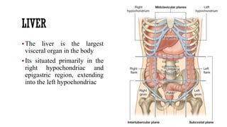
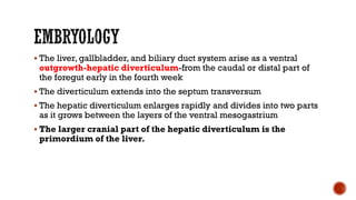
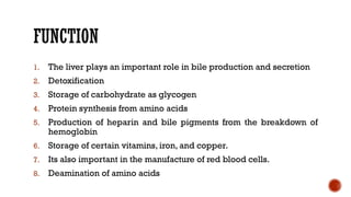
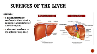
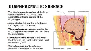
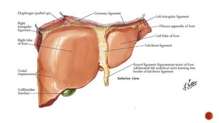
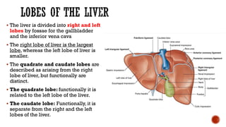
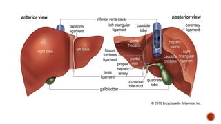
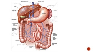
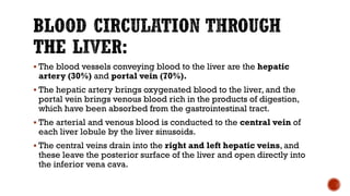
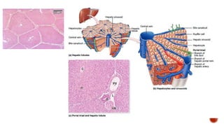
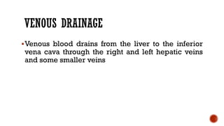
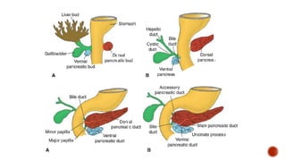
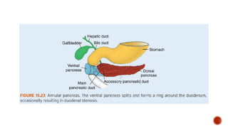
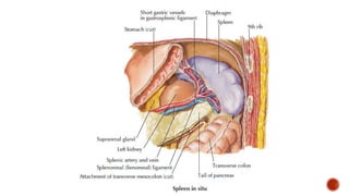
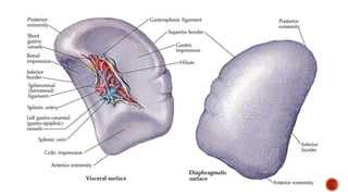
Mrs. Sichela
LIVER
The liver is the largest
visceral organ in the body
Its situated primarily in the
right hypochondriac and
epigastric region, extending
into the left hypochondriac
 The liver, gallbladder, and biliary duct system arise as a ventral
outgrowth-hepatic diverticulum-from the caudal or distal part of
the foregut early in the fourth week
 The diverticulum extends into the septum transversum
 The hepatic diverticulum enlarges rapidly and divides into two parts
as it grows between the layers of the ventral mesogastrium
 The larger cranial part of the hepatic diverticulum is the
primordium of the liver.
 The proliferating endodermal cells give rise to hepatocytes and to
the epithelial lining of the intrahepatic part of the biliary apparatus.
 The fibrous and hematopoietic tissue and Kupffer cells of the
liver are derived from mesenchyme in the septum transversum
 The small caudal part of the hepatic diverticulum becomes the
gallbladder, and the stalk of the diverticulum forms the cystic duct
 The stalk connecting the hepatic and cystic ducts to the duodenum
becomes the bile duct
FUNCTION
1. The liver plays an important role in bile production and secretion
2. Detoxification
3. Storage of carbohydrate as glycogen
4. Protein synthesis from amino acids
5. Production of heparin and bile pigments from the breakdown of
hemoglobin
6. Storage of certain vitamins, iron, and copper.
7. Its also important in the manufacture of red blood cells.
8. Deamination of amino acids
Include:
 a diaphragmatic
surface in the anterior,
superior, and posterior
directions; and
 a visceral surface in
the inferior direction
 The diaphragmatic surface of the liver,
which is smooth and domed, lies
against the inferior surface of the
diaphragm
 Associated with it are the subphrenic
and hepatorenal recesses:
 The subphrenic recess separates the
diaphragmatic surface of the liver from
the diaphragm
 The hepatorenal recess is between
the liver and the right kidney and right
suprarenal gland.
 The subphrenic and hepatorenal
recesses are continuous anteriorly
Visceral surface
 The visceral surface of the liver is covered with visceral peritoneum except
in the fossa for the gallbladder and at the porta hepatis
 The porta hepatis serves as the point of entry into the liver for the hepatic
arteries and the portal vein, and the exit point for the hepatic ducts
Structures related to the liver include the following:
 esophagus;
 right anterior part of the stomach;
 superior part of the duodenum;
 lesser omentum;
 gallbladder;
 right colic flexure;
 right transverse colon;
 right kidney; and
 right suprarenal gland.
 The liver is attached to the
anterior abdominal wall by the
falciform ligament
 Additional folds of peritoneum
connect the liver to
 the stomach (hepatogastric
ligament),
 the duodenum
(hepatoduodenal ligament),
and
 the diaphragm (right and left
triangular ligaments and
anterior and posterior
coronary ligaments).
 The liver is divided into right and left
lobes by fossae for the gallbladder
and the inferior vena cava
 The right lobe of liver is the largest
lobe, whereas the left lobe of liver is
smaller.
 The quadrate and caudate lobes are
described as arising from the right
lobe of liver, but functionally are
distinct.
 The quadrate lobe: functionally it is
related to the left lobe of the liver.
 The caudate lobe: Functionally, it is
separate from the right and the left
lobes of the liver.
BLOOD SUPPLY
Arteries:
 The hepatic artery, a branch of the
celiac artery, divides into right and left
terminal branches that enter the porta
hepatis.
Veins:
 The portal vein divides into right and
left terminal branches that enter the
porta hepatis behind the arteries.
 The hepatic veins (three or more)
emerge from the posterior surface of
the liver and drain into the inferior
vena cava.
 The blood vessels conveying blood to the liver are the hepatic
artery (30%) and portal vein (70%).
 The hepatic artery brings oxygenated blood to the liver, and the
portal vein brings venous blood rich in the products of digestion,
which have been absorbed from the gastrointestinal tract.
 The arterial and venous blood is conducted to the central vein of
each liver lobule by the liver sinusoids.
 The central veins drain into the right and left hepatic veins, and
these leave the posterior surface of the liver and open directly into
the inferior vena cava.
VENOUS DRAINAGE
Venous blood drains from the liver to the inferior
vena cava through the right and left hepatic veins
and some smaller veins
NERVE SUPPLY
 Nerve supply to the liver is derived from the coeliac
plexus and gastric branches of the vagal trunks
LYMPHATICS
 Lymph vessels emerge either through the porta hepatis
to the nodes of the lesser omentum or pass from the
bare area through the diaphragm to the thoracic nodes
 Hepatic lymph forms half of the lymph transmitted by
the thoracic duct
GALL BLADDER
 The gallbladder is a pear-shaped sac lying on
the visceral surface of the right lobe of the liver
in a fossa between the right and quadrate
lobes.
It has:
 Fundus: a rounded end, which may project from
the inferior border of the liver
 Body:a major part in the fossa, which may be
against the transverse colon and the superior
part of the duodenum
 A neck: a narrow part with mucosal folds
forming the spiral fold.
 The gallbladder receives, concentrates, and
stores bile from the liver.
 The arterial supply to the gallbladder
is the cystic artery from the right
hepatic artery (a branch of the
hepatic artery proper).
CYSTOHEPATIC TRIANGLE OF
CALOT
 In the angle between the common
hepatic duct and the cystic duct.
 Cystohepatic triangle (of Calot)
which is formed by;
 Superiorly - visceral surface of the
liver
 Inferior-laterally - cystic duct
 Medially - common hepatic duct.
VENOUS DRAINAGE
 The cystic veins, draining the neck of the gallbladder and cystic duct:
enter the liver directly or drain through the portal vein to the liver,
after joining the veins draining the hepatic ducts and upper bile
duct.
 The veins from the fundus and body of the gallbladder pass directly
into the visceral surface of the liver and drain into the hepatic
sinusoids.
BILIARY SYSTEM
 A system of vessels and ducts that collect and deliver bile from the
liver parenchyma to the second part of the duodenum.
 It is divided into intrahepatic and extrahepatic biliary trees.
 The intrahepatic ducts are formed from the larger bile canaliculi
which come together to form segmental ducts.
 These fuse close to the porta hepatis into right and left hepatic ducts.
 The extrahepatic biliary tree consists of the right and left hepatic
ducts, the common hepatic duct, the cystic duct and gallbladder and
the common bile duct.
 The pancreatic duct joins the bile duct.
 The joining of these two structures forms the hepatopancreatic
ampulla (ampulla ofVater),
 The ampulla enters the descending (second) part of the duodenum at
the major duodenal papilla.
 Surrounding the ampulla is the sphincter of ampulla (sphincter of
Oddi), which is a collection of smooth muscle.
 The ampulla opens into the lumen of the duodenum by means of a small
papilla, the major duodenal papilla.
 The accessory pancreatic duct empties into the duodenum just above
the major duodenal papilla at the minor duodenal papilla
 Occasionally, the bile and pancreatic ducts open separately into the
duodenum.
PORTAL VENOUS SYSTEM
 A portal venous system occurs when a capillary bed pools into
another capillary bed through veins without first going through
the heart.
 Both capillary beds and the blood vessels that connect them are
considered part of the portal venous system
 Portal venous systems are considered veins because the blood
vessels that join them are either veins or venules
 Examples of such include the hepatic portal system and hypophyseal
portal system
 The portal system carries venous blood (rich in nutrients that have
been extracted from food) to the liver for processing.
 The major vessel of the portal system is the portal vein.
 It is the point of convergence for the venous drainage of the spleen,
pancreas, gallbladder and the abdominal part of the gastrointestinal
tract.
 The portal vein is formed by the union of the splenic vein and the
superior mesenteric vein, posterior to the neck of the pancreas, at
the level of L2.
PORTO-SYSTEMIC ANASTOMOSIS
 Under normal conditions, the portal venous blood traverses the liver
and drains into the inferior vena cava of the systemic venous
circulation through the hepatic veins.
 This is the direct route.
 However, other, smaller communications exist between the portal and
systemic systems, and they become important when the direct route
becomes blocked.
 Gastroesophageal junction - Left gastric vein (tributaries) form a
portosystemic anastomosis with tributaries of the azygos system of
veins
 Anus – superior rectal vein of the portal system anastomoses with
the middle and inferior rectal veins of the systemic venous
system
 Anterior Abdominal wall around umbilicus – para-umbilical
veins anastomoses with veins on the anterior abdominal wall
PANCREAS
Lies largely in the floor of the lesser sac in the epigastric
and left hypochondriac regions, where it forms a major
portion of the stomach bed.
Its a retroperitoneal organ except for a small portion of
its tail, which lies in the lienorenal ligament.
EMBRYOLOGY
 The pancreas develops from two pancreatic diverticula (buds), which
envaginated from the endodermal lining of the gut tube in the region
of the second portion of the duodenum.
 The dorsal pancreatic bud grows into the dorsal mesentery.
 The ventral pancreatic bud initially grows into the ventral mesentery
but subsequently leaves the ventral mesentery, rotates around the gut
tube to enter the dorsal mesentery.
 The two pancreatic buds fuse together to form a single pancreas but
retain two separate ducts that enter the gut tube
EMBRYOLOGY
 The ventral bud forms the uncinate process and the inferior part of
the head of pancreas
 The remaining part forms from the dorsal bud
 The main or major pancreatic duct (of Wirsung) is formed by the
distal part of the dorsal pancreatic duct and the entire ventral
pancreatic duct.
 The proximal part of the dorsal pancreatic duct becomes the
minor pancreatic duct (of Santorini) or obliterates
 The pancreas is both an exocrine and endocrine gland.
 Endocrine part is made up of islets of Langerhans,
 It secretes two major hormones, insulin and glucagon,
which lower and raise blood sugar levels.
 Its exocrine function is to produce most of the
enzymes that digest foodstuffs in the small
intestine.
 Provides alkaline medium for activity of pancreatic
enzymes
 Has two ducts;
1. Main pancreatic duct (duct of Wirsung)
 Begins in the tail, runs to the right along the entire pancreas, and
carries pancreatic juice containing enzymes.
 Joins the bile duct to form the hepatopancreatic ampulla (ampulla of
Vater) before entering the second part of the duodenum at the
greater papilla.
2. Accessory pancreatic duct (Santorini's duct)
 Begins in the lower portion of the head and drains a small portion of
the head and body.
 Empties at the lesser duodenal papilla about 2 cm above the greater
papilla.
BLOOD SUPPLY AND DRAINAGE
 The head is supplied by the anastomotic arcades between the superior and inferior
pancreaticoduodenal arteries.
 The neck, body and tail are supplied by the splenic artery
 The arterial supply to the pancreas includes the:
 Branches from the gastroduodenal artery from the common hepatic
artery (a branch of the celiac trunk):
 anterior superior pancreaticoduodenal artery
 posterior superior pancreaticoduodenal artery
 Branches from inferior pancreatic artery (a branch of the splenic
artery);
 dorsal pancreatic artery
 great pancreatic artery
 Branches from the inferior pancreaticoduodenal artery (a branch
of the superior mesenteric artery);
 anterior inferior pancreaticoduodenal artery
 posterior inferior pancreaticoduodenal artery
Nerve Supply
 Sympathetic and parasympathetic nerve fibres reach the pancreas
along the arteries from the coeliac and superior mesenteric plexus
Lymph Drainage
 Lymphnodes of the pancreas lie along the superior border of the
pancreas (pancreaticoduodenal nodes) and on the
pancreaticoduodenal arteries
 The spleen is a large hemolymphoid organ consisting of vascular and
lymphatic tissue.
Location
 The spleen is located in the left hypogastrium between the fundus of the
stomach and the diaphragm
 It is found opposite the left ninth to eleventh ribs.
Ligaments
 The spleen is connected to the:
 greater curvature of the stomach by the gastrosplenic ligament, which
contains the short gastric and gastro-omental vessels
 left kidney by the splenorenal ligament, which contains the splenic
vessels.
 Both these ligaments are parts of the greater omentum.
Surfaces
 diaphragmatic
 visceral surfaces
Borders
 superior
 inferior borders
Poles
 anterior (lateral)
 posterior (medial) ends or poles
 The spleen is surrounded by visceral peritoneum except in the area of the hilum
 The splenic hilum is the entry point for the splenic vessels
The arterial supply to the spleen is the splenic artery from the celiac trunk
Thank you...

Accessory Digestive Organs. Liver, spleen

  • 1.
  • 2.
    LIVER The liver isthe largest visceral organ in the body Its situated primarily in the right hypochondriac and epigastric region, extending into the left hypochondriac
  • 3.
     The liver,gallbladder, and biliary duct system arise as a ventral outgrowth-hepatic diverticulum-from the caudal or distal part of the foregut early in the fourth week  The diverticulum extends into the septum transversum  The hepatic diverticulum enlarges rapidly and divides into two parts as it grows between the layers of the ventral mesogastrium  The larger cranial part of the hepatic diverticulum is the primordium of the liver.
  • 4.
     The proliferatingendodermal cells give rise to hepatocytes and to the epithelial lining of the intrahepatic part of the biliary apparatus.  The fibrous and hematopoietic tissue and Kupffer cells of the liver are derived from mesenchyme in the septum transversum  The small caudal part of the hepatic diverticulum becomes the gallbladder, and the stalk of the diverticulum forms the cystic duct  The stalk connecting the hepatic and cystic ducts to the duodenum becomes the bile duct
  • 5.
    FUNCTION 1. The liverplays an important role in bile production and secretion 2. Detoxification 3. Storage of carbohydrate as glycogen 4. Protein synthesis from amino acids 5. Production of heparin and bile pigments from the breakdown of hemoglobin 6. Storage of certain vitamins, iron, and copper. 7. Its also important in the manufacture of red blood cells. 8. Deamination of amino acids
  • 6.
    Include:  a diaphragmatic surfacein the anterior, superior, and posterior directions; and  a visceral surface in the inferior direction
  • 7.
     The diaphragmaticsurface of the liver, which is smooth and domed, lies against the inferior surface of the diaphragm  Associated with it are the subphrenic and hepatorenal recesses:  The subphrenic recess separates the diaphragmatic surface of the liver from the diaphragm  The hepatorenal recess is between the liver and the right kidney and right suprarenal gland.  The subphrenic and hepatorenal recesses are continuous anteriorly
  • 9.
    Visceral surface  Thevisceral surface of the liver is covered with visceral peritoneum except in the fossa for the gallbladder and at the porta hepatis  The porta hepatis serves as the point of entry into the liver for the hepatic arteries and the portal vein, and the exit point for the hepatic ducts
  • 10.
    Structures related tothe liver include the following:  esophagus;  right anterior part of the stomach;  superior part of the duodenum;  lesser omentum;  gallbladder;  right colic flexure;  right transverse colon;  right kidney; and  right suprarenal gland.
  • 12.
     The liveris attached to the anterior abdominal wall by the falciform ligament  Additional folds of peritoneum connect the liver to  the stomach (hepatogastric ligament),  the duodenum (hepatoduodenal ligament), and  the diaphragm (right and left triangular ligaments and anterior and posterior coronary ligaments).
  • 14.
     The liveris divided into right and left lobes by fossae for the gallbladder and the inferior vena cava  The right lobe of liver is the largest lobe, whereas the left lobe of liver is smaller.  The quadrate and caudate lobes are described as arising from the right lobe of liver, but functionally are distinct.  The quadrate lobe: functionally it is related to the left lobe of the liver.  The caudate lobe: Functionally, it is separate from the right and the left lobes of the liver.
  • 17.
    BLOOD SUPPLY Arteries:  Thehepatic artery, a branch of the celiac artery, divides into right and left terminal branches that enter the porta hepatis. Veins:  The portal vein divides into right and left terminal branches that enter the porta hepatis behind the arteries.  The hepatic veins (three or more) emerge from the posterior surface of the liver and drain into the inferior vena cava.
  • 19.
     The bloodvessels conveying blood to the liver are the hepatic artery (30%) and portal vein (70%).  The hepatic artery brings oxygenated blood to the liver, and the portal vein brings venous blood rich in the products of digestion, which have been absorbed from the gastrointestinal tract.  The arterial and venous blood is conducted to the central vein of each liver lobule by the liver sinusoids.  The central veins drain into the right and left hepatic veins, and these leave the posterior surface of the liver and open directly into the inferior vena cava.
  • 21.
    VENOUS DRAINAGE Venous blooddrains from the liver to the inferior vena cava through the right and left hepatic veins and some smaller veins
  • 23.
    NERVE SUPPLY  Nervesupply to the liver is derived from the coeliac plexus and gastric branches of the vagal trunks LYMPHATICS  Lymph vessels emerge either through the porta hepatis to the nodes of the lesser omentum or pass from the bare area through the diaphragm to the thoracic nodes  Hepatic lymph forms half of the lymph transmitted by the thoracic duct
  • 24.
    GALL BLADDER  Thegallbladder is a pear-shaped sac lying on the visceral surface of the right lobe of the liver in a fossa between the right and quadrate lobes. It has:  Fundus: a rounded end, which may project from the inferior border of the liver  Body:a major part in the fossa, which may be against the transverse colon and the superior part of the duodenum  A neck: a narrow part with mucosal folds forming the spiral fold.  The gallbladder receives, concentrates, and stores bile from the liver.
  • 25.
     The arterialsupply to the gallbladder is the cystic artery from the right hepatic artery (a branch of the hepatic artery proper).
  • 26.
    CYSTOHEPATIC TRIANGLE OF CALOT In the angle between the common hepatic duct and the cystic duct.  Cystohepatic triangle (of Calot) which is formed by;  Superiorly - visceral surface of the liver  Inferior-laterally - cystic duct  Medially - common hepatic duct.
  • 27.
    VENOUS DRAINAGE  Thecystic veins, draining the neck of the gallbladder and cystic duct: enter the liver directly or drain through the portal vein to the liver, after joining the veins draining the hepatic ducts and upper bile duct.  The veins from the fundus and body of the gallbladder pass directly into the visceral surface of the liver and drain into the hepatic sinusoids.
  • 28.
    BILIARY SYSTEM  Asystem of vessels and ducts that collect and deliver bile from the liver parenchyma to the second part of the duodenum.  It is divided into intrahepatic and extrahepatic biliary trees.  The intrahepatic ducts are formed from the larger bile canaliculi which come together to form segmental ducts.  These fuse close to the porta hepatis into right and left hepatic ducts.  The extrahepatic biliary tree consists of the right and left hepatic ducts, the common hepatic duct, the cystic duct and gallbladder and the common bile duct.
  • 30.
     The pancreaticduct joins the bile duct.  The joining of these two structures forms the hepatopancreatic ampulla (ampulla ofVater),  The ampulla enters the descending (second) part of the duodenum at the major duodenal papilla.  Surrounding the ampulla is the sphincter of ampulla (sphincter of Oddi), which is a collection of smooth muscle.  The ampulla opens into the lumen of the duodenum by means of a small papilla, the major duodenal papilla.  The accessory pancreatic duct empties into the duodenum just above the major duodenal papilla at the minor duodenal papilla  Occasionally, the bile and pancreatic ducts open separately into the duodenum.
  • 33.
    PORTAL VENOUS SYSTEM A portal venous system occurs when a capillary bed pools into another capillary bed through veins without first going through the heart.  Both capillary beds and the blood vessels that connect them are considered part of the portal venous system  Portal venous systems are considered veins because the blood vessels that join them are either veins or venules  Examples of such include the hepatic portal system and hypophyseal portal system
  • 34.
     The portalsystem carries venous blood (rich in nutrients that have been extracted from food) to the liver for processing.  The major vessel of the portal system is the portal vein.  It is the point of convergence for the venous drainage of the spleen, pancreas, gallbladder and the abdominal part of the gastrointestinal tract.  The portal vein is formed by the union of the splenic vein and the superior mesenteric vein, posterior to the neck of the pancreas, at the level of L2.
  • 36.
    PORTO-SYSTEMIC ANASTOMOSIS  Undernormal conditions, the portal venous blood traverses the liver and drains into the inferior vena cava of the systemic venous circulation through the hepatic veins.  This is the direct route.  However, other, smaller communications exist between the portal and systemic systems, and they become important when the direct route becomes blocked.
  • 37.
     Gastroesophageal junction- Left gastric vein (tributaries) form a portosystemic anastomosis with tributaries of the azygos system of veins  Anus – superior rectal vein of the portal system anastomoses with the middle and inferior rectal veins of the systemic venous system  Anterior Abdominal wall around umbilicus – para-umbilical veins anastomoses with veins on the anterior abdominal wall
  • 40.
    PANCREAS Lies largely inthe floor of the lesser sac in the epigastric and left hypochondriac regions, where it forms a major portion of the stomach bed. Its a retroperitoneal organ except for a small portion of its tail, which lies in the lienorenal ligament.
  • 42.
    EMBRYOLOGY  The pancreasdevelops from two pancreatic diverticula (buds), which envaginated from the endodermal lining of the gut tube in the region of the second portion of the duodenum.  The dorsal pancreatic bud grows into the dorsal mesentery.  The ventral pancreatic bud initially grows into the ventral mesentery but subsequently leaves the ventral mesentery, rotates around the gut tube to enter the dorsal mesentery.  The two pancreatic buds fuse together to form a single pancreas but retain two separate ducts that enter the gut tube
  • 43.
    EMBRYOLOGY  The ventralbud forms the uncinate process and the inferior part of the head of pancreas  The remaining part forms from the dorsal bud  The main or major pancreatic duct (of Wirsung) is formed by the distal part of the dorsal pancreatic duct and the entire ventral pancreatic duct.  The proximal part of the dorsal pancreatic duct becomes the minor pancreatic duct (of Santorini) or obliterates
  • 47.
     The pancreasis both an exocrine and endocrine gland.  Endocrine part is made up of islets of Langerhans,  It secretes two major hormones, insulin and glucagon, which lower and raise blood sugar levels.  Its exocrine function is to produce most of the enzymes that digest foodstuffs in the small intestine.  Provides alkaline medium for activity of pancreatic enzymes
  • 48.
     Has twoducts; 1. Main pancreatic duct (duct of Wirsung)  Begins in the tail, runs to the right along the entire pancreas, and carries pancreatic juice containing enzymes.  Joins the bile duct to form the hepatopancreatic ampulla (ampulla of Vater) before entering the second part of the duodenum at the greater papilla. 2. Accessory pancreatic duct (Santorini's duct)  Begins in the lower portion of the head and drains a small portion of the head and body.  Empties at the lesser duodenal papilla about 2 cm above the greater papilla.
  • 50.
    BLOOD SUPPLY ANDDRAINAGE  The head is supplied by the anastomotic arcades between the superior and inferior pancreaticoduodenal arteries.  The neck, body and tail are supplied by the splenic artery
  • 51.
     The arterialsupply to the pancreas includes the:  Branches from the gastroduodenal artery from the common hepatic artery (a branch of the celiac trunk):  anterior superior pancreaticoduodenal artery  posterior superior pancreaticoduodenal artery  Branches from inferior pancreatic artery (a branch of the splenic artery);  dorsal pancreatic artery  great pancreatic artery  Branches from the inferior pancreaticoduodenal artery (a branch of the superior mesenteric artery);  anterior inferior pancreaticoduodenal artery  posterior inferior pancreaticoduodenal artery
  • 52.
    Nerve Supply  Sympatheticand parasympathetic nerve fibres reach the pancreas along the arteries from the coeliac and superior mesenteric plexus Lymph Drainage  Lymphnodes of the pancreas lie along the superior border of the pancreas (pancreaticoduodenal nodes) and on the pancreaticoduodenal arteries
  • 53.
     The spleenis a large hemolymphoid organ consisting of vascular and lymphatic tissue. Location  The spleen is located in the left hypogastrium between the fundus of the stomach and the diaphragm  It is found opposite the left ninth to eleventh ribs. Ligaments  The spleen is connected to the:  greater curvature of the stomach by the gastrosplenic ligament, which contains the short gastric and gastro-omental vessels  left kidney by the splenorenal ligament, which contains the splenic vessels.  Both these ligaments are parts of the greater omentum.
  • 55.
    Surfaces  diaphragmatic  visceralsurfaces Borders  superior  inferior borders Poles  anterior (lateral)  posterior (medial) ends or poles  The spleen is surrounded by visceral peritoneum except in the area of the hilum  The splenic hilum is the entry point for the splenic vessels The arterial supply to the spleen is the splenic artery from the celiac trunk
  • 57.