The document discusses two types of detectors used in spectrophotometric instruments: photomultiplier tubes and photodiode arrays. Photomultiplier tubes use a cascade of electrons to amplify light signals, making them very sensitive, while photodiode arrays use integrated circuits containing many photodiodes to detect light across a spectrum. The document also discusses applications of UV-VIS spectroscopy like quantitative measurements of concentration and qualitative analysis of substance structure from absorption spectra. Immunoassays are described using antibodies to detect antigens, with discussion of monoclonal vs polyclonal antibodies, test sensitivity and specificity, and qualitative vs quantitative testing methods like agglutination.
Presenting a topic which is entitled: Detectors
Above topic includes:
Types of detector
phototube detector
photomultiplier tubes
silicon photodiodes
photovoltaic cells
advantages
multi-channel photon detectors
linear photodiode arrays
photodiode array
with basics of instrumentation and science technology
https://www.linkedin.com/in/preeti-choudhary-266414182/
https://www.instagram.com/chaudharypreeti1997/
https://www.facebook.com/profile.php?id=100013419194533
https://twitter.com/preetic27018281
Please like, share, comment and follow.
stay connected
If any query then contact:
chaudharypreeti1997@gmail.com
Thanking-You
Preeti Choudhary
This presentation include the detailed explanation of various parts of a UV-Visible spectrophotometer and two types of UV-Visible spectrophotometers-Single beam and Doube beam. It also include the comparison between single beam and double beam spectrophotometers.
In this slide contains Interference In Atomic Absorption Spectroscopy and applications.
Presented by: Shaik Gouse ul azam. ( department of pharmaceutical analysis.)
RIPER, anantpur.
In this slide contains principle, instrumentation, methodology, and application of gel chromatography.
Presented by: SATHEES CHANDRA (Department of pharmaceutical analysis).
RIPER, anantapur
Gas chromatography and its instrumentationArgha Sen
Gas chromatography is an unique technology which helps us in separating volatile analytes. Its is an easy and reproduciple method for detecting residual solvents found in APIs.
ELISA or Enzyme-linked Immunosorbent Assay is a qualitative and quantitative assay for detecting the presence of antigens (virus, hormones, enzymes, etc.) in a sample.
General principle of immunoassay Theoretical basis and optimization of immun...Ashish Gadage
Unlock the mysteries of immunoassays with this comprehensive PowerPoint presentation. Delve into the fundamental principles that underpin immunoassay techniques, exploring the theoretical foundations and key concepts. From antigen-antibody interactions to signal amplification strategies, this presentation provides valuable insights into the world of immunoassay science.
Key Topics:
Basics of Immunoassay: Antigen-Antibody Interactions
Types of Immunoassays: ELISA, Western Blot, and More
Signal Detection and Amplification Techniques
Factors Affecting Assay Sensitivity and Specificity
Optimization Strategies for Enhanced Performance
Emerging Trends in Immunoassay Technology
Who Should View:
Designed for scientists, researchers, and students in the fields of immunology, biochemistry, and medical diagnostics. Whether you're new to immunoassays or seeking advanced insights, this presentation caters to a broad audience.
Presenter: Mr. Gadage Ashish Rambhau
(M Pharm Pharmacology)
Pravara Rural Education Society pravaranagar,Loni .
Since antigen and antibody reactions are specific, they can be used to identify each other.
These diagnostic tests are particularly useful in diagnosing for examples: infectious diseases, autoimmune diseases, and in typing of blood and tissues prior to transplantation.
serology presentation
Serology is the scientific study of blood serum and other bodily fluids such as semen and saliva.
In practical immunological terms, serology is the diagnostic identification of antibodies in the serum.
Antibodies are typically formed in response to;
An infection, (against a given microorganism),
Other foreign proteins (blood transfusion)
Or to one’s own proteins (autoimmune disease).
Nucleic Acids
DNA
Eukaryotic Chromosomes
The Histones
Deoxynucleic acid ( DNA )
Importance of Nucleotides
Base pairing
Denaturation and Renaturation
Determination GC content
Prokaryotic DNA synthesis
Prokaryotic DNA Replication
Transcription
Coding Strand and Template Strand
Steps of RNA synthesize
Macromolecules of life (Nucleic acids & Proteins)Amany Elsayed
Macromolecules of life (Nucleic acids & Proteins)
The Fibrous Proteins
The Collagens
The Globular Proteins
Structure and Function of Myoglobin
Minor Hemoglobin’s
Biological value of proteins
Nitrogen Balance
Protein Deficiency
Professional air quality monitoring systems provide immediate, on-site data for analysis, compliance, and decision-making.
Monitor common gases, weather parameters, particulates.
(May 29th, 2024) Advancements in Intravital Microscopy- Insights for Preclini...Scintica Instrumentation
Intravital microscopy (IVM) is a powerful tool utilized to study cellular behavior over time and space in vivo. Much of our understanding of cell biology has been accomplished using various in vitro and ex vivo methods; however, these studies do not necessarily reflect the natural dynamics of biological processes. Unlike traditional cell culture or fixed tissue imaging, IVM allows for the ultra-fast high-resolution imaging of cellular processes over time and space and were studied in its natural environment. Real-time visualization of biological processes in the context of an intact organism helps maintain physiological relevance and provide insights into the progression of disease, response to treatments or developmental processes.
In this webinar we give an overview of advanced applications of the IVM system in preclinical research. IVIM technology is a provider of all-in-one intravital microscopy systems and solutions optimized for in vivo imaging of live animal models at sub-micron resolution. The system’s unique features and user-friendly software enables researchers to probe fast dynamic biological processes such as immune cell tracking, cell-cell interaction as well as vascularization and tumor metastasis with exceptional detail. This webinar will also give an overview of IVM being utilized in drug development, offering a view into the intricate interaction between drugs/nanoparticles and tissues in vivo and allows for the evaluation of therapeutic intervention in a variety of tissues and organs. This interdisciplinary collaboration continues to drive the advancements of novel therapeutic strategies.
Introduction:
RNA interference (RNAi) or Post-Transcriptional Gene Silencing (PTGS) is an important biological process for modulating eukaryotic gene expression.
It is highly conserved process of posttranscriptional gene silencing by which double stranded RNA (dsRNA) causes sequence-specific degradation of mRNA sequences.
dsRNA-induced gene silencing (RNAi) is reported in a wide range of eukaryotes ranging from worms, insects, mammals and plants.
This process mediates resistance to both endogenous parasitic and exogenous pathogenic nucleic acids, and regulates the expression of protein-coding genes.
What are small ncRNAs?
micro RNA (miRNA)
short interfering RNA (siRNA)
Properties of small non-coding RNA:
Involved in silencing mRNA transcripts.
Called “small” because they are usually only about 21-24 nucleotides long.
Synthesized by first cutting up longer precursor sequences (like the 61nt one that Lee discovered).
Silence an mRNA by base pairing with some sequence on the mRNA.
Discovery of siRNA?
The first small RNA:
In 1993 Rosalind Lee (Victor Ambros lab) was studying a non- coding gene in C. elegans, lin-4, that was involved in silencing of another gene, lin-14, at the appropriate time in the
development of the worm C. elegans.
Two small transcripts of lin-4 (22nt and 61nt) were found to be complementary to a sequence in the 3' UTR of lin-14.
Because lin-4 encoded no protein, she deduced that it must be these transcripts that are causing the silencing by RNA-RNA interactions.
Types of RNAi ( non coding RNA)
MiRNA
Length (23-25 nt)
Trans acting
Binds with target MRNA in mismatch
Translation inhibition
Si RNA
Length 21 nt.
Cis acting
Bind with target Mrna in perfect complementary sequence
Piwi-RNA
Length ; 25 to 36 nt.
Expressed in Germ Cells
Regulates trnasposomes activity
MECHANISM OF RNAI:
First the double-stranded RNA teams up with a protein complex named Dicer, which cuts the long RNA into short pieces.
Then another protein complex called RISC (RNA-induced silencing complex) discards one of the two RNA strands.
The RISC-docked, single-stranded RNA then pairs with the homologous mRNA and destroys it.
THE RISC COMPLEX:
RISC is large(>500kD) RNA multi- protein Binding complex which triggers MRNA degradation in response to MRNA
Unwinding of double stranded Si RNA by ATP independent Helicase
Active component of RISC is Ago proteins( ENDONUCLEASE) which cleave target MRNA.
DICER: endonuclease (RNase Family III)
Argonaute: Central Component of the RNA-Induced Silencing Complex (RISC)
One strand of the dsRNA produced by Dicer is retained in the RISC complex in association with Argonaute
ARGONAUTE PROTEIN :
1.PAZ(PIWI/Argonaute/ Zwille)- Recognition of target MRNA
2.PIWI (p-element induced wimpy Testis)- breaks Phosphodiester bond of mRNA.)RNAse H activity.
MiRNA:
The Double-stranded RNAs are naturally produced in eukaryotic cells during development, and they have a key role in regulating gene expression .
This pdf is about the Schizophrenia.
For more details visit on YouTube; @SELF-EXPLANATORY;
https://www.youtube.com/channel/UCAiarMZDNhe1A3Rnpr_WkzA/videos
Thanks...!
A brief information about the SCOP protein database used in bioinformatics.
The Structural Classification of Proteins (SCOP) database is a comprehensive and authoritative resource for the structural and evolutionary relationships of proteins. It provides a detailed and curated classification of protein structures, grouping them into families, superfamilies, and folds based on their structural and sequence similarities.
Observation of Io’s Resurfacing via Plume Deposition Using Ground-based Adapt...Sérgio Sacani
Since volcanic activity was first discovered on Io from Voyager images in 1979, changes
on Io’s surface have been monitored from both spacecraft and ground-based telescopes.
Here, we present the highest spatial resolution images of Io ever obtained from a groundbased telescope. These images, acquired by the SHARK-VIS instrument on the Large
Binocular Telescope, show evidence of a major resurfacing event on Io’s trailing hemisphere. When compared to the most recent spacecraft images, the SHARK-VIS images
show that a plume deposit from a powerful eruption at Pillan Patera has covered part
of the long-lived Pele plume deposit. Although this type of resurfacing event may be common on Io, few have been detected due to the rarity of spacecraft visits and the previously low spatial resolution available from Earth-based telescopes. The SHARK-VIS instrument ushers in a new era of high resolution imaging of Io’s surface using adaptive
optics at visible wavelengths.
Seminar of U.V. Spectroscopy by SAMIR PANDASAMIR PANDA
Spectroscopy is a branch of science dealing the study of interaction of electromagnetic radiation with matter.
Ultraviolet-visible spectroscopy refers to absorption spectroscopy or reflect spectroscopy in the UV-VIS spectral region.
Ultraviolet-visible spectroscopy is an analytical method that can measure the amount of light received by the analyte.
Richard's entangled aventures in wonderlandRichard Gill
Since the loophole-free Bell experiments of 2020 and the Nobel prizes in physics of 2022, critics of Bell's work have retreated to the fortress of super-determinism. Now, super-determinism is a derogatory word - it just means "determinism". Palmer, Hance and Hossenfelder argue that quantum mechanics and determinism are not incompatible, using a sophisticated mathematical construction based on a subtle thinning of allowed states and measurements in quantum mechanics, such that what is left appears to make Bell's argument fail, without altering the empirical predictions of quantum mechanics. I think however that it is a smoke screen, and the slogan "lost in math" comes to my mind. I will discuss some other recent disproofs of Bell's theorem using the language of causality based on causal graphs. Causal thinking is also central to law and justice. I will mention surprising connections to my work on serial killer nurse cases, in particular the Dutch case of Lucia de Berk and the current UK case of Lucy Letby.
Spectrophotometric Instruments (Detector) and Application of UV – VIS spectroscopy and Immunoassays
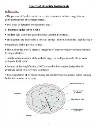
1. 1
Spectrophotometric Instruments
4- Detector :
• The purpose of the detector is convert the transmitted radiant energy into an
equivalent amount of electrical energy .
• Two types of detectors are frequently used :-
1- Photomultiplier tube ( PMT ) :
• Incident light strikes the coated cathode , emitting electrons .
• The electrons are attracted to a series of anodes , known as dynodes , each having a
Successively higher positive voltage .
• Theses dynodes are of a material that gives off many secondary electrons when hit
by single electrons .
• Initial electron emission at the cathods triggers a multiple cascade of electrons
within the PMT itself .
• Because of this amplification , PMT are used in instruments designed to be
extremely sensitive to very low light levels .
• the accumulation of electrons striking the anode produces a current signal that can
be fed into a meter or recorder .
2. 2
2- Photodiode arrays ( PDA ) :
• are new detectors being used in modern
spectrometers .
• photodiodes are composed of silicon crystals
that are sensitive to light in the wavelength range
170-1100 nm .
• Upon photon absorption by the doiode , a current is generated in the photodiode
that is proportional to the number of photons .
• Although photodiodes are not as sensitive as PMT because of the lack of internal
amplification , their excellent linearity , speed , and small size make them useful in
applications where light levels are adequate .
• PDA detectors are available in integrated circuits containing 256 to 2,048
photodiodes in a linear arrangement .
• Each photodiode responds to a specific wavelength, and as a result, a complete
UV/visible spectrum can be obtained in less than 1 second .
3. 3
◘ Application of UV – VIS spectroscopy :
• Although many different types of operations can be carries out on a
spectrophotometer, all applications fall in one of two categories.
1- Measurement of absorbance at a fixed wavelength :-
• Are most often used to obtain quantitative information , such as the concentration
of a solute in solution or the absorption coefficient of a chromophore .
• For fixed-wavelength measurements with a single-beam instrument, a cuvette
containing solvent only is placed in the sample beam and the instrument is adjusted
to read “Zero” absorbance .
• A matched cuvette containing sample plus solvent is then placed in the sample
chamber and the absorbance is read directly from the display .
• The adjustment to zero absorbance with only solvent in the sample chamber allows
the operator to obtain a direct reading of absorbance for the sample .
• Fixed-wavelength measurements using a double-beam spectrophotometer are made
by first zeroing the instrument with no cuvette in either the sample or reference
holder .
• Alternatively , the spectrophotometer can be balanced by placing matched cuvettes
containing water or solvent in both sample chambers .
• Then, a cuvette containing pure solvent is placed in the reference position and a
matched cuvette containing solvent plus sample is set in the sample position .
• The absorbance reading given by the instrument is that of the samples; that is, the
absorbance due to solvent is subtracted by the instrument.
2- Absorbance measurement as a function of wavelength :-
• It provides qualitative information that
assists in solving the identity and structure
of a pure substance by detecting
characteristic grouping of atoms in a
molecule.
4. 4
Immunoassays
◘ General considerations :
• Immunoassays are available for analysis of over 100 different analytes .
• Most immunoassay methods used specimens without any pretreatment and used
very small sample volumes ( 10 µl-50 µl ) .
• Antibodies ( Abs ) are incorporated into many clinical laboratory tests .
• They are useful because of their unique properties in recognizing and distinguishing
among closely related antigens ( Ags ) .
• Ab molecules is an immunoglobulin protein binds to a site in the Ag .
• An Ag is relatively large and complex and usually has multiple sites that can bind
to Abs with different specificities; each site on the Ag referred to as an antigenic
determinant or epitope .
• Some tests aid in diagnosis of infections through the detection of Abs to infectious
agents in patient specimens; examples are HIV , influenza , hepatitis , and rubella
tests .
• Other laboratory tests use the Ag-Ab reaction to measure or detect a substance not a
part of the immune system, such as using Abs to measure drug or hormone levels .
5. 5
◘ Monoclonal and polyclonal antibodies :
• Abs used in immunological testes can be monoclonal or polyclonal .
• Monoclonal Abs are of one class and one specificity ( react with only one epitope )
and are derived from one ( mono ) clone, or cell line .
• Monoclonal Abs are produced in laboratories and used as reagents in many
immunodiagnostic kits .
• polyclonal Abs are mixtures of antibodies produced by more than one ( poly ) cell
line .
• For instance , one bacterial infection will stimulate
many plasma cells ( poly clones) to respond, each
producing and secreting antibodies to a different
bacterial epitope .
• this results in a mixture of Abs in plasma that taken
together can react with multiple Ags .
6. 6
◘ Test sensitivity and specificity
• Sensitivity refers to the lower limit of
detection, or the lowest concentration
capable of being detected by a test method .
• Failure to detect small amounts of a
substance in a test will result in a false-
negative result.
• Specificity refers to the ability to detect only the substance for which the test is
designed .
• Reaction with other substances ( cross-reactivity ) decreases the specificity of the
test and can cause false positive results .
7. 7
◘ Qualitative, Semi-qualitative and quantitative tests :-
• Many immunological procedures are reported only as negative or positive
; these are called qualitative tests .
• Other procedures are semi-quantitative or quantitative .
• Semi-quantitative procedures usually estimate concentrations of
antibodies or sometimes antigen .
• The antibodies concentration can be
estimated by making serial dilutions
and determining the maximum
dilution still capable of causing a
visible reaction in the test procedure .
• Semi-quantitative estimates of
antibody concentration can be
expressed as a titer , the reciprocal of
the highest dilution showing a
reaction
• Quantitative immunological tests are less frequently performed .
• Examples of some quantitative tests are drug assays .
8. 8
◘ Principles of Ag-Ab tests :-
♠ Examples of tests that incorporate the Ag-Ab reaction include :
1- Agglutination :
• It is the visible clumping or aggregation of cells or particles as a result of reaction
with a specific antibody .
• IgM is the antibody class that reacts best in agglutination reactions because of its
large size and multivalent binding capacity .
• Antigen-coated cells or particles , such as red blood cells or latex beads, become
linked together and form visible clumps when reacted with sufficient antibody .
• In most agglutination test designs, the presence of agglutination indicates a positive
test.
• Blood typing, bacterial identification , and the classic latex test for rheumatoid
arthritis are tests based on the agglutination reaction .
• Agglutination tests can be qualitative or semi-quantitative .
• Semi-quantitative tests can performed on slides, in tubes, or in special microtiter
plates having wells with rounded bottoms .
• Serial dilutions of serum are made and
tested to determine the maximum dilution
capable of causing agglutination , which is
reported as a titer .
• When agglutination tests are performed in
microtiter plates , a negative test indicated
by the presence of non-agglutinated
particles concentrated in a small dot in the
bottom of the well .
• A diffuse pattern of cells spread over the
bottom of a round-bottomed well indicates a
positive test .