ELISA- Principle, Types and
Applications
• ELISA is an antigen antibody reaction.
• ELISA is a common laboratory technique
which is usually used to measure the
concentration of antibodies or antigens
proteins and hormones in blood.
• In 1971, ELISA was introduced by Peter
Perlmann and Eva Engvall at Stockholm
University in Sweden.
ELISA, (Enzyme-Linked
Immunoassay)
• ELISA is a plate based assay technique which is used for
detecting and quantifying substances such as peptides,
proteins, antibodies and hormones.
• An enzyme conjugated with an antibody reacts with
colorless substrate to generate a colored product. Such
substrate is called chromogenic substrate.
• A number of enzymes have been used for ELISA such as
• alkaline phosphatase,
• horse radish peroxidase and
• beta galactosidase.
Specific substrate such as
• ortho-phenyldiamine dihydrochloride (for peroxidase),
• Paranitrophenyl phosphate (for alkaline phosphatase) are used
which are hydrolysed by above enzymes to give colored end
product.
Principle
• ELISAs are typically performed in 96-well polystyrene plates. The serum is
incubated in a well, and each well contains a different serum.
• A positive control serum and a negative control serum would be included
among the 96 samples being tested.
• Antibodies or antigens present in serum are captured by corresponding
antigen or antibody coated on to the solid surface.
• After some time, the plate is washed to remove serum and unbound
antibodies or antigens with a series of wash buffer.
• To detect the bound antibodies or antigens, a secondary antibodies that
are attached to an enzyme such as peroxidase or alkaline phosphatase are
added to each well.
• After an incubation period, the unbound secondary antibodies are washed
off. When a suitable substrate is added, the enzyme reacts with it to
produce a color.
• This color produced is measurable as a function or quantity of antigens or
antibodies present in the given sample. The intensity of color/ optical
density is measured at 450nm.
• The intensity of the color gives an indication of the amount of antigen or
antibody.
Types of ELISA
• Frequently there are 3 types of ELISA on the
basis of binding structure between the
Antibody and Antigen.
– Indirect ELISA
– Sandwich ELISA
– Competitive ELISA
1. Indirect ELISA
• Antibody can be detected or quantitatively determined by
indirect ELISA.
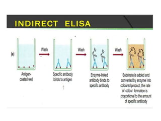
• In this technique, antigen is coated on the microtiter well.
• Serum or some other sample containing primary antibody
is added to the microtiter well and allowed to react with
the coated antigen.
• Any free primary antibody is washed away and the bound
antibody to the antigen is detected by adding an enzyme
conjugated secondary antibody that binds to the primary
antibody.
• Unbound secondary antibody is then washed away and a
specific substrate for the enzyme is added.
• Enzyme hydrolyzes the substrate to form colored products.
The amount of colored end product is measured by
spectrophotometric plate readers that can measure the
absorbance of all the wells of 96-well plate.
Procedure of Indirect ELISA
1. Coat the micro titer plate wells with antigen.
2. Block all unbound sites to prevent false positive results.
3. Add sample containing antibody (e.g. rabbit monoclonal
antibody) to the wells and incubate the plate at 37°c.
4. Wash the plate, so that unbound antibody is removed.
5. Add secondary antibody conjugated to an enzyme (e.g.
anti- mouse IgG).
6. Wash the plate, so that unbound enzyme-linked
antibodies are removed.
7. Add substrate which is converted by the enzyme to
produce a colored product.
8. Reaction of a substrate with the enzyme to produce a
colored product.
Advantages
1. Increased sensitivity, since more than one labeled antibody is bound
per primary antibody.
2. A wide variety of labeled secondary antibodies are available
commercially.
3. Maximum immunoreactivity of the primary antibody is retained
because it is not labeled.
4. Versatile because many primary antibodies can be made in one
species and the same labeled secondary antibody can be used for
detection.
5. Flexibility, since different primary detection antibodies can be used
with a single labeled secondary antibody.
6. Cost savings, since fewer labeled antibodies are required.
7. Different visualization markers can be used with the same primary
antibody.
Disadvantages
1. Cross-reactivity might occur with the secondary antibody, resulting in
nonspecific signal.
2. An extra incubation step is required in the procedure.
2. Sandwich ELISA
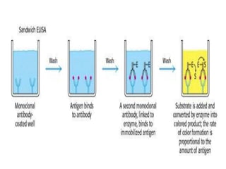
• Antigen can be detected by sandwich ELISA. In this
technique, antibody is coated on the microtiter well.
• A sample containing antigen is added to the well
and allowed to react with the antibody attached to
the well, forming antigen-antibody complex.
• After the well is washed, a second enzyme-linked
antibody specific for a different epitope on the
antigen is added and allowed to react with the
bound antigen.
• Then after unbound secondary antibody is removed
by washing. Finally substrate is added to the plate
which is hydrolyzed by enzyme to form colored
products.
Procedure of sandwich ELISA
1. Prepare a surface to which a known quantity of
antibody is bound.
2. Add the antigen-containing sample to the plate and
incubate the plate at 37°c.
3. Wash the plate, so that unbound antigen is removed.
4. Add the enzyme-linked antibodies which are also
specific to the antigen and then incubate at 37°c.
5. Wash the plate, so that unbound enzyme-linked
antibodies are removed.
6. Add substrate which is converted by the enzyme to
produce a colored product.
7. Reaction of a substrate with the enzyme to produce a
colored product.
Advantages
• High specificity, since two antibodies are used
the antigen is specifically captured and
detected.
• Suitable for complex samples, since the
antigen does not require purification prior to
measurement.
• Flexibility and sensitivity, since both direct and
indirect detection methods can be used.
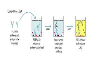
3. Competitive ELISA
• This test is used to measure the concentration of an
antigen in a sample.
• In this test, antibody is first incubated in solution with a
sample containing antigen.
• The antigen-antibody mixture is then added to the
microtitre well which is coated with antigen.
• The more the antigen present in the sample, the less
free antibody will be available to bind to the antigen-
coated well.
• After the well is washed, enzyme conjugated secondary
antibody specific for isotype of the primary antibody is
added to determine the amount of primary antibody
bound to the well.
• The higher the concentration of antigen in the sample,
the lower the absorbance.
Procedure
1. Antibody is incubated with sample containing
antigen.
2. Antigen-antibody complex are added to the
microtitre well which are pre-coated with the
antigen.
3. Wash the plate to remove unbound antibody.
4. Enzyme linked secondary antibody which is
specific to the primary antibody is added.
5. Wash the plate, so that unbound enzyme-linked
antibodies are removed.
6. Add substrate which is converted by the enzyme
into a fluorescent signal.
Advantages
1. High specificity, since two antibodies are used.
2. High sensitivity, since both direct and indirect detection
methods can be used.
3. Suitable for complex samples, since the antigen does not
require purification prior to measurement.
Application of ELISA
1. Presence of antigen or the presence of antibody in a
sample can be evaluated.
2. Determination of serum antibody concentrations in a virus
test.
3. Used in food industry when detecting potential food
allergens.
4. Applied in disease outbreaks- tracking the spread of
disease e.g. HIV, bird flu, common, colds, cholera, STD etc.