Downloaded 237 times

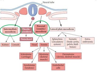
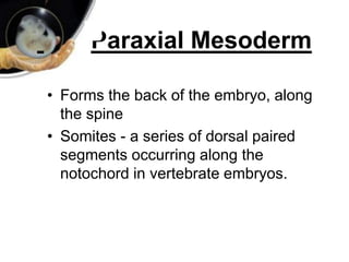
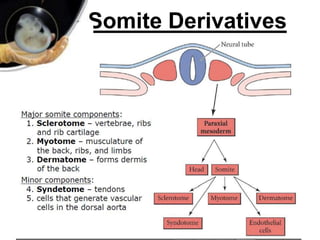
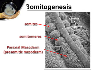

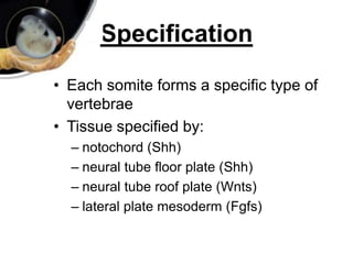
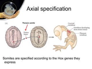
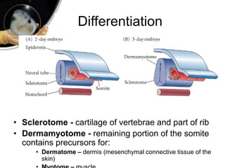
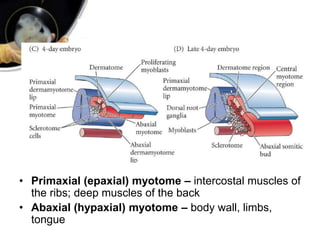
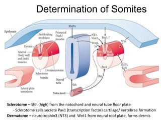
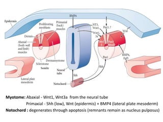
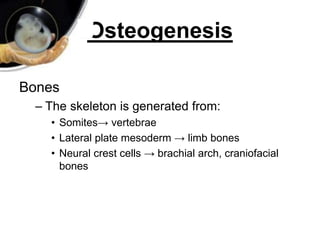
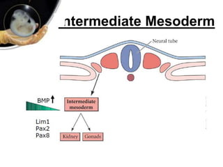
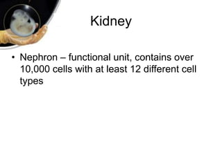
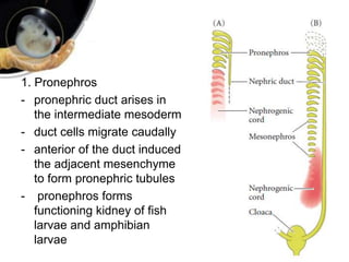
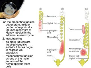
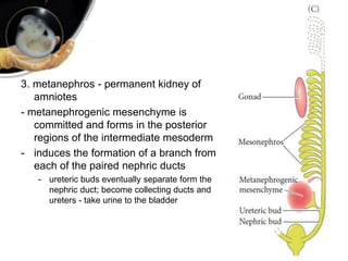
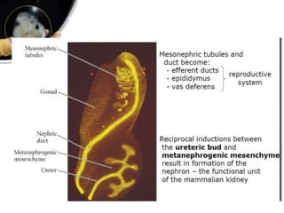


Paraxial and intermediate mesoderm form important structures. Paraxial mesoderm forms somites through a "clock and wave" mechanism, with each somite giving rise to vertebrae, muscle, and dermis. Somites are specified by surrounding tissues to develop appropriately. Intermediate mesoderm forms the kidney, with the pronephros, mesonephros, and metanephros appearing sequentially in vertebrate development. The metanephros becomes the permanent kidney in amniotes.


