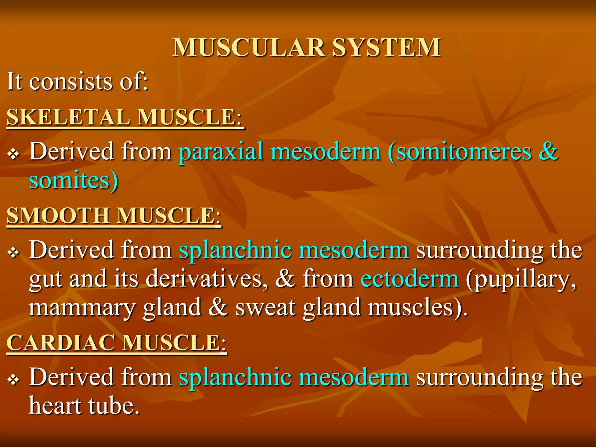
The muscular system develops primarily from the mesodermal germ layer. It consists of skeletal, smooth, and cardiac muscle. Skeletal muscle develops from paraxial mesoderm and forms the musculature of the axial skeleton, body wall, and limbs. Smooth muscle develops from splanchnic mesoderm surrounding organs and the ectoderm of certain structures. Cardiac muscle develops from splanchnic mesoderm surrounding the heart tube. Muscle tissue develops from embryonic muscle cells called myoblasts that fuse to form long muscle fibers containing myofibrils. Precise molecular signaling regulates the development of each muscle type from precursor tissues and cells.
Development of the musculoskeletal systemSahar Hafeez
The document summarizes the development of the musculoskeletal system from early embryonic stages. It discusses how the skeletal and muscular systems originate from mesoderm and the processes of ossification and myogenesis. Key events include the formation of somites which differentiate into sclerotome and dermomyotome, the development of limb buds and their rotation, and the segmentation of axial musculature into epimere and hypomere. The document provides an overview of the embryonic development of the skeletal and muscular systems.
Most muscles arise from paraxial mesoderm in the 3rd week of development. Skeletal muscles derive from paraxial mesoderm, with progenitor cells coming from the dorsolateral and dorsomedial portions of somites. By the 5th week, muscle precursor cells are divided into a small dorsal epimere and larger ventral hypomere. Cardiac muscle develops from splanchnic mesoderm surrounding the heart tube, while smooth muscle arises from surrounding splanchnic mesoderm of the gut and vasculature.
This document summarizes the development of the musculoskeletal system from early embryogenesis through the formation of major structures like bones, muscles, and limbs. It describes the formation of somites from the paraxial mesoderm and their differentiation into sclerotome, dermomyotome and myotome tissues. Key events covered include segmentation of the paraxial mesoderm into somites, somite differentiation, development of the vertebral column from sclerotome tissues, and muscle formation from dermomyotome and myotome tissues. Limb buds form from lateral plate mesoderm by the fourth week and develop a mesenchymal core and apical ectodermal ridge.
The skeletal system develops from mesenchyme tissue which originates from the mesoderm. Mesenchyme cells migrate and form cartilage and bone. Bones develop through two types of ossification - membranous and endochondral. The axial skeleton develops from somites which form vertebrae and ribs. Limbs develop from limb buds which are outgrowths that form the bones of the arms and legs. Joints form as cartilage develops between bone ends. Developmental anomalies can occur in bones, vertebrae, ribs and limbs.
Skeletal muscle tissue functions include movement, posture maintenance, joint stabilization, and heat generation. The main types of muscle tissue are skeletal, cardiac, and smooth muscle. Skeletal muscle is striated and voluntary, attaching to bones and moving the skeleton. Cardiac muscle is only found in the heart walls and has involuntary, rhythmic contractions. Smooth muscle lacks striations and controls involuntary functions like digestion and blood flow. All muscle tissues contain contractile filaments that slide past each other to cause shortening, but the tissues differ in organization, fiber type, and control.
The document discusses the histology of the three main types of muscle tissue - skeletal, cardiac, and smooth muscle. Skeletal muscle is striated and voluntary. It consists of long multinucleated fibers bundled together. Cardiac muscle is also striated but less so. It branches and has intercalated discs. Smooth muscle is not striated and consists of spindle-shaped cells. Microscopic examination can identify features like striations and nuclear placement that distinguish the three muscle types.
This document provides an overview of the pleura and lung. It begins by introducing the lung and its parts, including lobes and borders. It then describes the pleural layers in detail, including the parietal pleura layers of cervical, costal, mediastinal and diaphragmatic pleura. Relations and blood supply of the pleura are discussed. Bronchopulmonary segments and clinical significance are summarized. Common pleural conditions like pleurisy, pleural effusion and pneumothorax are briefly mentioned.
Paraxial mesoderm forms somites along the spine through somitogenesis. Somites differentiate into sclerotome, dermamyotome and myotome tissues. Sclerotome forms cartilage and bone of the vertebrae. Dermamyotome forms dermis and muscle lineages. Myotome separates into epaxial and hypaxial regions that form back muscles and body wall muscles. Intermediate mesoderm forms the urogenital system including pronephros, mesonephros and metanephros kidneys as well as gonads. The metanephros is the permanent kidney in amniotes.
This is a presentation on the suboccipital triangle. The objectives of this presentation are to provide an overview of the deepest set of pre-vertebral muscles. The presentation gives an overview of the suboccipital triangle, the borders, contents and it's clinical importance.
For further reading please refer to Keith Moore - Clinically Oriented Anatomy and Snell's Clinical Anatomy by Regions.
The document summarizes the joints of the thoracic wall, including the intervertebral joints between vertebrae, costovertebral joints between ribs and vertebrae, costochondral joints between ribs and costal cartilages, interchondral joints between costal cartilages, sternocostal joints between the sternum and costal cartilages, and sternoclavicular joints between the sternum and clavicle. It describes the specific articulations, ligaments, and movements at each of these joints. It also provides details on the manubriosternal joint between the manubrium and sternum, the xiphisternal joint between the xiphoid process and ster
Developmet of Integumentary System (Special Embryology)Dr. Sherif Fahmy
The skin has two origins - the epidermis develops from surface ectoderm while the dermis develops from mesoderm. Melanocytes invade the epidermis from the neural crest at the third month. Hair develops as epidermal proliferation penetrates the dermis, with the terminal end invaginated by mesoderm forming blood vessels and nerves. Proliferation of hair follicle cells forms sebaceous glands. At birth, the skin is covered in a whitish paste called vernix caseosa formed from secretions that protects the skin. Congenital skin and hair anomalies include ichthyosis, atrichia, and hypertrichosis. The mammary gland develops from a mammary line
The knee joint is the largest synovial joint in the body. It is composed of three bones: the femur, tibia, and patella. The knee joint has two articulations: the tibiofemoral joint between the femur and tibia, and the patellofemoral joint between the patella and femur. The knee joint is a compound synovial joint, with the tibiofemoral joint being a hinge type and the patellofemoral joint being a plane type. The knee joint is surrounded by ligaments such as the ACL and PCL, and contains a synovial fluid within its capsule. It also contains menisci that act as cushions and increase stability.
This document provides an overview of the anatomy of the upper limb. It begins by listing the learning objectives which are to describe the bones, joints, muscles, arteries, veins, and spaces of the upper limb. It then proceeds to discuss the bones of the upper limb including the scapula, clavicle, humerus, radius, ulna, carpals, metacarpals, and phalanges. It also describes the joints between these bones. Next, it covers the major muscles of the upper limb grouped into those attached to the axial skeleton and those of the upper limb itself. Finally, it briefly discusses the brachial plexus nerves and blood vessels of the axilla and upper limb.
Sesamoid bones are small, seed-shaped bones that form within certain tendons where they rub against bone. They develop to minimize friction, act as pulleys for muscle contraction, and alter the direction of muscle pull. The largest sesamoid bone is the patella at the front of the knee. Others form at the back of the knee, in the hand, and below the big toe. Sesamoid bones lack periosteum and ossify after birth, differing from true long bones.
1. The vertebral column develops from mesenchymal tissue that migrates and condenses to form the centrums and neural arches. Cartilage replaces the mesenchymal tissue before ossification begins.
2. Primary ossification centers form in the centrums and neural arches by 8 weeks, and secondary centers form after puberty. The notochord degenerates and forms the nucleus pulposus of intervertebral discs.
3. Costal processes in the cervical region form parts of the neck, in the thoracic region form ribs, and in lower regions fuse with transverse processes or the sacrum. Anomalies can include spina bifida from incomplete fusion or hem
Lecture 12 the skeleton embryology pdfMBBS IMS MSU
1. The vertebral column is derived from sclerotomes of somites, with each vertebra formed by fusion of portions from two adjacent somites.
2. The ribs are derived from ventral extensions of sclerotomal mesenchyme. The sternum is formed by fusion of right and left sternal bars.
3. The skull develops from mesenchyme around the brain, with some bones forming in membrane and some in cartilage. The limbs first appear as outgrowths from the body wall that get subdivided to form parts.
The shoulder joint is formed by the articulation of the humerus with the scapula. It includes the glenohumeral joint as well as the acromioclavicular and sternoclavicular joints. The glenohumeral joint is the most mobile joint in the body and is stabilized by static structures like ligaments and the labrum as well as dynamic structures like the four rotator cuff muscles that surround it.
The document discusses the epiphysis, which is one of the three parts of a long bone. It develops through enchondral ossification from secondary ossification centers. Lesions can occur at the epiphysis, including tumors and infections. Injuries to the epiphyseal plate in children are classified using the Salter-Harris system from Type I to V based on the fracture line. Rare types also exist. The epiphysis is an important part of long bone anatomy and development in the body.
This document provides information on the histology of cartilage. It discusses the different types of cartilage - hyaline, elastic, and fibrocartilage. It describes their microscopic appearance, including the arrangement of cells and matrix. Key points are highlighted for each cartilage type. Locations of different cartilages in the body are also listed. The document concludes with some clinical applications and references.
The neural tube develops from the ectoderm and forms the central nervous system. Neurulation involves the formation of the neural plate which elevates and fuses to form the neural tube. Neural crest cells dissociate and give rise to many structures. The brain develops from three primary vesicles-the prosencephalon, mesencephalon, and rhombencephalon. The spinal cord arises from the lower neural tube. Neurons and glia differentiate and migrate within the neural tube. Fusion of the neural folds and closure of neuropores must occur properly to prevent neural tube defects.
The document summarizes the embryological development of the skeletal system. It describes how the axial skeleton, including the vertebral column and ribs, and appendicular skeleton develop from somites in the embryo. It explains the three stages of development - blastemal/membranous, cartilaginous, and bony. It provides details on the formation of individual bones and joints, such as the development of vertebrae, ribs, sternum, skull, and others from sclerotomes, notochord, and cartilage models.
The document discusses various types and classifications of glands. It begins by defining glands as organs composed of specialized secretory cells derived from epithelial tissue. Glands are classified based on their site of secretion (exocrine, endocrine, paracrine), cell number (unicellular, multicellular), duct structure (simple, compound), secretory end piece shape (tubular, alveolar, etc.), secretion type (serous, mucous, mixed), secretion mode (merocrine, apocrine, holocrine, cytocrine), and developmental origin (ectodermal, mesodermal, endodermal). Key exocrine gland features and the development of both exocrine and endocrine glands are also
The document describes the anatomy of the axilla and brachial plexus. The axilla is a pyramid-shaped space bounded by bones and muscles that contains neurovascular structures passing from the neck to the upper limb. The brachial plexus, located in the posterior neck and axilla, is formed by the union of cervical and thoracic spinal nerves and divides into cords and branches that innervate the upper limb. Injuries to different parts of the brachial plexus can cause weakness or loss of sensation in specific areas due to disruption of the corresponding nerves.
Here are the answers to the short answer questions:
1. Scoliosis is caused by vertebral asymmetry or half a vertebra missing.
Lordosis is caused by failure of posterior segmentation in the presence of anterior active growth.
Spina bifida is caused by imperfect fusion or nonunion of the vertebral arches.
Kyphosis occurs when the two ossific centers which appear in the body of the vertebra fail to develop adequately.
2. The notochord in the centrum (body) of the vertebra degenerates. The notochord in the intervertebral disc persists and forms the nucleus pulposus, which is later surrounded by circular fibers of the annulus fib
2nd week of intrauterine developement of embryoReda Cheema
During the second week of development:
1. Implantation of the blastocyst is completed, with trophoblast cells differentiating into cytotrophoblast and syncytiotrophoblast.
2. Syncytiotrophoblast produces human chorionic gonadotropin (hCG) hormone, which can be detected in pregnancy tests by the end of the second week.
3. An amniotic cavity forms within the embryoblast, and the epiblast and hypoblast layers develop to form the embryonic disc surrounded by extraembryonic tissues.
The fascia lata is a deep fascial investment that encloses the thigh muscles like a stocking. It is attached superiorly to the inguinal ligament and pubic bone, laterally to the iliac crest, and posteriorly to the sacrum and ischial tuberosity. It is modified to form the iliotibial tract and separates the thigh into three compartments by intermuscular septa. The sartorius opening contains the saphenous vein and lymphatics covered by the cribriform fascia. The tensor fascia lata muscle tightens the iliotibial band and braces the knee during movement.
The document summarizes the lateral and posterior compartments of the leg. The lateral compartment contains the peroneus longus and brevis muscles, innervated by the superficial peroneal nerve. The posterior compartment contains several muscles - the gastrocnemius, soleus, plantaris, popliteus, flexor hallucis longus, and flexor digitorum longus - innervated by the tibial nerve. The document describes the origins, insertions, actions and clinical relevance of the muscles in these compartments.
Development of the Muscular System (Human Embryology, Zoo 404)Hilton Kollie
This is a PowerPoint presentation undertaken by Fasama H. Kollie and Antoinette H. Wright. This presentation gives a clue about how the muscular system develop during embryonic development.
Muscular system embryology for medical studentsymusa1334
Muscular development begins with the formation of somites from paraxial mesoderm, which give rise to skeletal muscle fibers. Smooth muscle develops from splanchnic mesoderm and some ectoderm-derived tissues. Cardiac muscle originates from splanchnic mesoderm surrounding the heart tube. Key regulatory genes like MyoD and Myf5 activate muscle-specific genes that promote the differentiation of myoblasts into myofibers. Precise molecular signaling controls the patterning and migration of myoblasts. Abnormalities can occur if muscles fail to develop properly.
Development of the musculoskeletal systemSahar Hafeez
In this presentation development of the Musculoskeletal system which is one of the largest systems of human body has been described. The viewer would be able to learn about the concept of Intrauterine bone formation in general and the role of embryonic connective tissue. Also, the origin of the two muscle groups of the , Extensors & Flexors along with their motor innervation pattern has been described in this presentation.
(Lec 2) Embryology - Embryologic derivation of oral and dental structures 2Hamzeh AlBattikhi
1) The embryo develops three main layers - ectoderm, endoderm, and mesoderm. The mesoderm separates the endoderm and ectoderm and forms somites through segmentation.
2) Somites differentiate into dermatome, sclerotome, and myotome. The myotome forms muscles, sclerotome forms bones/connective tissue, and dermatome forms skin. Head somites are "atypical" and contribute to facial muscles and skin innervation.
3) There are controversy around the number of occipital somites, classically described as 4 pairs, that contribute to tongue and skull base muscles/bones and are innervated
The skeletal and muscular systems develop from paraxial mesoderm, lateral plate mesoderm, and neural crest cells. Mesoderm forms somites that differentiate into sclerotome and dermomyotome, with sclerotome cells becoming mesenchyme that migrates and forms cartilage models through endochondral ossification or membraneous bone, while dermomyotome forms myoblasts that fuse into muscle fibers. Limb buds develop from lateral plate mesoderm and rotate as cartilage and bone form through endochondral ossification while surrounding musculature develops from dermomyotome and surrounding nerves
The document summarizes the structure and function of the muscular system. It describes the three main types of muscle tissue - skeletal, smooth, and cardiac muscle - and their distinguishing characteristics. It also details the structure of skeletal muscle from the organ level down to the contractile proteins that enable muscle contraction in response to neural stimulation.
The document summarizes the musculoskeletal system, including skeletal muscles, joints, and the skeleton. It describes the three main types of muscle - skeletal, cardiac, and smooth muscle. It discusses the composition of skeletal muscle fibers and their ultrastructure. Key details are provided on the sliding filament model of muscle contraction, including the roles of actin, myosin, tropomyosin, and troponin. Excitation-contraction coupling and the neuro-muscular junction are also summarized. Principal skeletal muscles of the face, neck, trunk, shoulder, and limbs are briefly outlined.
This document provides an overview of the histology of the muscular system for medical science students. It begins with an introduction to muscle tissues and contractile cells, outlining the three main types of muscle tissues: skeletal, cardiac, and smooth muscle. It then discusses the distinguishing characteristics, structures, and functions of each type of muscle tissue in more detail. The document also covers the contractile elements within muscle tissues, as well as the processes of injury, repair, and regeneration of skeletal and cardiac muscle fibers.
This document provides an overview of muscle tissues and contractile cells. It discusses the three main types of muscle tissue - skeletal, cardiac, and smooth muscle - and their distinguishing characteristics. Skeletal and cardiac muscle are striated due to their arrangement of thin actin and thick myosin filaments into sarcomeres. Smooth muscle is non-striated. Each muscle tissue has a specific location, control mechanism, and function. The document also examines the cellular components that enable muscle contraction, including myofibrils composed of sarcomeres containing overlapping actin and myosin filaments.
This document discusses embryology and the development of the musculoskeletal system. It covers the following key points:
1. Embryology is the study of developmental events during prenatal stages, specifically the embryonic and fetal periods. The embryonic period is the first 8 weeks when the basic body plan takes shape, and the fetal period is the remaining 30 weeks when structures continue growing.
2. Musculoskeletal development begins with the formation of somites from paraxial mesoderm, which give rise to bones, cartilage, and muscles. Bones develop through membranous or endochondral ossification. Long bones are examples of endochondral ossification, forming cartilage models that are later replaced with bone.
3. Lim
This document discusses embryonic and fetal development from 3-8 weeks (embryonic period) and 9 weeks to birth (fetal period). During the embryonic period, the three germ layers give rise to specific tissues and organs as the main organ systems are established. Neurulation occurs as the neural tube forms from the neural plate. Neural crest cells migrate throughout the body. The mesoderm forms somites which differentiate into muscle, bone and skin tissues. Blood islands form and later hematopoietic stem cells arise. The endoderm forms the gastrointestinal tract. During the fetal period, organs mature and the fetus grows rapidly in the third, fourth and fifth months.
The skeletal system develops from mesoderm and neural crest cells. Paraxial mesoderm forms somites which differentiate into sclerotome and dermomyotome. Sclerotome cells form the mesenchyme which can become bone, cartilage or connective tissue. Bones form through intramembranous or endochondral ossification, where cartilage templates are replaced by bone. The axial skeleton includes the skull, vertebrae, ribs and sternum, while the appendicular skeleton comprises the shoulder and pelvic girdles and limb bones.
During the embryonic period from 3-8 weeks, organogenesis occurs where organs develop from embryonic tissue. The three germ layers (ectoderm, mesoderm, endoderm) give rise to specific tissues and organs. The ectoderm forms the neural tube which becomes the central nervous system. It also forms the neural crest cells which migrate and develop into many structures. The mesoderm separates into three masses - paraxial mesoderm forms the skeleton, intermediate mesoderm forms the genitourinary system, and lateral plate mesoderm forms body wall structures. Somites develop from the paraxial mesoderm and form skeletal muscle, bone and dermis. Blood vessels also develop throughout the mesoderm
Embryology and Development of Spinal Dysraphism and Tethered Spinal Cord Synd...AnthonyGokYan
This document discusses the development of the spinal cord and vertebral column as well as spina bifida. It begins by describing how the neural tube forms from ectoderm and influences vertebral column development. It then details the layers that form in the spinal cord - ventricular zone, mantle zone that forms gray matter, and marginal zone that forms white matter. Next, it discusses the formation of the vertebral column from somites and changes in spinal cord positioning. Finally, it describes spina bifida where the vertebral arches fail to fuse, occurring as occulta or cystica with varying neurological involvement.
Gastrulation occurs during weeks 2-4 of gestation and involves the inward migration of cells from the blastula to form the three germ layers - ectoderm, mesoderm, and endoderm. Each germ layer then gives rise to different tissues and organs. Neurulation follows gastrulation and occurs during weeks 3-4, forming the neural tube from the ectoderm. Somites form from the paraxial mesoderm on either side of the neural tube and differentiate into structures like bone, cartilage, muscle and tendons. Neural crest cells form at the border of the neural plate and migrate to form parts of the nervous system, face and pigmentation.
The document provides an overview of the muscular system including the three types of muscle tissues - skeletal, cardiac, and smooth muscle. It describes the microscopic anatomy of skeletal muscle fibers and their sarcomere structure. The sliding filament theory of muscle contraction is explained, involving the interaction of the thick myosin and thin actin filaments through ATP hydrolysis. Contraction is triggered by an action potential causing calcium release and the binding of myosin heads to actin, pulling the Z-lines inward.
8 Surprising Reasons To Meditate 40 Minutes A Day That Can Change Your Life.pptxHolistified Wellness
We’re talking about Vedic Meditation, a form of meditation that has been around for at least 5,000 years. Back then, the people who lived in the Indus Valley, now known as India and Pakistan, practised meditation as a fundamental part of daily life. This knowledge that has given us yoga and Ayurveda, was known as Veda, hence the name Vedic. And though there are some written records, the practice has been passed down verbally from generation to generation.
10 Benefits an EPCR Software should Bring to EMS Organizations Traumasoft LLC
The benefits of an ePCR solution should extend to the whole EMS organization, not just certain groups of people or certain departments. It should provide more than just a form for entering and a database for storing information. It should also include a workflow of how information is communicated, used and stored across the entire organization.
The skin is the largest organ and its health plays a vital role among the other sense organs. The skin concerns like acne breakout, psoriasis, or anything similar along the lines, finding a qualified and experienced dermatologist becomes paramount.
Travel vaccination in Manchester offers comprehensive immunization services for individuals planning international trips. Expert healthcare providers administer vaccines tailored to your destination, ensuring you stay protected against various diseases. Conveniently located clinics and flexible appointment options make it easy to get the necessary shots before your journey. Stay healthy and travel with confidence by getting vaccinated in Manchester. Visit us: www.nxhealthcare.co.uk
Adhd Medication Shortage Uk - trinexpharmacy.comreignlana06
The UK is currently facing a Adhd Medication Shortage Uk, which has left many patients and their families grappling with uncertainty and frustration. ADHD, or Attention Deficit Hyperactivity Disorder, is a chronic condition that requires consistent medication to manage effectively. This shortage has highlighted the critical role these medications play in the daily lives of those affected by ADHD. Contact : +1 (747) 209 – 3649 E-mail : sales@trinexpharmacy.com
Clinic ^%[+27633867063*Abortion Pills For Sale In Tembisa Central19various
Clinic ^%[+27633867063*Abortion Pills For Sale In Tembisa Central Clinic ^%[+27633867063*Abortion Pills For Sale In Tembisa CentralClinic ^%[+27633867063*Abortion Pills For Sale In Tembisa CentralClinic ^%[+27633867063*Abortion Pills For Sale In Tembisa CentralClinic ^%[+27633867063*Abortion Pills For Sale In Tembisa Central
Travel Clinic Cardiff: Health Advice for International TravelersNX Healthcare
Travel Clinic Cardiff offers comprehensive travel health services, including vaccinations, travel advice, and preventive care for international travelers. Our expert team ensures you are well-prepared and protected for your journey, providing personalized consultations tailored to your destination. Conveniently located in Cardiff, we help you travel with confidence and peace of mind. Visit us: www.nxhealthcare.co.uk
Does Over-Masturbation Contribute to Chronic Prostatitis.pptxwalterHu5
In some case, your chronic prostatitis may be related to over-masturbation. Generally, natural medicine Diuretic and Anti-inflammatory Pill can help mee get a cure.
2. The Muscular System develops from the
mesodermal germ layer, except some smooth
muscle tissue.
3. MUSCULAR SYSTEM
It consists of:
SKELETAL MUSCLE:
Derived from paraxial mesoderm (somitomeres &
somites)
SMOOTH MUSCLE:
Derived from splanchnic mesoderm surrounding the
gut and its derivatives, & from ectoderm (pupillary,
mammary gland & sweat gland muscles).
CARDIAC MUSCLE:
Derived from splanchnic mesoderm surrounding the
heart tube.
4. Muscle tissue develops from myoblasts,
embryonic muscle cells that are derived from
mesenchyme (embryonic connective tissue).
5. STRIATED SKELETAL
MUSCULATURE
Somites and somitomeres
form the musculature of the
axial skeleton, body wall,
limbs and head.
From the occipital region
caudally, somites
differentiate into
sclerotome, dermatome &
two muscle forming
regions.
6. Dorsolateral region of the
somite expresses the muscle-
specific gene MYO-D and
migrates to provide progenitor
cells for limb and body wall
(hypomeric) musculature.
Dorsomedial region,
migrates ventral to cells that
form the dermatome, and
forms the myotome. This
region, which expresses the
muscle-specific gene MYF5,
forms epimeric musculature.
7. Border between somite and the parietal layer
of the lateral plate mesoderm called lateral
somatic frontier.
Separate primaxial domain( around neural tube
---somite derived paraxial mesoderm
Abaxial domain (lateral plate mesoderm +cells
of somite cross the frontier)
Lateral somatic frontier define as border
between dermis derived from dermatomes in
the back and dermis derived from lateral plate
mesoderm in body wall
Border of rib priaxial and cartilage is abaxial.
8. Origin of muscle from abaxial &
primaxial precursors
primaxial Abaxail
Cervical scalenes infrahyoid
Geniohyoid, prevertebral
Thoracoabdominal region intercostal Pectoralis major and minor
External and internal
abdominus
Transversus abdominus
Sternalis, rectus
abdominus,pelvic diaphragm
Upper limb Rhomboid, levator scapulae Distal limb muscles
Lower limb All lower limb muscles
10. STRIATED SKELETAL
MUSCULATURE (cont’d)
During differentiation, precursor cells, the
myoblasts, fuse and form long, multinucleated
muscle fibers.
Myofibrils soon appear in the cytoplasm, and by the
end of the third month, cross-striations typical of
skeletal muscle appear.
A similar process occurs in the seven somitomeres in
the head region rostral to the occipital somites.
Somitomeres remain loosely organized structures,
however, never segregating into sclerotome and
dermomyotome segments.
11. MOLECULAR REGULATION OF
SKELETAL MUSCLE DEVELOPMENT
BMP4 and probably FGFs from lateral plate mesoderm, together with
WNT proteins from adjacent ectoderm, signal the dorsolateral cells
of the somite to express the muscle-specific gene MYO-D.
BMP4 secreted by overlying ectoderm induces production of WNT
proteins by the dorsal neural tube, and these proteins cause
dorsomedial cells of the somite to activate MYF5, another muscle
specific gene.
13. PATTERNING OF MUSCLES
Patterns of muscle formation are controlled
by connective tissue into which myoblasts
migrate.
In the head region, C.T. are derived from
neural crest cells.
In cervical and occipital regions, C.T.
differentiate from somitic mesoderm.
In the body wall and limbs, C.T. originate
from lateral plate mesoderm.
14. DERIVATIVES OF PRECURSOR
MUSCLE CELLS
By the end of 5th week,
prospective muscle cells are
collected into 2 parts:
a small dorsal portion, the epimere
(formed from dorsomedial cells of
somite), and
A large ventral part, the hypomere
(formed from dorsolateral cells of
somite)
Nerves innervating segmental
muscles are also divided into a
dorsal primary ramus for the
epimere and a ventral primary
ramus for the hypomere.
15. Myoblasts of the epimeres form the extensor
muscles of the vertebral column, & those of the
hypomeres give rise to muscles of limbs & body
wall;
Myoblasts from cervical hypomeres form the
scalene, geniohyoid, and prevertebral muscles.
Myoblasts from the thoracic segments split into
three layers (external, internal, innermost
intercostal)
Myoblasts of abdominal wall form 3 layers
Myoblasts from the hypoblast of the lumbar
segments form the quadratus lumborum muscle.
Myoblasts from sacral and coccygeal regions form
the pelvic diaphragm & striated muscles of the anus.
16. A ventral longitudinal muscle
column arises at the ventral tip
of the hypomeres.
This column is represented by
the rectus abdominis muscle
in the abdominal region and
by the infrahyoid
musculature in the cervical
region.
In the thorax, the longitudinal
muscle normally disappears
but is occasionally represented
by the sternalis muscle.
17. HEAD MUSCULATURE
All voluntary muscles of the head region are derived from
paraxial mesoderm (somitomeres and somites) including
the:
Musculature of the tongue,
Eye (except that of iris which is derived from the optic
cup ectoderm)
Musculature of pharyngeal arches, innervated by the
pharyngeal arch nerves.
Pattern of muscle formation in head is directed by C.T
elements derived from neural crest cells.
19. LIMB MUSCULATURE
Ist indication of limb
musculature is observed in
7th week of development as
a condensation of
mesenchyme near the base
of limb buds.
Mesenchyme is derived
from dorsolateral cells of
somites (C4 to T2) that
migrate into the limb bud to
form the muscles.
Limb buds with their segments of origin
indicated. With further development
the segmental pattern disappears; however, an
orderly sequence in the dermatome
pattern can still be recognized in the adult.
20.
21. With elongation of the limb buds,
the muscular tissue splits into
flexor and extensor components
The upper limb buds lie opposite
the lower five cervical and upper
two thoracic segments.
The lower limb buds lie opposite
the lower four lumbar and upper
two sacral segments.
As in other regions, connective
tissue dictates the pattern of
muscle formation, and this tissue
is derived from somatic
mesoderm, which also gives rise
to the bones of the limb.
Limb buds at 7 weeks
22. 6 week embryo showing
myotome regions of somites
that give rise to skeletal
muscles
8 week embryo showing the
developing trunk & limb
musculature
23. As soon as the buds are formed, the appropriate
spinal nerves penetrate into the mesenchyme.
At first, they enter with isolated dorsal and ventral
branches, but soon these branches unite to form
large dorsal and ventral nerves.
The radial nerve, which supplies the extensor
musculature, is formed by a combination of the
dorsal segmental branches.
The ulnar and median nerves, which supply the
flexor musculature, are formed by a combination
of ventral branches.
24. Immediately after the nerves have entered
the limb buds, they establish an intimate
contact with differentiating mesodermal
condensations.
The early contact between the nerve and
muscle cells is a pre-requisite for their
complete functional differentiation.
Spinal nerves not only play an important
role in differentiation and motor innervation
of limb musculature but also provide
sensory innervation for dermatomes.
25. CLINICAL CORRELATES
Poland anomaly: it
is the total or partial
absence of pectoralis
major muscle.
Similarly palmaris
longus, serratus
anterior & quadratus
femoris muscles may
be partially or totally
absent.
26. Prune belly syndrome
Partial or complete
absence of abdominal
musculature
Usually the abdominal
wall is so thin that organs
are visible and easily
palpated.
Often associated with
malformations of the
urinary tract and bladder.
27. DEVELOPMENT OF CARDIAC
MUSCLES
From splanchnic mesoderm surrounding the
endothelial heart tube.
Myoblasts adhere to one another by special
attachments that later form intercalated discs (but
the intervening cell membranes do not disintegrate).
Myofibrils develop as in skeletal muscles but
myoblasts do not fuse.
During later development, few special bundles of
muscle cells with irregularly distributed myofibrils
(future purkinjie fibers) form the conducting system
of heart.
28. DEVELOPMENT OF SMOOTH
MUSCLES
Smooth muscle for the dorsal aorta and large
arteries is derived from lateral plate mesoderm and
neural crest cells
In the coronary arteries, smooth muscle originates
from pro-epicardial cells and neural crest cells
(proximal segments).
Smooth muscle in the wall of gut and gut
derivatives is derived from splanchnic layer of
lateral plate mesoderm
29. DEVELOPMENT OF SMOOTH
MUSCLES (cont’d)
The musculature of the iris (sphincter and dilator
pupillae) and the myoepithelial cells in the
mammary and sweat glands are thought to be
derived from the mesenchymal cells that originate
from the ectoderm.
As smooth muscle fibers develop into sheets or
bundles, they receive autonomic innervation.
30. MOLECULAR REGULATION OF SMOOTH
MUSCLE DIFFERENTIATION
Serum response factor (SRF) is a transcription factor
responsible for smooth muscle cell differentiation. This
factor is upregulated by growth factors through kinase
phosphorylation pathways.
Myocardin and myocardin-related transcription factors
(MRTFs) then act as co-activators to enhance the activity
of SRF, thereby initiating the genetic cascade responsible
for smooth muscle development.
32. The integumentary system consists of:
The skin
Appendages of skin
Sweat glands
Nails
Hair
Sebaceous glands
Arrector muscles of hairs
Mammary gland
33. SKIN HAS A TWO-FOLD ORIGIN:
EPIDERMIS: develops from surface ectoderm
DERMIS: develops from underlying mesenchyme
(derived from mesoderm)
34. EPIDERMIS
Initially, the embryo is
covered by a single layer
of ectodermal cells.
In the beginning of the
2nd month, this
epithelium divides and
form a layer of squamous
cells, the periderm or
epitrichium, on the
surface.
35. Continuous keratinization
& desquamation of
periderm cells.
Continuous replacement
of periderm cells by
underlying basal cells.
With further proliferation
of cells in the basal layer
(stratum germinativum), a
third intermediate zone is
formed by 11 weeks.
36. Replacement of periderm
cells continues until about
21st week;
Thereafter, the periderm
disappears & the stratum
corneum forms.
Proliferation of cells in
stratum germinativum also
forms epidermal ridges,
which extend into developing
dermis begin to
appear at 10 weeks &
permanently established by
17th week Finger
prints.
37. The transformation of surface ectoderm into
multilayered epidermis results from continuing
Inductive interactions with the dermis (i.e.
ectodermal / mesenchymal interaction ).
38. Skin is classified as THICK or THIN SKIN, based
on the thickness of the epidermis.
Thick skin: palms and soles; lacks hair follicles,
arrector pili muscles, & sebaceous glands BUT has
sweat glands.
Thin skin: most of the rest of the body; contains hair
follicles, arrector pili muscles, sebaceous & sweat
glands.
39. Vernix caseosa
Vernix (Latin., varnish)
White greasy substance that covers the fetal skin.
Contains
exfoliated periderm cells
Sebum
Protects developing skin from constant exposure to
amniotic fluid (with its urine content)
Facilitates birth of the fetus, because of slippery
nature.
40. Development of melanocytes
During the first 3 months of development,
dermis is invaded by neural crest cells.
These cells differentiate into melanoblasts.
They then migrate to dermo-epidermal
junction & differentiate into melanocytes (with
formation of pigment granules, at 40-50 days )
41. The cell bodies of
melanocytes are usually
confined to basal layers of
epidermis; however their
dendritic processes extend
between the epidermal cells.
melanocyte begin producing
melanin (Gr, melas black )
pigment , before birth which
can be transferred
intercellularly to
keratinocytes of skin & hair
bulb by way of dendritic
processes.
42. Development of melanocytes (cont’d)
Pigment formation can be observed prenatally
in the epidermis of dark skinned fetuses.
Increased amounts of melanin are produced in
response to UV light.
Relative content of melanin in melanocytes
accounts for different colours of skin.
43. PIGMENTARY DISORDERS
Defects arise because of faulty
migration or proliferation of
neural crest cells. Some types
of WS result from mutations in
PAX3, including WS1 and
WS3
PIEBALDISM
rare autosomal dominant disorder of
melanocyte development
Patchy absence of hair pigment.
patches of hypopigmentation and
depigmentation on the mid-forehead,
chest, abdomen, and upper and lower
extremities. A white forelock and
pigment loss of the medial eyebrows
WAARDENBURG-KLEIN
SYNDROME
a rare genetic disorder most often
characterized by varying degrees of
deafness, minor defects in structures
arising from the neural crest, and
pigmentation anomalies.
Patches of white hair
Heterochromia irides
White patches of skin
Deafness
44. ALBINISM
Autosomal recessive trait
Disease of melanocyte function
Characterized by globally reduced or absence of pigmentation in
skin, hair and eyes
VITILIGO
Result from loss of melanocytes due to an autoimmune disorder.
Patchy loss of pigment from effected areas including skin and
overlying hair and oral mucosa
Associated with other autoimmune diseases, particularly of thyroid
DERMATOGLYPHICS
scientific study of fingerprints.
In humans and animals, dermatoglyphs are present on fingers,
palms, toes, and soles.
45. DERMIS
The dermis is derived from
somatic layer of lateral plate
mesoderm & some from the
dermatomes of the somites
During the 3rd and 4th
months, this tissue, the
corium, form many irregular
papillary structures, the
dermal papillae/ dermal
ridges , which project upward
into the epidermis.
46. Most of these papillae usually contain a
small capillary (providing nourishment to
epidermis) or sensory nerve end organ.
The deeper layer of the dermis, the
subcorium, contains large amounts of fatty
tissue.
47. KERATINIZATION OF SKIN
ICHTHYOSIS
It is excessive keratinization of
the skin
is characteristic of a group of
hereditary disorders that are
usually inherited as autosomal
recessive trait but may also be
X-linked.
49. HAIR
Hairs appear as solid
epidermal proliferations
penetrating the underlying
dermis.
At their terminal ends, hair
buds invaginate.
The invaginations, hair
papillae are rapidly filled with
mesoderm in which vessels
and nerve endings develop.
50. Soon, cells in the center of the
hair buds become spindle
shaped and keratinized forming
the hair shaft, while peripheral
cells become cuboidal giving
rise to the epithelial hair
sheath.
The dermal root sheath is
formed by the surrounding
mesenchyme.
A small smooth muscle attaches
to the dermal root sheath. This
muscle is known as an arrector
pili muscle (derived from
mesenchyme).
51. Contraction of arrector pili muscles cause
‘goose bumps’ on the surface of the skin.
The hair forming the eyebrows and the cilia
forming the eyelashes have no arrector pili
muscles.
52. Continuous proliferation of
epithelial cells at the base of the
shaft pushes the hair upward.
By the end of 3rd month, the
first hair appear on the surface, in
the region of the eye brow and
upper lip.
The first hair that appears, lanugo
hair, is shed at about the time of
the birth and is later replaced by
coarser hairs arising from new
hair follicles.
54. ATRICHIA
It is the congenital absence of hair and
is usually associated with abnormalities
of other ectodermal derivatives such as
teeth and nails.
55. DEVELOPMENT OF SEBACEOUS
GLANDS
Epithelial wall of hair follicle
usually show a small bud
penetrating surrounding
mesoderm. The buds branch to
form alveoli & associated ducts.
Central cells of alveoli break
down, forming oily secretion –
sebum, that is released on hair
shaft & passed to the skin
surface, where it mixes with
desquamated peridermal cells to
form vernix caseosa.
Sebaceous glands independent of
hair follicles (glans penis, labia
minora) develop in a similar
manner, to bud from the
epidermis.
56.
57. DEVELOPMENT OF ECCRINE
SWEAT GLANDS
As epidermal downgrowths into
underlying mesenchyme
Its end coils – primordium of
the secretory part.
Epithelial attachment to
epidermis – primordium of the
duct
Central cells of primordial ducts
degenerate, forming a lumen.
The peripheral cells of secretory
part differentiate into
myoepithelial & secretory cells.
Eccrine sweat glands begin to
function shortly after birth.
58. DEVELOPMENT OF APOCRINE
SWEAT GLANDS
Confined mostly to axilla, pubic, perineal
regions.
Develops from downgrowths of the stratum
germinativum of the epidermis that gives rise
to hair follicles.
As a result, the ducts of these glands open not
onto the skin surface but into the upper part of
hair follicles , superficial to the openings of
sebaceous glands.
They secrete only after puberty.
59. DEVELOPMENT OF NAILS
Toenails and fingernails begin to develop at
the tips of the digits at about 10 weeks.
Development of fingernails precedes that of
toenails by about 4 weeks.
60. The primordia of nails appear as thickened areas of
epidermis at the tip of each digit.
Later these nail fields migrate onto the dorsal surface
carrying their innervation from the ventral surface.
The nail fields are surrounded laterally and
proximally by folds of epidermis, the nail folds.
Cells from the proximal nail fold grow over the nail
field and become keratinized to form the nail plate.
61. At first the developing nail is covered by
superficial layers of epidermis, the
eponychium.
This later degenerates exposing the nail except
at its base where it persists as the cuticle.
The fingernails reach the fingertips by about
32 weeks; the toenails reach the toetips by
about 36 weeks.
62. CONGENITAL ANONYCHIA
Absence of nails at birth – very rare
Anonychia results from failure of the nail folds to
form or from failure of the proximal nail folds to
form nail plates.
63. MAMMARY GLANDS
First indication of mammary
glands is found in the form of a
band like thickening of
epidermis, the mammary line or
mammary ridge
In a seven week embryo---extent
of line—base of forelimb to
hindlimb.
Major part of the mammary line
disappears; a small portion in the
thoracic region persists and
penetrates the underlying
mesenchyme.
64. Here, it forms 16 – 24 sprouts
which in turn give rise to small
solid buds.
By the end of the prenatal life, the
epithelial sprouts are canalized and
form the lactiferous ducts,
whereas the buds form small ducts
and alveoli of the gland.
Initially, the lactiferous ducts open
into a small epithelial pit
Shortly after birth, this pit is
transformed into the nipple by
proliferation of the underlying
mesenchyme.
67. Polymastia– remnant of mammary line
develops into complete breast
Inverted nipple – the lactiferous ducts
open into the original epithelial pit that
has failed to evert, due to failure of
underlying mesenchymal proliferation.