This document discusses embryology and the development of the musculoskeletal system. It covers the following key points:
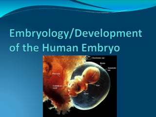
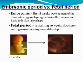
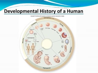
1. Embryology is the study of developmental events during prenatal stages, specifically the embryonic and fetal periods. The embryonic period is the first 8 weeks when the basic body plan takes shape, and the fetal period is the remaining 30 weeks when structures continue growing.
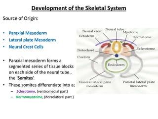
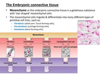
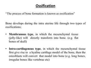
2. Musculoskeletal development begins with the formation of somites from paraxial mesoderm, which give rise to bones, cartilage, and muscles. Bones develop through membranous or endochondral ossification. Long bones are examples of endochondral ossification, forming cartilage models that are later replaced with bone.
3. Lim


![Development of the Muscular System [Human Embryology]](https://cdn.slidesharecdn.com/ss_thumbnails/developmentofthemuscularsystemhumanembryology-190416145251-thumbnail.jpg?width=640&height=640&fit=bounds)


