Download to read offline


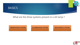
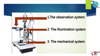


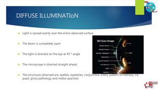

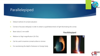
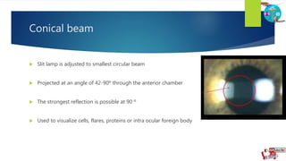
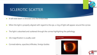


The document discusses the three main systems of a slit lamp: the observation system, illumination system, and mechanical system. It describes different illumination techniques used with a slit lamp including diffuse, direct, optic section, parallelepiped, conical beam, and sclerotic scatter illumination. Each technique illuminates the eye in a different way to examine different structures like the cornea, anterior chamber, or cells and proteins in the eye.


