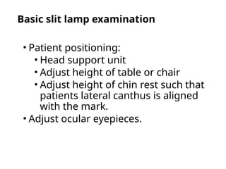
Basic slit lampexamination
• Patient positioning:
• Head support unit
• Adjust height of table or chair
• Adjust height of chin rest such that
patients lateral canthus is aligned
with the mark.
• Adjust ocular eyepieces.
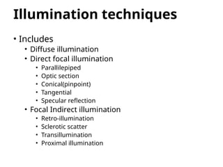
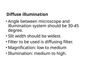
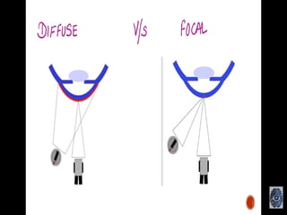
Diffuse illumination
• Anglebetween microscope and
illumination system should be 30-45
degree.
• Slit width should be widest.
• Filter to be used is diffusing filter.
• Magnification: low to medium
• Illumination: medium to high.
6.
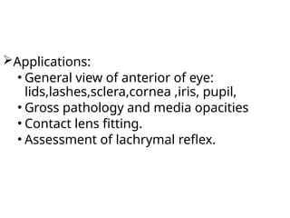
Applications:
• General viewof anterior of eye:
lids,lashes,sclera,cornea ,iris, pupil,
• Gross pathology and media opacities
• Contact lens fitting.
• Assessment of lachrymal reflex.
7.
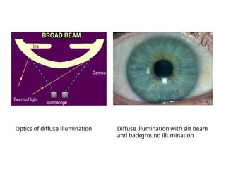
Optics of diffuseillumination Diffuse illumination with slit beam
and background illumination
p
8.
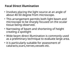
Focal Direct illumination
•Involves placing the light source at an angle of
about 40-50 degree from microscope.
• This arrangement permits both light beam and
microscope to be sharply focused on the ocular
tissue being observed.
• Narrowing of beam and shortening of height
creating a spotlight
• Wide beam direct illumination is commonly used
as a preliminary technique to evaluate large area.
• it is particularly suitable for assessment of
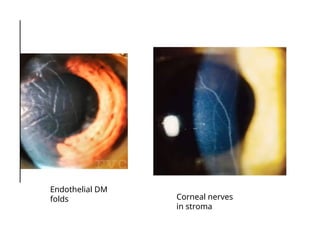
cataracts,scars,nerves,vessels etc.
10.
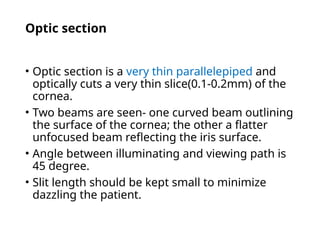
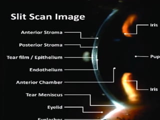
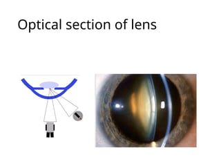
Optic section
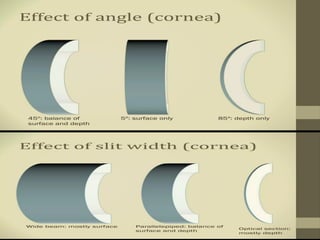
• Opticsection is a very thin parallelepiped and
optically cuts a very thin slice(0.1-0.2mm) of the
cornea.
• Two beams are seen- one curved beam outlining
the surface of the cornea; the other a flatter
unfocused beam reflecting the iris surface.
• Angle between illuminating and viewing path is
45 degree.
• Slit length should be kept small to minimize
dazzling the patient.
11.
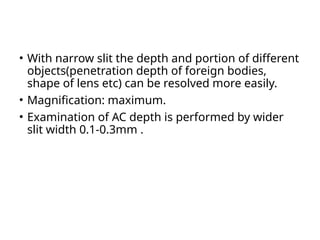
• With narrowslit the depth and portion of different
objects(penetration depth of foreign bodies,
shape of lens etc) can be resolved more easily.
• Magnification: maximum.
• Examination of AC depth is performed by wider
slit width 0.1-0.3mm .
12.
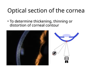
Optical section ofthe cornea
• To determine thickening, thinning or
distortion of corneal contour
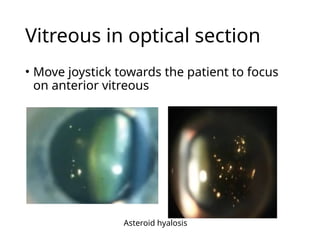
Vitreous in opticalsection
• Move joystick towards the patient to focus
on anterior vitreous
Asteroid hyalosis
16.
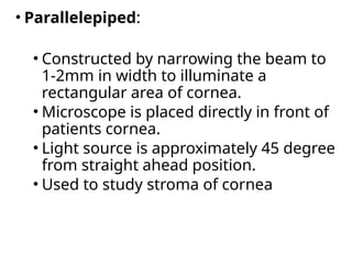
• Parallelepiped:
• Constructedby narrowing the beam to
1-2mm in width to illuminate a
rectangular area of cornea.
• Microscope is placed directly in front of
patients cornea.
• Light source is approximately 45 degree
from straight ahead position.
• Used to study stroma of cornea
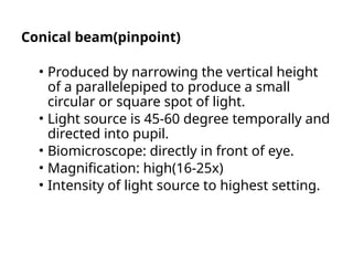
Conical beam(pinpoint)
• Producedby narrowing the vertical height
of a parallelepiped to produce a small
circular or square spot of light.
• Light source is 45-60 degree temporally and
directed into pupil.
• Biomicroscope: directly in front of eye.
• Magnification: high(16-25x)
• Intensity of light source to highest setting.
20.
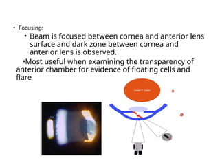
• Focusing:
• Beamis focused between cornea and anterior lens
surface and dark zone between cornea and
anterior lens is observed.
•Most useful when examining the transparency of
anterior chamber for evidence of floating cells and
flare seen in anterior uveitis.
21.
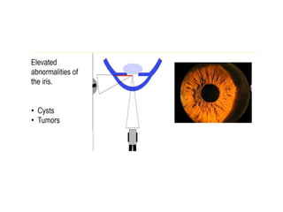
Tangential illumination
• Requiresthat the illumination arm and
the viewing arm be separated by 90
degree.
• Medium –wide beam of moderate height
is used.
• Microscope is pointing straight ahead.
• Magnification of 10x,16x,or 25x are
used.
22.
• Observe:
• Anteriorand posterior cornea
• Iris is best viewed without dilation
by this method.
• Anterior lens (especially useful for
viewing pseudoexfolation).
24.
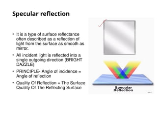
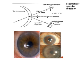
Specular reflection
• Itis a type of surface reflectance
often described as a reflection of
light from the surface as smooth as
mirror.
• All incident light is reflected into a
single outgoing direction (BRIGHT
DAZZLE)
• PRINCIPLE- Angle of incidence =
Angle of reflection
• Quality Of Reflection = The Surface
Quality Of The Reflecting Surface
25.
• Established byseparating the microscope and
slit beam by equal angles from normal to
cornea.
• Position of illuminator about 30 degree to one
side and the microscope 30 degree to otherside.
• Angle of illuminator to microscope must be
equal and opposite.
• Angle of light should be moved until a very
bright reflex obtained from corneal surface
which is called zone of specular reflection.
26.
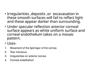
• Irregularities ,deposits,or excavasation in
these smooth surfaces will fail to reflect light
and these appear darker than surrounding.
• Under specular reflection anterior corneal
surface appears as white uniform surface and
corneal endothelium takes on a mosaic
pattern.
• Uses-
1. Movement of the lipid layer of the cornea
2. Tear meniscus
3. Irregularities on anterior cornea
4. Corneal endothelium
27.
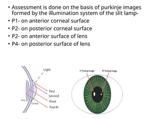
• Assessment isdone on the basis of purkinje images
formed by the illumination system of the slit lamp-
• P1- on anterior corneal surface
• P2- on posterior corneal surface
• P2- on anterior surface of lens
• P4- on posterior surface of lens