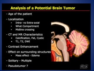
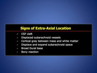
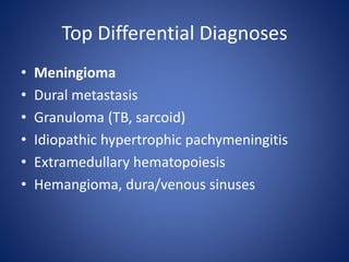
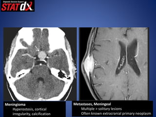
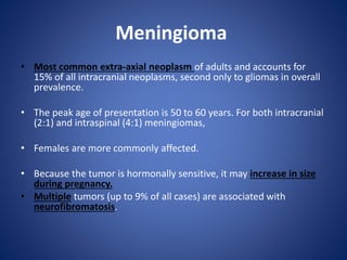
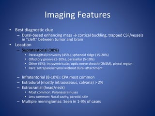
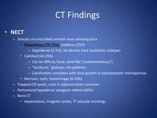
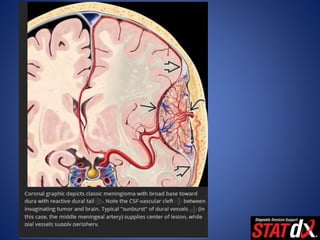
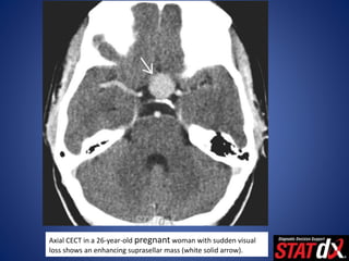
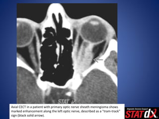
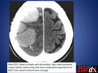
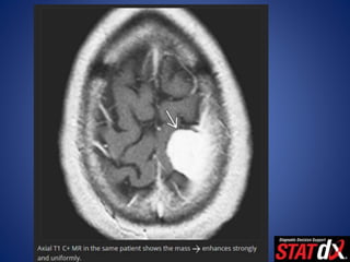
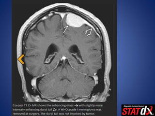
EXTRA-AXIAL MASSES
( TYPICAL MENINGIOMA )
BY:
DR. HASSAN ALQARNI
R1 RADIOLOGY RESIDENT
SAUDI BOARD
Top Differential Diagnoses
• Meningioma
• Dural metastasis
• Granuloma (TB, sarcoid)
• Idiopathic hypertrophic pachymeningitis
• Extramedullary hematopoiesis
• Hemangioma, dura/venous sinuses
•
Meningioma
Hyperostosis, cortical
irregularity, calcification
Metastases, Meningeal
Multiple > solitary lesions
Often known extracranial primary neoplasm
Tuberculosis
• Abnormal CXR, lab values
• Travel history to endemic areas,
immunocompromised
Hypertrophic Pachymeningitis
• localised inflammatory thickening of
the dura
Extramedullary Hematopoiesis
Patients with chronic anemia or marrow depletion
Multiple > solitary
Diagnosis :
Meningioma
Meningioma
• Most common extra-axial neoplasm of adults and accounts for
15% of all intracranial neoplasms, second only to gliomas in overall
prevalence.
• The peak age of presentation is 50 to 60 years. For both intracranial
(2:1) and intraspinal (4:1) meningiomas,
• Females are more commonly affected.
• Because the tumor is hormonally sensitive, it may increase in size
during pregnancy.
• Multiple tumors (up to 9% of all cases) are associated with
neurofibromatosis.
Pathology
• Arises from arachnoid cap cells with some probable
contribution from dural fi broblasts and pial cells.
• It is believed that intraventricular meningiomas arise
from arachnoidal cap cell rests buried within the
choroid plexus.
Clinical Presentation
• Most common signs/symptoms
– < 10% of all meningiomas are symptomatic
– Symptoms depend on tumor site
• Convexity/parasagittal = seizures, hemiparesis
• Basisphenoid, diaphragma sellae = visual field defects
• Cavernous sinus = cranial nerve deficit(s)
• Frontal = anosmia
Imaging Features
• Best diagnostic clue
– Dural-based enhancing mass → cortical buckling, trapped CSF/vessels
in "cleft" between tumor and brain
• Location
– Supratentorial (90%)
• Parasagittal/convexity (45%), sphenoid ridge (15-20%)
• Olfactory groove (5-10%), parasellar (5-10%)
• Other (5%): Intraventricular, optic nerve sheath (ONSM), pineal region
• Rare: Intraparenchymal without dural attachment
– Infratentorial (8-10%): CPA most common
– Extradural (mostly intraosseous, calvaria) > 2%
– Extracranial (head/neck)
• Most common: Paranasal sinuses
• Less common: Nasal cavity, parotid, skin
– Multiple meningiomas: Seen in 1-9% of cases
CT Findings
• NECT
– Sharply circumscribed smooth mass abutting dura
• Hyperdense (70-75%), isodense (25%)
– Hypodense (1-5%), fat density (rare lipoblastic subtype)
• Calcified (20-25%)
– Can be diffuse, focal, sand-like ("psammomatous")
– "Sunburst," globular, rim patterns
– Calcification correlates with slow growth in asymptomatic meningiomas
• Necrosis, cysts, hemorrhage (8-23%)
– Trapped CSF pools, cysts in adjacent brain common
– Peritumoral hypodense vasogenic edema (60%)
– Bone CT
• Hyperostosis, irregular cortex, ↑ vascular markings
• CECT
– > 90% enhance homogeneously, intensely
• CTA
– May be helpful prior to DSA, embolization
– Delineates arterial supply, venous drainage
Axial CECT in a 26-year-old pregnant woman with sudden visual
loss shows an enhancing suprasellar mass (white solid arrow).
Axial CECT in a patient with primary optic nerve sheath meningioma shows
marked enhancement along the left optic nerve, described as a "tram-track"
sign (black solid arrow).
Axial CECT shows multiple well-delineated, right-sided extraaxial
masses (white solid arrow) that were moderately hyperdense on
NECT (not shown) and enhance strongly.
MR Findings
• T1WI
– Typically iso- to slightly hypointense with cortex
– Necrosis, cysts, hemorrhage (8-23%)
– Look for gray matter "buckling"
• T2WI
– Variable ("sunburst" pattern may be evident)
– 8-23% of intratumoral cysts (common; can be almost microcystic),
hemorrhage (rare)
– Best sequence for
• Visualizing CSF/vascular cleft between tumor, brain (80%)
• Identifying vascular flow voids (80%)
• Predicting "hard" meningioma at surgery (hypointense)
• FLAIR
– Hyperintense peritumoral edema, dural tail
• DWI
– DWI, ADC maps for TM variable in appearance
– Lower ADC in MM and AM compared to TM
• T1WI C+
– > 95% enhance homogeneously, intensely
– Dural tail (35-80% of cases) nonspecific
• Other neoplasms (schwannoma, adenoma, metastases), nonneoplastic
dural-based masses
– En plaque: Sessile thickened enhancing dura
• MRV
– Evaluate sinus involvement
• MRS
– Elevated levels of alanine at short TE
• Triplet-like spectral pattern at 1.3-1.5 ppm (overlapping of Ala, Lac)
• Elevated Glx alfa/glutathione
Angiographic Findings
– "Sunburst" or radial appearance
• Dural vessels supply lesion core
• Pial vessels may be parasitized, supply periphery
– Prolonged vascular "stain"
– Venous phase vital to evaluate sinus involvement
Thank you

extra axial Meningioma

  • 1.
    EXTRA-AXIAL MASSES ( TYPICALMENINGIOMA ) BY: DR. HASSAN ALQARNI R1 RADIOLOGY RESIDENT SAUDI BOARD
  • 5.
    Top Differential Diagnoses •Meningioma • Dural metastasis • Granuloma (TB, sarcoid) • Idiopathic hypertrophic pachymeningitis • Extramedullary hematopoiesis • Hemangioma, dura/venous sinuses
  • 6.
    • Meningioma Hyperostosis, cortical irregularity, calcification Metastases,Meningeal Multiple > solitary lesions Often known extracranial primary neoplasm
  • 7.
    Tuberculosis • Abnormal CXR,lab values • Travel history to endemic areas, immunocompromised Hypertrophic Pachymeningitis • localised inflammatory thickening of the dura
  • 8.
    Extramedullary Hematopoiesis Patients withchronic anemia or marrow depletion Multiple > solitary
  • 9.
  • 10.
    Meningioma • Most commonextra-axial neoplasm of adults and accounts for 15% of all intracranial neoplasms, second only to gliomas in overall prevalence. • The peak age of presentation is 50 to 60 years. For both intracranial (2:1) and intraspinal (4:1) meningiomas, • Females are more commonly affected. • Because the tumor is hormonally sensitive, it may increase in size during pregnancy. • Multiple tumors (up to 9% of all cases) are associated with neurofibromatosis.
  • 11.
    Pathology • Arises fromarachnoid cap cells with some probable contribution from dural fi broblasts and pial cells. • It is believed that intraventricular meningiomas arise from arachnoidal cap cell rests buried within the choroid plexus.
  • 12.
    Clinical Presentation • Mostcommon signs/symptoms – < 10% of all meningiomas are symptomatic – Symptoms depend on tumor site • Convexity/parasagittal = seizures, hemiparesis • Basisphenoid, diaphragma sellae = visual field defects • Cavernous sinus = cranial nerve deficit(s) • Frontal = anosmia
  • 13.
    Imaging Features • Bestdiagnostic clue – Dural-based enhancing mass → cortical buckling, trapped CSF/vessels in "cleft" between tumor and brain • Location – Supratentorial (90%) • Parasagittal/convexity (45%), sphenoid ridge (15-20%) • Olfactory groove (5-10%), parasellar (5-10%) • Other (5%): Intraventricular, optic nerve sheath (ONSM), pineal region • Rare: Intraparenchymal without dural attachment – Infratentorial (8-10%): CPA most common – Extradural (mostly intraosseous, calvaria) > 2% – Extracranial (head/neck) • Most common: Paranasal sinuses • Less common: Nasal cavity, parotid, skin – Multiple meningiomas: Seen in 1-9% of cases
  • 14.
    CT Findings • NECT –Sharply circumscribed smooth mass abutting dura • Hyperdense (70-75%), isodense (25%) – Hypodense (1-5%), fat density (rare lipoblastic subtype) • Calcified (20-25%) – Can be diffuse, focal, sand-like ("psammomatous") – "Sunburst," globular, rim patterns – Calcification correlates with slow growth in asymptomatic meningiomas • Necrosis, cysts, hemorrhage (8-23%) – Trapped CSF pools, cysts in adjacent brain common – Peritumoral hypodense vasogenic edema (60%) – Bone CT • Hyperostosis, irregular cortex, ↑ vascular markings
  • 15.
    • CECT – >90% enhance homogeneously, intensely • CTA – May be helpful prior to DSA, embolization – Delineates arterial supply, venous drainage
  • 17.
    Axial CECT ina 26-year-old pregnant woman with sudden visual loss shows an enhancing suprasellar mass (white solid arrow).
  • 18.
    Axial CECT ina patient with primary optic nerve sheath meningioma shows marked enhancement along the left optic nerve, described as a "tram-track" sign (black solid arrow).
  • 19.
    Axial CECT showsmultiple well-delineated, right-sided extraaxial masses (white solid arrow) that were moderately hyperdense on NECT (not shown) and enhance strongly.
  • 20.
    MR Findings • T1WI –Typically iso- to slightly hypointense with cortex – Necrosis, cysts, hemorrhage (8-23%) – Look for gray matter "buckling" • T2WI – Variable ("sunburst" pattern may be evident) – 8-23% of intratumoral cysts (common; can be almost microcystic), hemorrhage (rare) – Best sequence for • Visualizing CSF/vascular cleft between tumor, brain (80%) • Identifying vascular flow voids (80%) • Predicting "hard" meningioma at surgery (hypointense)
  • 21.
    • FLAIR – Hyperintenseperitumoral edema, dural tail • DWI – DWI, ADC maps for TM variable in appearance – Lower ADC in MM and AM compared to TM
  • 22.
    • T1WI C+ –> 95% enhance homogeneously, intensely – Dural tail (35-80% of cases) nonspecific • Other neoplasms (schwannoma, adenoma, metastases), nonneoplastic dural-based masses – En plaque: Sessile thickened enhancing dura • MRV – Evaluate sinus involvement • MRS – Elevated levels of alanine at short TE • Triplet-like spectral pattern at 1.3-1.5 ppm (overlapping of Ala, Lac) • Elevated Glx alfa/glutathione
  • 28.
    Angiographic Findings – "Sunburst"or radial appearance • Dural vessels supply lesion core • Pial vessels may be parasitized, supply periphery – Prolonged vascular "stain" – Venous phase vital to evaluate sinus involvement
  • 30.