Downloaded 29 times
![Radiology for Radiation Oncologists
Skull base radiology
Dr Kanhu Charan Patro
MD,DNB[RADIATION ONCOLOGY],MBA,CEPC,PDCR
HOD, Radiation Oncology
MGCHRI, Visakhapatnam, INDIA
1](https://image.slidesharecdn.com/skullbaseradiology-200709161512/85/Skull-base-radiology-FOR-RADIATION-ONCOLOGISTS-1-320.jpg)
![Radiology for Radiation Oncologists
Skull base radiology
Dr Kanhu Charan Patro
MD,DNB[RADIATION ONCOLOGY],MBA,CEPC,PDCR
HOD, Radiation Oncology
MGCHRI, Visakhapatnam, INDIA
1](https://image.slidesharecdn.com/skullbaseradiology-200709161512/75/Skull-base-radiology-FOR-RADIATION-ONCOLOGISTS-1-2048.jpg)

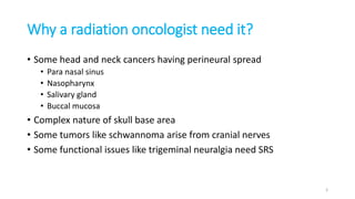
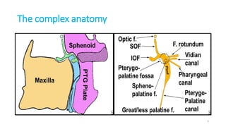
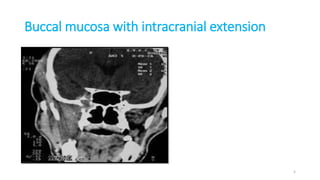
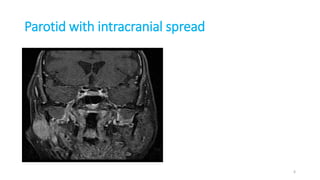
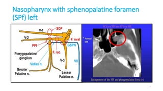
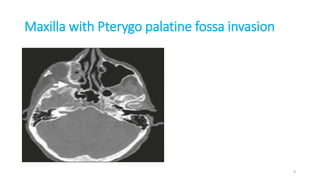

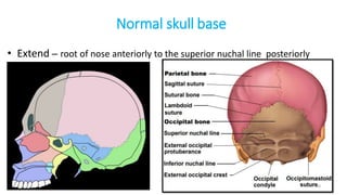
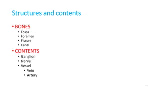


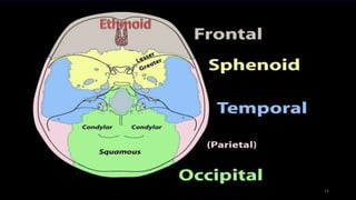
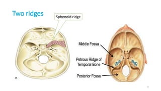
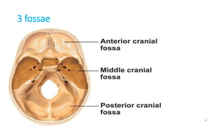
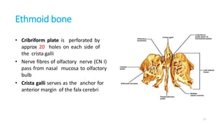
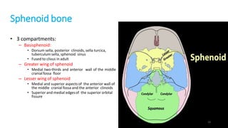
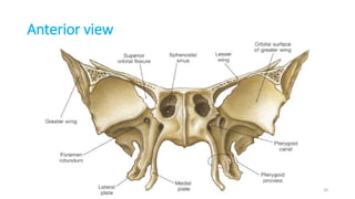
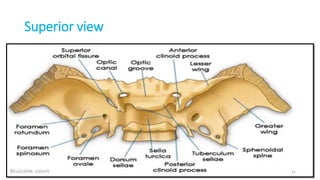
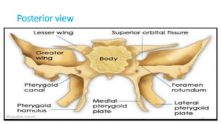
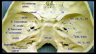
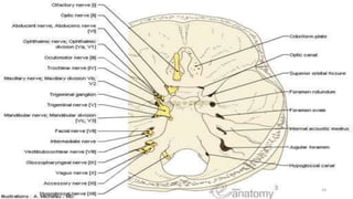
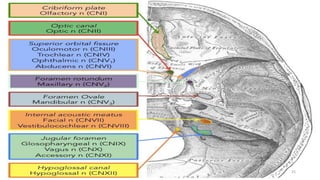
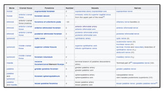
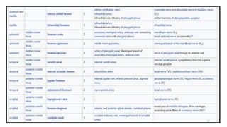
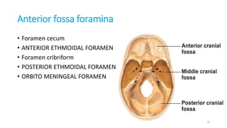
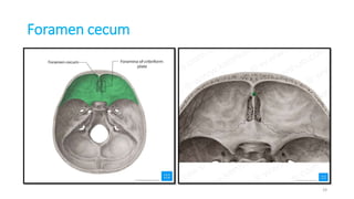
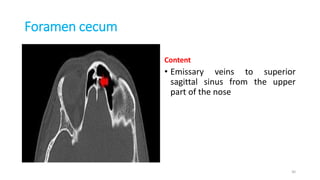
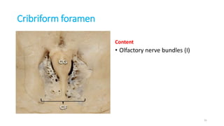
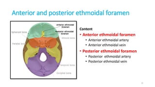
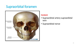
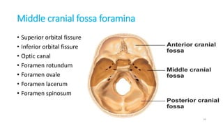
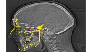
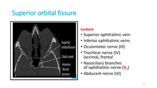
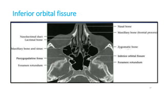
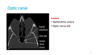
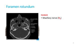
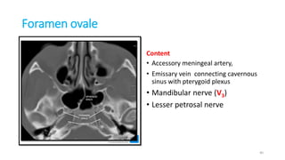
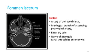
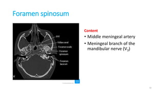
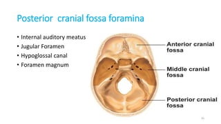
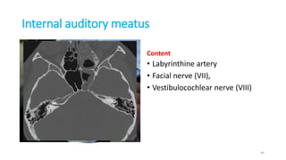
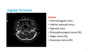
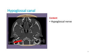
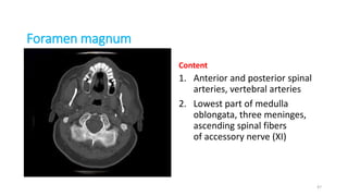
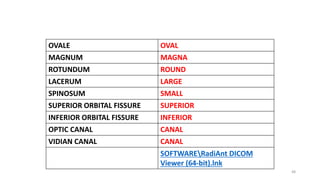
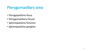
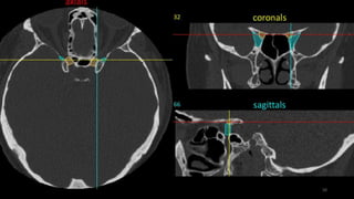
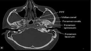
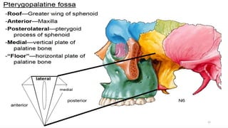
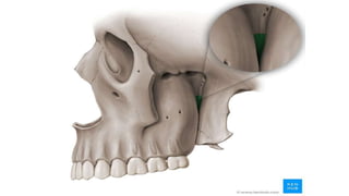
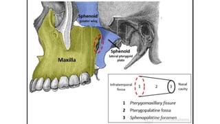
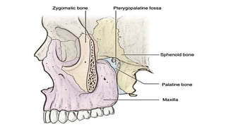
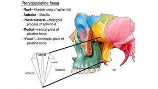
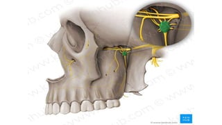
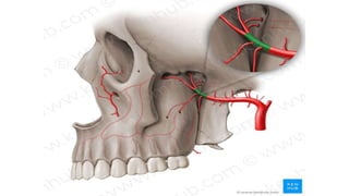
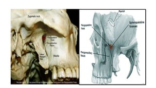
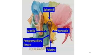
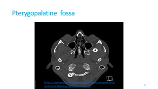
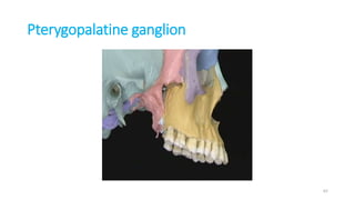
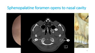
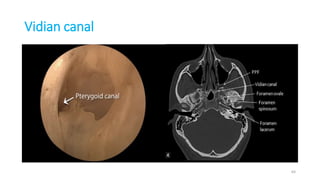
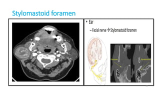
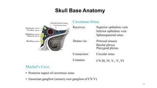
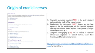
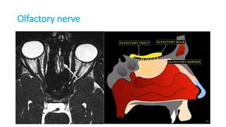
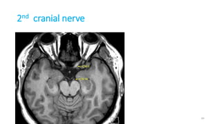
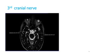
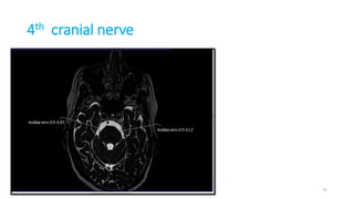
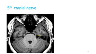
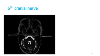
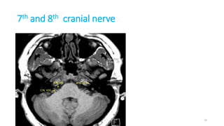
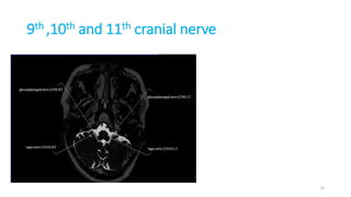
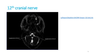

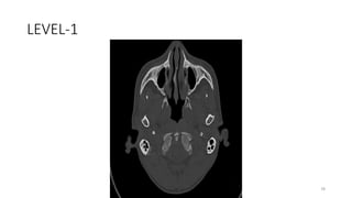
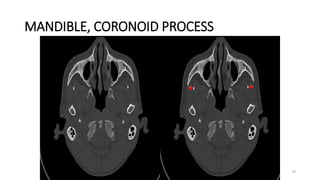
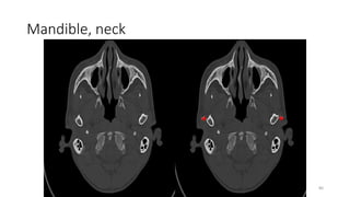
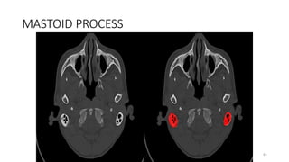
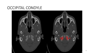
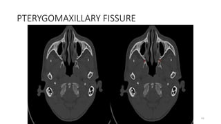
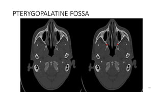
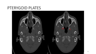
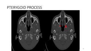
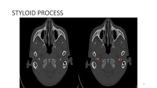
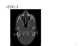

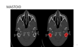
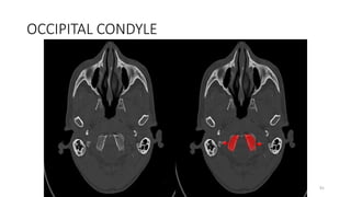
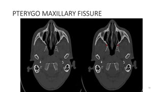
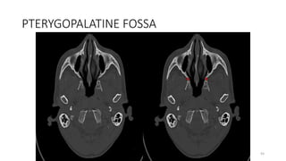
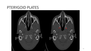
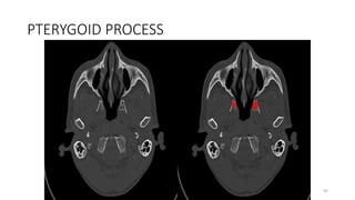
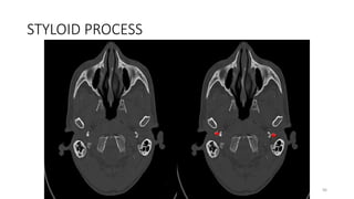
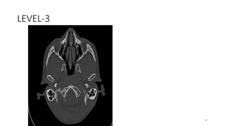
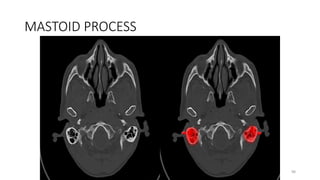
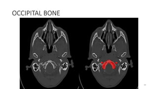
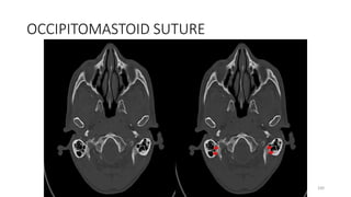
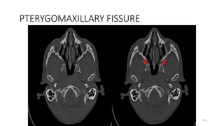
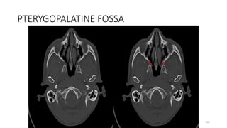
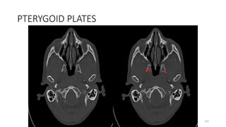

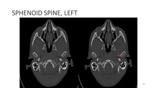
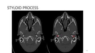
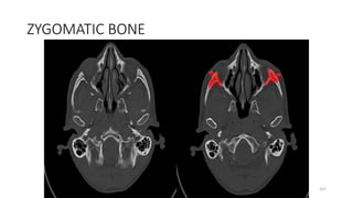
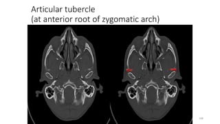
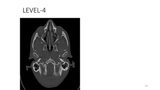
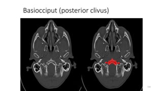
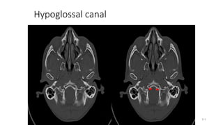
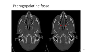
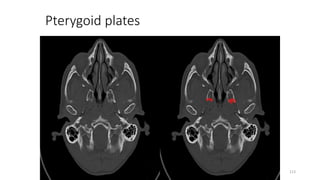
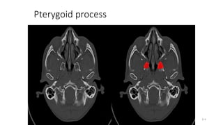

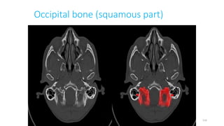

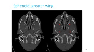

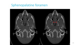
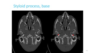
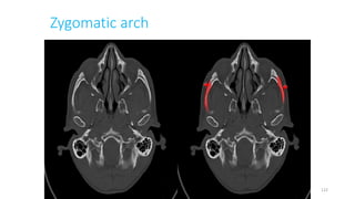
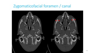
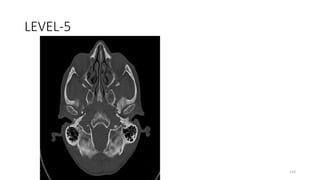
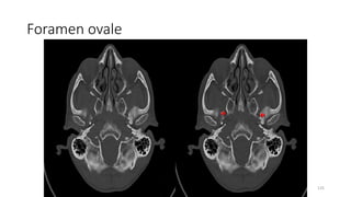
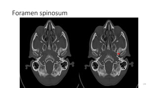
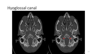
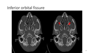
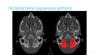
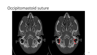
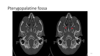
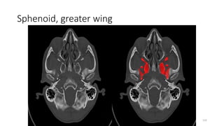
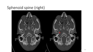
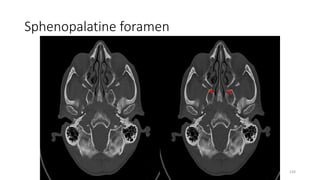
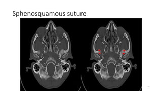
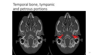
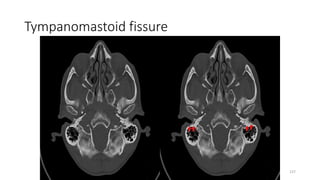

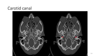
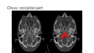
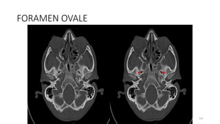

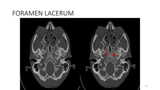
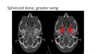

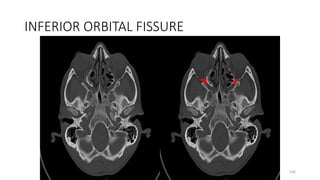
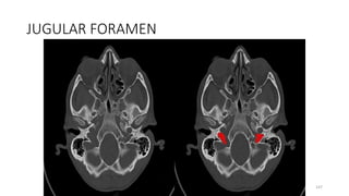
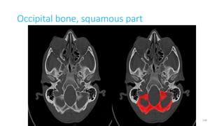
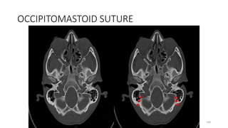
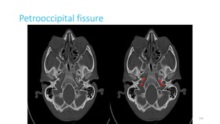
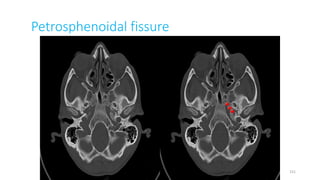
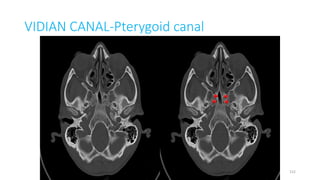
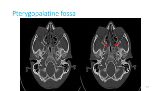
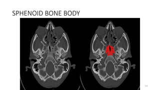
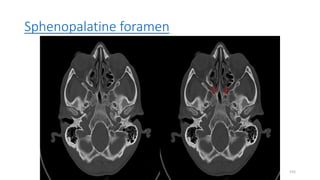

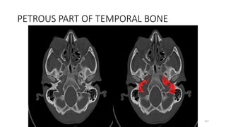
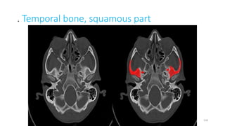
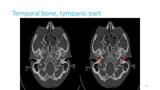
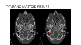
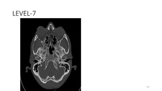
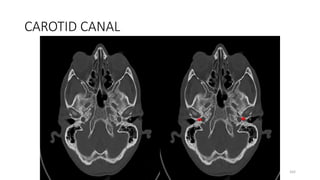
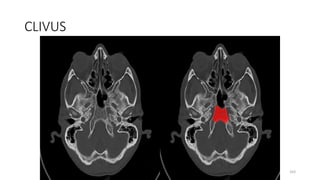
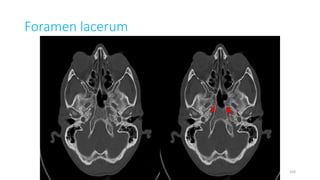
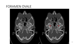
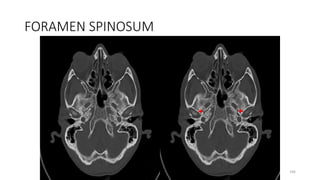
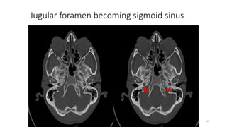
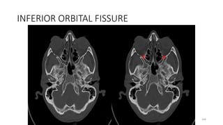
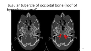
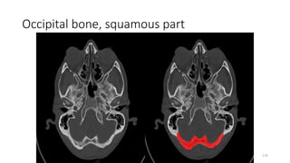
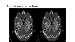


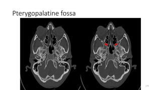
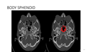
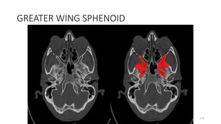
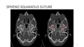
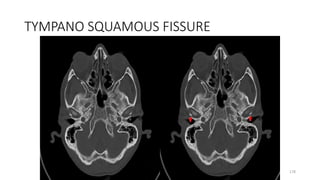
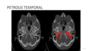
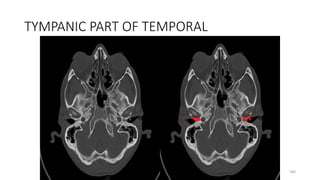
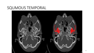


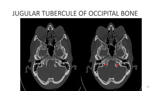
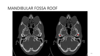

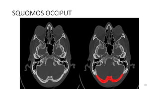
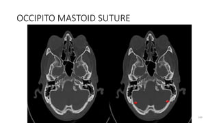
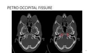
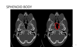
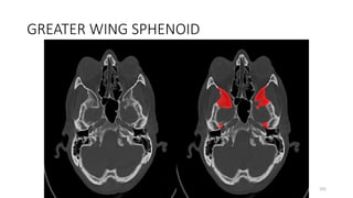
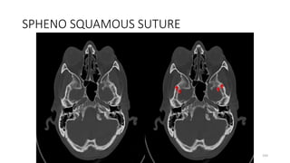
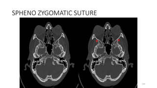
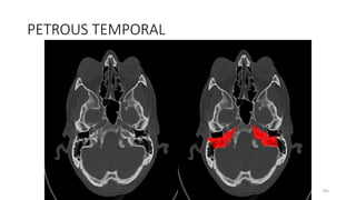

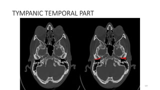
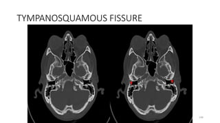
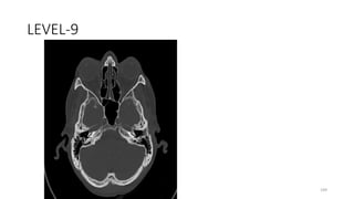
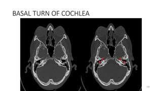
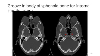
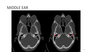
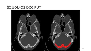
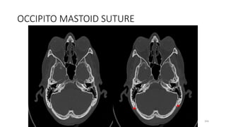
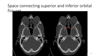
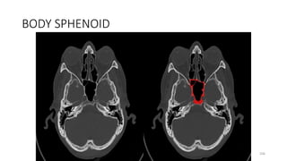


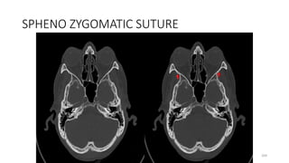
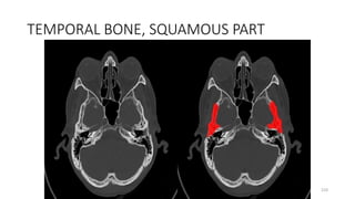
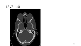
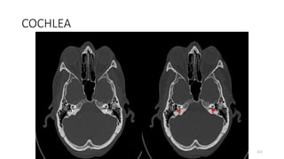
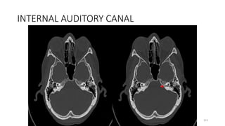
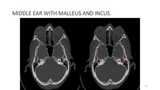
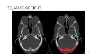
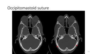
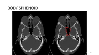
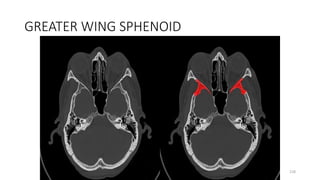
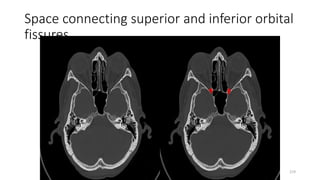
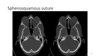
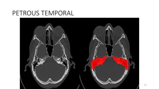

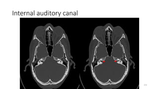

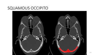
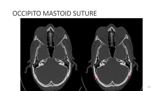
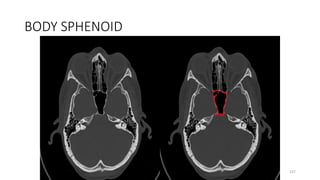
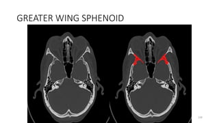
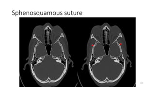
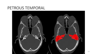
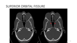
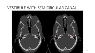
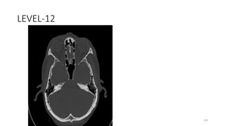
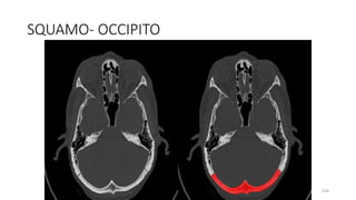
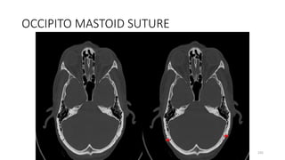
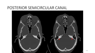
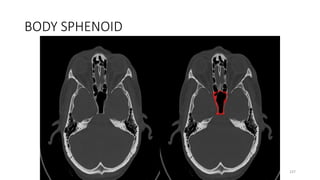
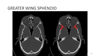
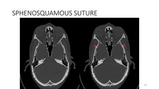


This document provides an overview of skull base radiology for radiation oncologists. It discusses the complex anatomy of the skull base area and lists the important structures and contents, including bones, foramina, fissures, canals, ganglia, nerves, vessels, and more. It highlights key areas like the anterior, middle, and posterior cranial fossae and provides labeled images to illustrate important structures like foramina, canals, and the origins and paths of cranial nerves. Slice-by-slice views from different skull base levels are also included to demonstrate the anatomy in detail.


![CTEV [ clubfoot] DR ARUN LAL ,DR MOHAMED ASHRAF travancore medical college k...](https://cdn.slidesharecdn.com/ss_thumbnails/ctevclubfootdrarunlaldrmohamedashraftravancoremedicalcollegekollamkeralaindia-260208063247-18fc466c-thumbnail.jpg?width=640&height=640&fit=bounds)
![ONFH[AVN HIP] -TRIPLE REGIME -A NOVAL SURGICAL CONCEPT .pptx](https://cdn.slidesharecdn.com/ss_thumbnails/onfhavnhip2026koaconcalicutdrgokuldevdrmashraf-260210064517-213ec005-thumbnail.jpg?width=640&height=640&fit=bounds)


