Downloaded 2,171 times
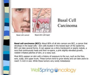
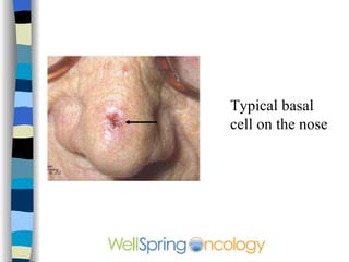
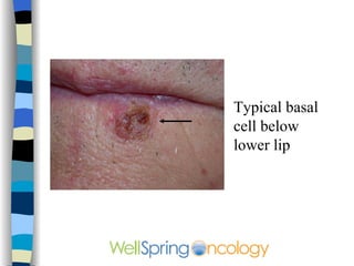
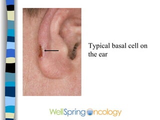
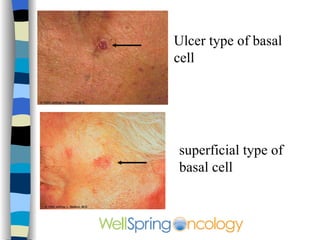
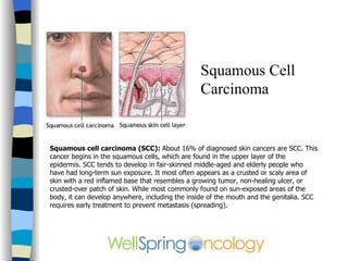
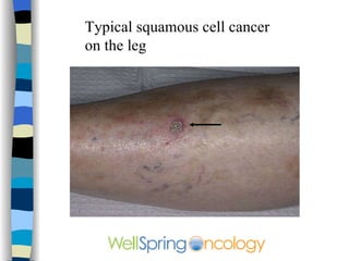

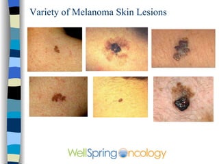
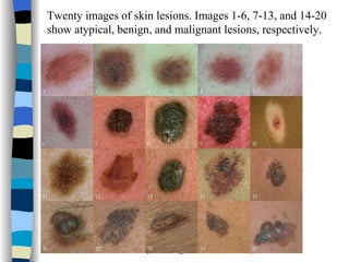
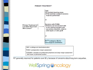
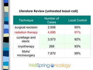
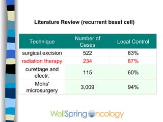
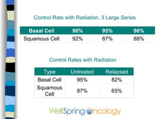
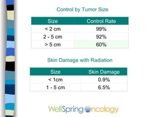
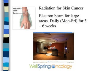
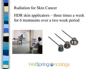
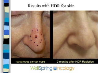
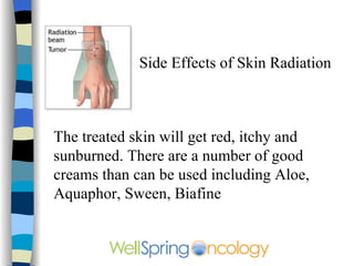
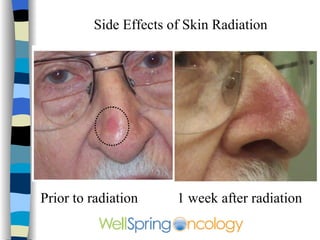


The document discusses different types of skin cancer including basal cell carcinoma, squamous cell carcinoma, and melanoma. It provides details on symptoms, treatment options like surgery, radiation therapy, and Mohs microsurgery. Radiation therapy is described as an effective non-surgical treatment option that results in mild sunburn-like side effects and healing of the skin within a few weeks. Images are included showing examples of different skin cancers and results after radiation treatment.


![CTEV [ clubfoot] DR ARUN LAL ,DR MOHAMED ASHRAF travancore medical college k...](https://cdn.slidesharecdn.com/ss_thumbnails/ctevclubfootdrarunlaldrmohamedashraftravancoremedicalcollegekollamkeralaindia-260208063247-18fc466c-thumbnail.jpg?width=640&height=640&fit=bounds)


