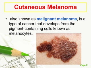
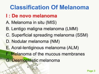
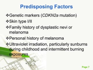
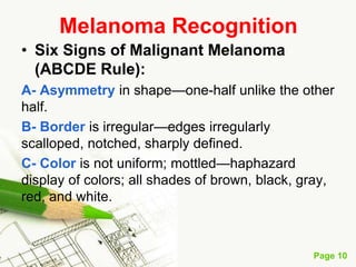
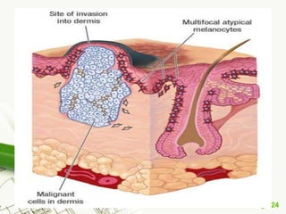
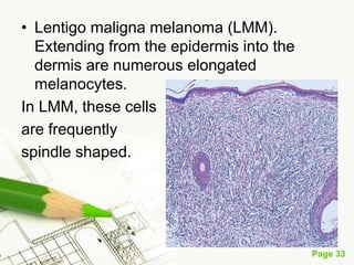
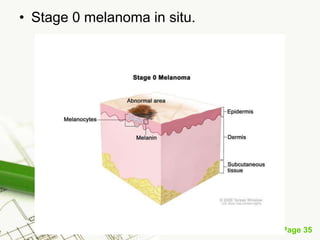
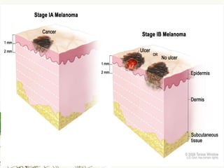
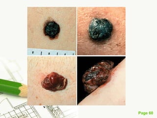
Melanoma
Cutaneous Melanoma
also known as malignant melanoma, is a type of cancer that develops from the pigment-containing cells known as melanocytes.
Classification Of Melanoma
I : De novo melanoma
A. Melanoma in situ (MIS)
B. Lentigo maligna melanoma (LMM)
C. Superficial spreading melanoma (SSM)
D. Nodular melanoma (NM)
E. Acral-lentiginous melanoma (ALM)
F. Melanoma of the mucous membranes
G. Desmoplastic melanoma
II Melanoma arising from precursors
Melanoma arising in dysplastic nevomelanocytic nevi
B. Melanoma arising in congenital nevomelanocytic nevi
C. Melanoma arising in common NMN
Etiology And Pathogenesis
The etiology and pathogenesis of cutaneous melanoma are unknown.
Epidemiologic studies demonstrate a role for genetic predisposition and sun exposure in melanoma development.
The major genes involved in melanoma development reside on chromosome 9p21.
Etiology
UVR, mostly of the UVB spectrum (290–320 nm) that induces mutations in suppressor genes. The propensity for multiple BCC may be inherited. Associated with mutations in the PTCH gene in many cases.
Predisposing Factors
Genetic markers (CDKN2a mutation)
Skin type I/II
Family history of dysplastic nevi or melanoma
Personal history of melanoma
Ultraviolet irradiation, particularly sunburns during childhood and intermittent burning exposures
Number (>50) and size (>5 mm) of melanocytic nevi
Congenital nevi
Number of dysplastic nevi (>5)
Dysplastic melanocytic nevus syndrome
Immune suppression (debatable)
Number (>50) and size (>5 mm) of melanocytic nevi
Congenital nevi
Number of dysplastic nevi (>5)
Dysplastic melanocytic nevus syndrome
Immune suppression (debatable)
Six Signs of Malignant Melanoma (ABCDE Rule):
A- Asymmetry in shape—one-half unlike the other half.
B- Border is irregular—edges irregularly scalloped, notched, sharply defined.
C- Color is not uniform; mottled—haphazard display of colors; all shades of brown, black, gray, red, and white.
D- Diameter is usually large.
E- Elevation is almost always present and is irregular—surface distortion is assessed by side-lighting. others use E for Enlargement— a history of an increase in the size of lesion is one of the most important signs of malignant melanoma.
Lentigo Maligna Melanoma (LMM)
2. Page 2
Cutaneous Melanoma
• also known as malignant melanoma, is a
type of cancer that develops from the
pigment-containing cells known as
melanocytes.
3. Page 3
Classification Of Melanoma
I : De novo melanoma
A. Melanoma in situ (MIS)
B. Lentigo maligna melanoma (LMM)
C. Superficial spreading melanoma (SSM)
D. Nodular melanoma (NM)
E. Acral-lentiginous melanoma (ALM)
F. Melanoma of the mucous membranes
G. Desmoplastic melanoma
4. Page 4
II Melanoma arising from precursors
A. Melanoma arising in dysplastic
nevomelanocytic nevi
B. Melanoma arising in congenital
nevomelanocytic nevi
C. Melanoma arising in common NMN
5. Page 5
Etiology And Pathogenesis
• The etiology and pathogenesis of
cutaneous melanoma are unknown.
• Epidemiologic studies demonstrate a role
for genetic predisposition and sun
exposure in melanoma development.
• The major genes involved in melanoma
development reside on chromosome 9p21.
6. Page 6
Etiology
• UVR, mostly of the UVB spectrum (290–320 nm)
that induces mutations in suppressor genes. The
propensity for multiple BCC may be inherited.
Associated with mutations in the PTCH gene in
many cases.
7. Page 7
Predisposing Factors
Genetic markers (CDKN2a mutation)
Skin type I/II
Family history of dysplastic nevi or
melanoma
Personal history of melanoma
Ultraviolet irradiation, particularly sunburns
during childhood and intermittent burning
exposures
8. Page 8
Predisposing Factors cont..
Number (>50) and size (>5 mm) of
melanocytic nevi
Congenital nevi
Number of dysplastic nevi (>5)
Dysplastic melanocytic nevus syndrome
Immune suppression (debatable)
9. Page 9
Data And Facts
• Melanoma represents 5% of all cancers by
incidence in males and 6% in females.
• Most frequent sites
Whites
• Male: back, upper extremities.
• Female: back, lower legs.
Blacks and Asians: soles, mucous
membranes, palms, nail beds.
10. Page 10
Melanoma Recognition
• Six Signs of Malignant Melanoma
(ABCDE Rule):
A- Asymmetry in shape—one-half unlike the other
half.
B- Border is irregular—edges irregularly
scalloped, notched, sharply defined.
C- Color is not uniform; mottled—haphazard
display of colors; all shades of brown, black, gray,
red, and white.
11. Page 11
D- Diameter is usually large.
E- Elevation is almost always present and
is irregular—surface distortion is assessed
by side-lighting. others use E for
Enlargement— a history of an increase in
the size of lesion is one of the most
important signs of malignant melanoma.
12. Page 12
Clinical Manifestation
• The clinical characteristics of the four
major types of melanoma are summarized
in Table ليجوه .
• Also discussed in this section are
melanoma in situ and desmoplastic
melanoma
14. Page 14
Melanoma In Situ (MIS)
• The clinical features of MIS are not always
clearly presented.
the term is used when melanoma cells are
• confined to the epidermis, above the basement
membrane;
• basilar melanocytic atypia, hyperplasia, and
spread either occur in single-file alignment along
the basal membrane or are distributed
throughout the epidermis (pagetoid spread)
17. Page 17
2- or a macule with barely perceptible
elevation with irregular borders and marked
variegation of color: brown, dark brown, and black
or reddish tones but without gray or blue
18. Page 18
• The clinical correlations of MIS are lentigo
maligna and flat superficial spreading
melanoma and these are discussed in
the respective sections below.
19. Page 19
Lentigo Maligna Melanoma
(LMM)
• The clinical features of MIS are not always
clearly presented.
the term is used when melanoma cells are
• confined to the epidermis, above the basement
membrane;
• basilar melanocytic atypia, hyperplasia, and
spread either occur in single-file alignment along
the basal membrane or are distributed
throughout the epidermis (pagetoid spread)
20. Page 20
Epidemiology
• Age of Onset: Median age 65.
• Sex Equal incidence in males and
females.
• Race Rare in brown- (e.g., Asians, East
• Indians) and extremely rare in black
skinned (African Americans and Africans)
persons.
• Highest incidence in whites and skin
phototypes I, II, and III.
21. Page 21
Epidemiology
• Incidence 5% of primary cutaneous
melanomas.
• Predisposing Factors:
Same factors as in sun induced non
melanoma skin cancer: older population,
outdoor occupations (farmers, sailors,
construction workers).
22. Page 22
Pathogenesis
• In contrast to SSM and NM, which appear
to be related to intermittent high-intensity
sun exposure and occur on the
intermittently exposed areas (back and
legs) of young or middle-aged adults, LM
and LMM occur on the face, neck, and
dorsa of the forearms or hands
23. Page 23
Pathogenesis
; furthermore, LM and LMM occur almost
always in older persons with evidence of
heavily sun damaged skin (dermatoheliosis).
The evolution of the lesion most clearly
reveals the transition from the radial to the
vertical growth phase and from a clinically
recognizable MIS to invasive melanoma
25. Page 25
Clinical Manifestation
• LMM very slowly evolves from LM over a
period of several years, sometimes up to
20 years. There is practically always a
background of dermatoheliosis.
26. Page 26
Skin Lesions Lentigo Maligna
• Uniformly flat, macule ; 0.5 cm or larger,
up to 20 cm.
27. Page 27
• Lentigo maligna A very large lentigo maligna on the right
cheek with the typical variegation in color (tan, brown, black)
and highly irregular shape. The lesion is flat, macular, and
thus represents in situ melanoma.
28. Page 28
• The classically macular lentigo maligna is highly
irregular in shape and variegated in color. However,
there is a bluish component and a large pink nodule in
the infraorbital region, indicating a switch from the radial
to the vertical growth phase and thus invasiveness: the
lesion is now called lentigo maligna melanoma.
29. Page 29
• Lentigo Maligna Melanoma The clinical change
that indicates the transition of LM to LMM is the
appearance of variegated red, white, and blue
and of papules, plaques, or nodules
30. Page 30
Thus LMM is the same as LM plus (1)
gray areas (indicate focal regression), and
blue areas [indicating dermal pigment
(melanocytes or melanin)], and
• (2) papules or nodules, which may be
blue, black, or pink
31. Page 31
Distribution
• Single isolated lesion on the sun exposed
areas:
Forehead,
Nose,
Cheeks,
Neck,
Forearms,
And dorsa of hands;
Rarely on lower legs.
32. Page 32
Laboratory Examination
• Dermatopathology : LM shows increased
numbers of atypical melanocytes distributed in a
single layer along the basal layer and above the
basement membrane of an epidermis that
shows elongation of rete ridges.
33. Page 33
• Lentigo maligna melanoma (LMM).
Extending from the epidermis into the
dermis are numerous elongated
melanocytes.
In LMM, these cells
are frequently
spindle shaped.
34. Page 34
• Atypical melanocytes are usually singly
dispersed but may also aggregate to small
nests and extend into the hair follicles,
reaching the middermis, even in the
preinvasive stage of LM. In LMM, they
invade the dermis (vertical growth phase)
and expand into the deeper tissues
37. Page 37
Stage I melanoma
• . In stage IA, the tumor is not more than 1
millimeter thick, with no ulceration (break
in the skin). In stage IB, the tumor is either
not more than 1 millimeter thick, with
ulceration, OR more than 1 but not more
than 2 millimeters thick, with no ulceration.
Skin thickness is different on different
parts of the body.
40. Page 40
Stage II melanoma
• In stage IIA, the tumor is either more than 1 but
not more than 2 millimeters thick, with ulceration
(break in the skin), OR it is more than 2 but not
more than 4 millimeters thick, with no ulceration.
In stage IIB, the tumor is either more than 2 but
not more than 4 millimeters thick, with ulceration,
OR it is more than 4 millimeters thick, with no
ulceration. In stage IIC, the tumor is more than 4
millimeters thick, with ulceration. Skin thickness
is different on different parts of the body.
42. Page 42
Stage III melanoma
The tumor may be any thickness, with or without
ulceration (a break in the skin), and (a) cancer has
spread to one or more lymph nodes; (b) lymph
nodes with cancer may be joined together
(matted); (c) cancer may be in a lymph vessel
between the primary tumor and nearby lymph
nodes; and/or (d) very small tumors may be found
on or under the skin, not more than 2 centimeters
away from the primary tumor.
43. Page 43
Stage IV melanoma. The cancer has
spread to other parts of the body,
such as the brain, lung, liver, lymph
nodes, small intestine, and bone.
44. Page 44
Stage IV melanoma
• In stage IV, the cancer has spread to other
places in the body, such as the lung, liver,
brain, bone, soft tissue, or gastrointestinal
(GI) tract. Cancer may have spread to
places in the skin far away from where it
first started.
45. Page 45
Management
1. Very early LM lesions: Imiquimod.
2. Excise with 1-cm beyond the clinically
visible lesion where possible and provided
the flat component does not involve a major
organ. Use of Wood lamp and dermoscopy
help in defining borders.
3. Sentinel node to be done in lesions >1.0
mm in terms of thickness.
47. Page 47
Epidemiology
• Age of Onset 30 to 50 (median, 37) years
of age.
• Sex Slightly higher incidence in females.
• Race In world surveys, white-skinned
persons overwhelmingly predominate.
48. Page 48
Epidemiology
• Incidence SSM constitutes 70% of all
melanomas arising in white persons.
• Predisposing Factors:
• In order of importance these are presence
of precursor lesions , family history,
light skin color (skin phototypes I and II);
and sunburns,
49. Page 49
Skin Lesions
• SSM is the lesion to which the ABCDE
rule .
• Initially a very flat plaque 5–12 mm or
smaller
50. Page 50
• Dark brown, black, with admixture of pink,
gray, and blue-gray hues—with marked
variegation and a haphazard pattern.
White areas indicate regressed portions
51. Page 51
• An SSM is thus a flat plaque with all
shades of brown to black plus the
American flag or the tricolore (red, blue,
white)
52. Page 52
• As the vertical growth phase progresses,
nodules appear; eventually erosions and
even superficial ulceration develop
53. Page 53
Distribution
• Back (males and females); legs (females,
between knees and ankles); anterior trunk
and legs in males lesions on covered
areas, e.g., buttocks, lower abdomen
54. Page 54
Laboratory Examination
• Dermatopathology
• Malignant melanocytes expand in a
pagetoid pattern Superficial papillary body
of the dermis—the radial growth phase.
• In the vertical growth phase, presenting
clinically as small nodules, they expand
further into the reticular dermis
55. Page 55
Course And Prognosis
• Left untreated, SSM develops deep
invasion (vertical growth) over months to
years
57. Page 57
Nodular Melanoma
Epidemiology
• Age of Onset Middle life.
• Sex Equal incidence in males and females
• Race NM occurs in all races,
• Incidence NM constitutes 15%
58. Page 58
Pathogenesis
• Both SSM and NM occur in approximately
the same sites (upper back in males, lower
legs in females),
59. Page 59
Clinical Manifestation
• Skin Lesions Uniformly elevated “blueberrylike”
nodule or ulcerated or “thick” plaque; may
become polypoid. Uniformly dark blue, black, or
“thundercloud”gray Early lesions are 1–3 cm in
size but may grow much larger if undetected.
Oval or round, usually with smooth, not irregular,
borders, as in all other types of melanoma.
Sharply defined, may be pedunculated
61. Page 61
Distribution
• Back (males and females); legs (females,
between knees and ankles); anterior trunk
and legs in males lesions on covered
areas, e.g., buttocks, lower abdomen
62. Page 62
Laboratory Examinations
• Dermatopathology Malignant
melanocytes,which appear as epithelioid,
spindle, or small atypical cells, show little
lateral (radial) growth within and below the
epidermis and invade vertically into the
dermis and underlying subcutaneous fat
64. Page 64
• Serology Serum levels of S-100 beta and
melanoma-inhibiting activity (MIA), S-
cysteinyldopa, and lactate dehydrogenase
(LDH) levels are markers for advanced
melanoma patie
65. Page 65
Differential Diagnosis
• Blue/Black Papule/Nodule NM can be
confusedwith hemangioma (long history)
and pyogenic granuloma (short history—
weeks) sometimes almost
indistinguishable from pigmented basal
cell carcinoma
66. Page 66
Desmoplastic Melanoma (Dm)
• The term desmoplasia refers to connective
tissue proliferation and, when applied to
malignant melanoma, describes (1) a dermal
fibroblastic component of melanoma with only
minimal melanocytic proliferation at the dermal-
epidermal junction; (2) nerve-centered
superficial malignant melanoma with or without
an atypical intraepidermal melanocytic
component; or (3) other lesions in which the
tumor appears to arise in lentigo maligna or,
rarely, in acral lentiginous melanoma or
superficial spreading melanoma
67. Page 67
• DM may be a variant of LMM in that most
lesions occur on the head and neck in
patients with dermatoheliosis. DM is more
likely to recur locally and metastasize than
LMM, however. DM is rare and occurs
more frequently in women and persons
>55 years old
68. Page 68
• At diagnosis DM lesions have been
present from months to years. DM is
asymptomatic, usually not pigmented and
is therefore overlooked by the patient.
Early lesions may appear as variegated
lentiginous macules or plaques, at times
with small blue-gray specks of color .
• Later lesions may appear as dermal
nodules, and although they commonly lack
any melanin
69. Page 69
• There are mixed views about the
prognosis of DM. In one series,
approximately 50% of patients
experienced a local recurrence after
primary excision of DM, usually within 3
years of excision; some patients
experienced multiple recurrences. Lymph
node metastasis occurs less often than
local recurrence. In one series, 20%
developed metastases, and DM was
regarded as a more aggressive tumor than
LMM.
71. Page 71
Acral Lentiginous Melanoma
Epidemiology
• Age of Onset Median age is 65.
• Incidence 7 to 9% of all melanomas; in
whites, 2 to 8%.
• Sex Male:female ratio, 3:1.
• Race ALM is the principal melanoma in
the Japanese (50–70%) and in American
and sub- Saharan African blacks.
72. Page 72
Clinical Manifestation
Skin Lesions
• Acral and Palm/Sole Macular or slightly
raised lesion in the radial growth phase
• Subungual Subungual macule beginning
at the nail matrix nail bed and nail plate.
Papules, nodules, and destruction of the
nail plate may occur in the vertical growth
phase
74. Page 74
Laboratory Examination
• Dermatopathology intense lymphocytic
• inflammation at the dermal-epidermal
junction. Characteristic large melanocytes
along the basal cell layer may extend as
large nests into the dermis, along eccrine
ducts.
75. Page 75
Prognosis
• Five-year survival rates are <50%. The
subungual type of ALM has a better 5-year
survival rate (80%) than does the volar
type,
76. Page 76
Management
• Subungual ALM and volar-type ALM:
amputation [toe(s), finger(s)]; volar and
plantar ALM: wide excision with split skin
grafting. Sentinel lymph node procedure
necessary in most cases
77. Page 77
Amelanotic Melanoma
• All types of melanoma can be amelanotic.
Since they don't have the characteristic
pigment marker they are a diagnostic
challenge often there are pigmented clones
in the tumor, which reveal its nature as a
melanoma In most cases only biopsy will
reveal the correct diagnosis
79. Page 79
Malignant Melanoma Of The
Mucosa
• Malignant melanomas arising in the
mucosal epithelial lining of the respiratory
tract and gastrointestinal and genitourinary
tracts are very rare, with an annual
incidence of 0.15% per 100,000
individuals.
80. Page 80
• Major sites of the mucosal melanomas are
the vulva and vagina (45%) and the nasal
and oral cavity (43%).
81. Page 81
Metastatic Melanoma
• Metastatic melanoma occurs in 15–26% of
stage I
• stage II melanoma .
• The spread of disease from the primary
site usually occurs in a stepwise
sequence: primary melanoma→ regional
metastasis → distant metastasis.
82. Page 82
• Distant metastasis can occur, skipping the
regional lymph nodes and indicating
hematogenous spread.
• Distant metastases occur anywhere but
usually in the following organs: lungs (18–
36%), liver (14–29%), brain (12–20%),
bone (11–17%), and intestines (1–7%).
83. Page 83
• Widespread metastasis can also lead to
singlem metastatic melanoma cell
lodgement in all organs with melanosis of
the skin ,mucous membranes, liver,
kidney, heart muscle and other tissues.
84. Page 84
• Melanoma may have a late recurrence
(≥10 years). The usual time is 14 years,
but there have been “very late”
recurrences (>15 years) Patients with a
solitary metastasis confined to the
subcutaneous, nonregional lymph nodes
or lung are most likely to benefit from
surgical intervention.
86. Page 86
Staging Of Melanoma
• Staging of melanoma depends on its TNM
Classification (primary tumor, regional n
odes, m etastases.
• Clinical staging of melanoma
differentiates between local, regional, and
distant disease and is based on
microstaging of the melanoma and clinical
and imaging evaluation for metastases.
87. Page 87
• Pathologic staging consists of
microstaging of the primary tumor and
pathologic evaluation of regional lymph
nodes . Staging of melanoma is strongly
correlated with survival.
88. Page 88
Prognosis Of Melanoma
• Prognosis of melanoma can be either
excellent or grave, depending on whether
the tumor is diagnosed early or late, when
regional or distant metastases have
occurred This emphasizes the importance
of early diagnosis, of questioning patients
for melanoma
89. Page 89
Management Of Melanoma
• The only curative treatment of melanoma
is early surgical excision.
90. Page 90
• SOURCE: From FITZPATRICK’S COLOR ATLAS
AND SYNOPSIS OF CLINICAL DERMATOLOGY
SIXTH EDITION