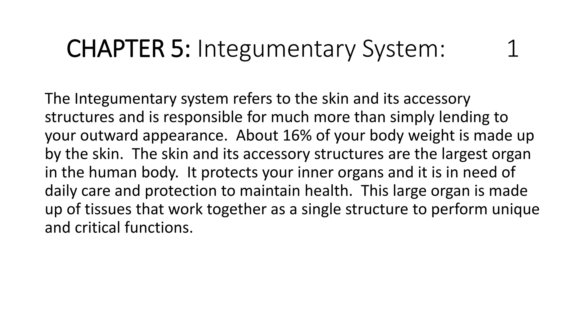
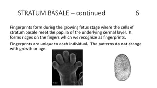
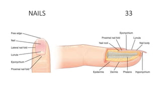
CHAPTER 5: Integumentary System: 1
The Integumentary system refers to the skin and its accessory
structures and is responsible for much more than simply lending to
your outward appearance. About 16% of your body weight is made up
by the skin. The skin and its accessory structures are the largest organ
in the human body. It protects your inner organs and it is in need of
daily care and protection to maintain health. This large organ is made
up of tissues that work together as a single structure to perform unique
and critical functions.
The skin is composed of two main layers: the epidermis, made of closely packed epithelial cells, and the dermis,
made of dense, irregular connective tissue that houses blood vessels, hair follicles, sweat glands, and other
structures. Beneath the dermis lies the hypodermis, which is composed mainly of loose connective and fatty
tissues. 2
EPIDERMIS 3
• Keratinized, stratified squamous epithelium
• Four to five layers of epithelial depending on its location in the body
• Avascular (no blood vessels)
• Four layers of skin is referred to as thin skin (most skin is thin)
• Thick skin is found only on the palms of the hands and the soles of the feet.
• Bedsores can be prevented by eliminating pressure points.
Layers of the Skin 4
From deep to superficial
Stratum basale
Stratum spinosum
Stratum granulosum
Stratum lucidum
Stratum corneum
Keratinocytes located in all layers except the stratum basale. Keratinocyte –
cell that manufactures and store protein keratin. Keratin- intracellular
fibrous protein, gives hair, nails, and skin their hardness and water-resistant
properties.
STRATUM BASALE 5
Stratum germinativum – another name.
Deepest epidermal layer, attaches the epidermis to basal lamina, below
which lie the layers of the dermis.
Made up of a single layer of primarily basal cells. Two cells that come from
this layer are Merkel cell and Melanocyte.
Merkel cell: functions as a receptor and responsible for stimulating sensory
nerves that the brain perceives as touch. Abundant on surfaces of the feet
and hands.
Melanocyte: cell that produces pigment melanin. Melanin gives hair and
skin its color and helps protect living cells in the epidermis from UV damage.
STRATUM BASALE – continued 6
Fingerprints form during the growing fetus stage where the cells of
stratum basale meet the papilla of the underlying dermal layer. It
forms ridges on the fingers which we recognize as fingerprints.
Fingerprints are unique to each individual. The patterns do not change
with growth or age.
STRATUM SPINOSUM 7
• Composed of eight to 10 layers of keratinocytes.
• Langerhans cells are located in this layer. These cells will help you to
rid your body of bacteria, if you cut yourself and bacteria enters the
wound.
• Begin synthesis of keratin and release water-repelling glycolipids that
helps prevent water loss from the body thus making the skin
relatively waterproof.
• New keratinocytes are produced atop the stratum basale and those in
the stratum spinosum are pushed to the stratum granulosum.
STRATUM GRANULOSUM 8
• Grainy appearance due to further changes that take place in the
keratinocytes as they are pushed into the stratum granulosum.
• Cells are three to five layers deep. They become flatter, cell
membranes thicken and they generate large amounts of protein
keratin which is fibrous. Also produces keratohyalin. These two
proteins make up the bulk of the keratinocyte mass in the stratum
granulosum and give the layer its grainy appearance. The nuclei and
other cell organelles disintegrate as the cells die, leaving behind the
keratin, keratohyalin, and cell membranes that will form the stratum
lucidum, the stratum corneum, and the accessory structures of hair
and nails.
Stratum Lucidum 9
• The stratum lucidum is a smooth, seemingly translucent layer of the
epidermis located just above the stratum granulosum and below the
stratum corneum. This thin layer of cells is found only in the thick skin
of the palms, soles, and digits. The keratinocytes that compose the
stratum lucidum are dead and flattened. These cells are densely
packed with eleiden, a clear protein rich in lipids, derived from
keratohyalin, which gives these cells their transparent appearance
and provides a barrier to water.
Stratum Corneum 10
• The stratum corneum is the most superficial layer of the epidermis and is
the layer exposed to the outside environment
• There are usually 15 to 30 layers of cells in the stratum corneum.
• This dry, dead layer helps prevent the penetration of microbes and the
dehydration of underlying tissues, and provides a mechanical protection
against abrasion for the more delicate, underlying layers. Keeps us from
drying up.
• Cells in this layer are shed periodically and are replaced by cells pushed up
from the stratum granulosum (or stratum lucidum in the case of the palms
and soles of feet).
• The entire layer is replaced during a period of about 4 weeks.
• Cosmetic procedures, such as microdermabrasion, help remove some of
the dry, upper layer and aim to keep the skin looking “fresh” and healthy.
DERMIS 11
• It contains blood and lymph vessels, nerves, and other structures,
such as hair follicles and sweat glands.
• The dermis is made of two layers of connective tissue that compose
an interconnected mesh of elastin and collagenous fibers, produced
by fibroblasts.
• Dermis is made up of two layers: Papillary and Reticular. Both of
these are composed mainly of connective tissue.
• The arrector pili is located in the dermis layer. It is responsible for the
formation of goose bumps when stimulated by the sympathetic
nervous system.
PAPILLARY LAYER 12
• Made of loose, areolar connective tissue, which means the collagen
and elastin fibers of this layer form a loose mesh.
• Superficial layer of the dermis projects into the stratum basale of the
epidermis to form finger-like dermal papillae.
• Within the papillary layer are fibroblasts, a small number of fat cells
(adipocytes), and an abundance of small blood vessels.
• The papillary layer contains phagocytes, defensive cells that help fight
bacteria or other infections that have breached the skin.
• This layer also contains lymphatic capillaries, nerve fibers, and touch
receptors called the Meissner corpuscles.
RETICULAR LAYER 13
• This layer is much thicker.
• Composed of dense, irregular connective tissue.
• Well vascularized and has a rich sensory and sympathetic nerve supply.
• The reticular layer appears reticulated (netlike) due to a tight meshwork of
fibers.
• Elastin fibers provide some elasticity to the skin, enabling movement.
Collagen fibers provide structure and tensile strength, with strands of
collagen extending into both the papillary layer and the hypodermis. In
addition, collagen binds water to keep the skin hydrated.
• Collagen injections and Retin-A creams help restore skin turgor by either
introducing collagen externally or stimulating blood flow and repair of the
dermis, respectively.
Papillary and Reticular Layer 14
HYPODERMIS 15
• Also called the subcutaneous layer or superficial fascia
• Layer directly below the dermis and serves to connect the skin to the
underlying fascia (fibrous tissue) of the bones and muscles.
• Not strictly a part of the skin, although the border between the
hypodermis and dermis can be difficult to distinguish.
• Consists of well vascularized, loose, areolar connective tissue and
adipose tissue
• Functions as a mode of fat storage and provides insulation and
cushioning for the integument
PIGMENTATION 16
• The color of skin is influenced by a number of pigments, including
melanin, carotene, and hemoglobin.
• melanin is produced by cells called melanocytes, found scattered
throughout the stratum basale of the epidermis. transferred into the
keratinocytes via a cellular vesicle called a melanosome
• Melanin occurs in two primary forms. Eumelanin exists as black and
brown, whereas pheomelanin provides a red color.
• Dark-skinned individuals produce more melanin than those with pale
skin.
PIGMENTATION 17
• The amount of melanin present in our skin is dependent on a balance
between available sunlight and folic acid destruction, and protection from
UV radiation and vitamin D production.
• It requires about 10 days after initial sun exposure for melanin synthesis to
peak, which is why pale-skinned individuals tend to suffer sunburns of the
epidermis initially. Dark-skinned individuals can also get sunburns, but are
more protected than are pale-skinned individuals.
• Melanosomes are temporary structures that are eventually destroyed by
fusion with lysosomes; this fact, along with melanin-filled keratinocytes in
the stratum corneum sloughing off, makes tanning impermanent.
PIGMENTATION 18
• Too much sun exposure can eventually lead to wrinkling due to the
destruction of the cellular structure of the skin, and in severe cases,
can cause sufficient DNA damage to result in skin cancer.
• When there is an irregular accumulation of melanocytes in the skin,
freckles appear.
• Moles are larger masses of melanocytes, and although most are
benign, they should be monitored for changes that might indicate the
presence of cancer
MOLES 19
ALBINISM 20
• Albinism is a genetic disorder that affects (completely or partially) the
coloring of skin, hair, and eyes. The defect is primarily due to the
inability of melanocytes to produce melanin. Individuals with albinism
tend to appear white or very pale due to the lack of melanin in their
skin and hair. Recall that melanin helps protect the skin from the
harmful effects of UV radiation. Individuals with albinism tend to
need more protection from UV radiation, as they are more prone to
sunburns and skin cancer. They also tend to be more sensitive to light
and have vision problems due to the lack of pigmentation on the
retinal wall. Treatment of this disorder usually involves addressing the
symptoms, such as limiting UV light exposure to the skin and eyes.
VITILIGO 21
• In vitiligo, the melanocytes in certain areas lose their ability to
produce melanin, possibly due to an autoimmune reaction. This leads
to a loss of color in patches.
• Neither albinism nor vitiligo directly affects the lifespan of an
individual.
VITILIGO 22
CHANGES IN THE APPEARANCE OF SKIN 23
• Other changes in the appearance of skin coloration can be indicative of diseases associated with
other body systems.
• Liver disease or liver cancer can cause the accumulation of bile and the yellow pigment bilirubin,
leading to the skin appearing yellow or jaundiced (jaune is the French word for “yellow”).
• Tumors of the pituitary gland can result in the secretion of large amounts of melanocyte-
stimulating hormone (MSH), which results in a darkening of the skin.
• Similarly, Addison’s disease can stimulate the release of excess amounts of adrenocorticotropic
hormone (ACTH), which can give the skin a deep bronze color.
• A sudden drop in oxygenation can affect skin color, causing the skin to initially turn ashen (white).
• With a prolonged reduction in oxygen levels, dark red deoxyhemoglobin becomes dominant in
the blood, making the skin appear blue, a condition referred to as cyanosis (kyanos is the Greek
word for “blue”). This happens when the oxygen supply is restricted, as when someone is
experiencing difficulty in breathing because of asthma or a heart attack. However, in these cases
the effect on skin color has nothing do with the skin’s pigmentation
ACCESSORY STRUCTURES 24
• Hair
• Nails
• Sweat glands
• Sebaceous glands
HAIR 25
HAIR – continued 26
• Keratinous filament growing out of the epidermis.
• Primarily made of dead, keratinized cells.
• Strands of hair originate in an epidermal penetration of the dermis called
the hair follicle.
• Hair shaft - part of the hair not anchored to the follicle, and much of this is
exposed at the skin’s surface.
• The rest of the hair, which is anchored in the follicle, lies below the surface
of the skin and is referred to as the hair root.
• The hair root ends deep in the dermis at the hair bulb. The hair bulb
surrounds the hair papilla, which is made of connective tissue and contains
blood capillaries and nerve endings from the dermis
HAIR – continued 27
• Hair matrix - layer of mitotically active basal cells
• As the dead skin on the surface sheds, the basal cells of the hair bulb divide
and push cells outward in the hair root and shaft as the hair grows.
• The medulla forms the central core of the hair, which is surrounded by the
cortex, a layer of compressed, keratinized cells that is covered by an outer
layer of very hard, keratinized cells known as the cuticle.
• Hair texture (straight, curly) is determined by the shape and structure of
the cortex, and to the extent that it is present, the medulla. The shape and
structure of these layers are, in turn, determined by the shape of the hair
follicle. Hair growth begins with the production of keratinocytes by the
basal cells of the hair bulb. As new cells are deposited at the hair bulb, the
hair shaft is pushed through the follicle toward the surface.
HAIR – continued 28
• Keratinization is completed as the cells are pushed to the skin surface
to form the shaft of hair that is externally visible.
• The external hair is completely dead and composed entirely of
keratin. For this reason, our hair does not have sensation.
Furthermore, you can cut your hair or shave without damaging the
hair structure because the cut is superficial.
• Most chemical hair removers also act superficially; however,
electrolysis and yanking both attempt to destroy the hair bulb so hair
cannot grow
HAIR – continued 29
• Hair serves a variety of functions, including protection, sensory input, thermoregulation, and
communication.
• For example, hair on the head protects the skull from the sun.
• The hair in the nose and ears, and around the eyes (eyelashes) defends the body by trapping and excluding
dust particles that may contain allergens and microbes.
• Hair of the eyebrows prevents sweat and other particles from dripping into and bothering the eyes.
• Hair also has a sensory function due to sensory innervation by a hair root plexus surrounding the base of
each hair follicle.
• Hair is extremely sensitive to air movement or other disturbances in the environment, much more so than
the skin surface. This feature is also useful for the detection of the presence of insects or other potentially
damaging substances on the skin surface.
• Each hair root is connected to a smooth muscle called the arrector pili that contracts in response to nerve
signals from the sympathetic nervous system, making the external hair shaft “stand up.”
• Humans as goose bumps and even more obvious in animals, such as when a frightened cat raises its fur.
HAIR GROWTH 30
• Hair grows and is eventually shed and replaced by new hair.
• This occurs in three phases.
• Anagen phase, during which cells divide rapidly at the root of the hair,
pushing the hair shaft up and out. The length of this phase is measured in
years, typically from 2 to 7 years. grows at the rate of 0.3 mm per day
• Catagen phase lasts only 2 to 3 weeks, and marks a transition from the hair
follicle’s active growth.
• Telogen phase, the hair follicle is at rest and no new growth occurs. At the
end of this phase, which lasts about 2 to 4 months, another anagen phase
begins.
HAIR GROWTH – continued 31
• On average, 50 hairs are lost and replaced per day.
• Hair loss occurs if there is more hair shed than what is replaced and
can happen due to hormonal or dietary changes.
• Hair loss can also result from the aging process, or the influence of
hormones
HAIR COLOR 32
• Hair gets its color from the pigment melanin, produced by
melanocytes in the hair papilla.
• Different hair color results from differences in the type of melanin,
which is genetically determined.
• As a person ages, the melanin production decreases, and hair tends
to lose its color and becomes gray and/or white.
NAILS 33
NAILS – continued 34
• Nail bed - specialized structure of the epidermis that is found at the
tips of our fingers and toes.
• Nail body is formed on the nail bed, and protects the tips of our
fingers and toes as they are the farthest extremities and the parts of
the body that experience the maximum mechanical stress. Forms a
back-support for picking up small objects with the fingers. Composed
of densely packed dead keratinocytes. The epidermis in this part of
the body has evolved a specialized structure upon which nails can
form. The nail body forms at the nail root which enables the nail to
grow continuously.
NAILS – continued 35
• The lateral nail fold overlaps the nail on the sides, helping to anchor
the nail body.
• Nail fold that meets the proximal end of the nail body forms the nail
cuticle, also called the eponychium.
• Nail bed is rich in blood vessels, making it appear pink, except at the
base, where a thick layer of epithelium over the nail matrix forms a
crescent-shaped region called the lunula (the “little moon”).
• Area beneath the free edge of the nail, furthest from the cuticle, is
called the hyponychium. It consists of a thickened layer of stratum
corneum.
SWEAT GLANDS 36
• When the body becomes warm, sudoriferous glands produce sweat
to cool the body. Sweat glands develop from epidermal projections
into the dermis and are classified as merocrine glands; that is, the
secretions are excreted by exocytosis through a duct without affecting
the cells of the gland.
• There are two types of sweat glands, each secreting slightly different
products. Eccrine sweat gland and Apocrine sweat gland
ECCRINE SWEAT GLAND 37
• Produces a hypotonic sweat for thermoregulation. These glands are
found all over the skin’s surface, but are especially abundant on the
palms of the hand, the soles of the feet, and the forehead. They are
coiled glands lying deep in the dermis, with the duct rising up to a
pore on the skin surface, where the sweat is released. This type of
sweat, released by exocytosis, is hypotonic and composed mostly of
water, with some salt, antibodies, traces of metabolic waste, and
dermicidin, an antimicrobial peptide.
• Eccrine glands are a primary component of thermoregulation in
humans and thus help to maintain homeostasis. These gland produce
a watery sweat.
APOCRINE SWEAT GLANDS 38
• Apocrine sweat gland associated with hair follicles in densely hairy areas,
such as armpits and genital regions.
• Larger than eccrine sweat glands and lie deeper in the dermis, sometimes
even reaching the hypodermis, with the duct normally emptying into the
hair follicle. In addition to water and salts, apocrine sweat includes organic
compounds that make the sweat thicker and subject to bacterial
decomposition and subsequent smell.
• The release of this sweat is under both nervous and hormonal control, and
plays a role in the poorly understood human pheromone response. Most
commercial antiperspirants use an aluminum-based compound as their
primary active ingredient to stop sweat. When the antiperspirant enters
the sweat gland duct, the aluminum-based compounds precipitate due to a
change in pH and form a physical block in the duct, which prevents sweat
from coming out of the pore
SEBACEOUS GLANDS 39
• Sebaceous gland - type of oil gland that is found all over the body and
helps to lubricate and waterproof the skin and hair. Most are
associated with hair follicles. They generate and excrete sebum, a
mixture of lipids, onto the skin surface, thereby naturally lubricating
the dry and dead layer of keratinized cells of the stratum corneum,
keeping it pliable. The fatty acids of sebum also have antibacterial
properties, and prevent water loss from the skin in low-humidity
environments. The secretion of sebum is stimulated by hormones,
many of which do not become active until puberty. Thus, sebaceous
glands are relatively inactive during childhood.
FUNCTIONS 40
• The skin and accessory structures:
• protecting the body from invasion by microorganisms, chemicals, and
other environmental factors
• preventing dehydration
• acting as a sensory organ
• modulating body temperature and electrolyte balance
• synthesizing vitamin D.
• Underlying hypodermis has important roles in storing fats, forming a
“cushion” over underlying structures, and providing insulation from
cold temperatures
PROTECTION 41
• Protects the rest of the body from the basic elements of nature such
as wind, water, and UV sunlight.
• Acts as a protective barrier against water loss, due to the presence of
layers of keratin and glycolipids in the stratum corneum.
• First line of defense against abrasive activity due to contact with grit,
microbes, or harmful chemicals.
• Sweat excreted from sweat glands deters microbes from over-
colonizing the skin surface by generating dermicidin, which has
antibiotic properties.
SENSORY FUNCTION 42
• The fact that you can feel an ant crawling on your skin, allowing you
to flick it off before it bites, is because the skin, and especially the
hairs projecting from hair follicles in the skin, can sense changes in
the environment.
• The skin acts as a sense organ because the epidermis, dermis, and the
hypodermis contain specialized sensory nerve structures that detect
touch, surface temperature, and pain. These receptors are more
concentrated on the tips of the fingers, which are most sensitive to
touch.
SENSORY FUNCTION – continued 43
• Meissner corpuscle - responds to light touch
• Pacinian corpuscle - responds to vibration.
• Merkel cells, seen scattered in the stratum basale, are touch
receptors.
• Sensory nerves connected to each hair follicle
• Pain and temperature receptors scattered throughout the skin
• Motor nerves innervate the arrector pili muscles and glands. This rich
innervation helps us sense our environment and react accordingly
THERMOREGULATION 44
• The integumentary system helps regulate body temperature through its
tight association with the sympathetic nervous system, the division of the
nervous system involved in our fight-or-flight responses. The sympathetic
nervous system is continuously monitoring body temperature and initiating
appropriate motor responses. Recall that sweat glands, accessory
structures to the skin, secrete water, salt, and other substances to cool the
body when it becomes warm. Even when the body does not appear to be
noticeably sweating, approximately 500 mL of sweat (insensible
perspiration) are secreted a day. If the body becomes excessively warm due
to high temperatures, vigorous activity , or a combination of the two, sweat
glands will be stimulated by the sympathetic nervous system to produce
large amounts of sweat, as much as 0.7 to 1.5 L per hour for an active
person. When the sweat evaporates from the skin surface, the body is
cooled as body heat is dissipated.
THERMOREGULATION- continued 45
• In addition to sweating, arterioles in the dermis dilate so that excess
heat carried by the blood can dissipate through the skin and into the
surrounding environment. This accounts for the skin redness that
many people experience when exercising.
• When body temperatures drop, the arterioles constrict to minimize
heat loss, particularly in the ends of the digits and tip of the nose.
This reduced circulation can result in the skin taking on a whitish hue.
Although the temperature of the skin drops as a result, passive heat
loss is prevented, and internal organs and structures remain warm. If
the temperature of the skin drops too much (such as environmental
temperatures below freezing), the conservation of body core heat can
result in the skin actually freezing, a condition called frostbite.
VITAMIN D SYNTHESIS 46
• The epidermal layer of human skin synthesizes vitamin D when exposed to
UV radiation (sun).
• Vitamin D is essential for normal absorption of calcium and phosphorous,
which are required for healthy bones.
• Absence of sun exposure can lead to a lack of vitamin D in the body,
leading to a condition called rickets, a painful condition in children where
the bones are misshapen due to a lack of calcium, causing bowleggedness.
• Elderly individuals who suffer from vitamin D deficiency can develop a
condition called osteomalacia, a softening of the bones.
• In present day society, vitamin D is added as a supplement to many foods,
including milk and orange juice, compensating for the need for sun
exposure.
SKIN CANCERS 47
• Basal cell carcinoma - most common of all cancers that occur in the
United States and is frequently found on the head, neck, arms, and
back, which are areas that are most susceptible to long-term sun
exposure. They begin to grow toward the surface and become an
uneven patch, bump, growth, or scar on the skin surface. Like most
cancers, basal cell carcinomas respond best to treatment when
caught early. Treatment options include surgery, freezing
(cryosurgery), and topical ointments.
• Limiting exposure to the sun can reduce skin cancers.
SQUAMOUS CELL CARCINOMA 48
• Squamous cell carcinoma is a cancer that affects the keratinocytes of
the stratum spinosum and presents as lesions commonly found on
the scalp, ears, and hands.
• It is the second most common skin cancer.
• The American Cancer Society reports that two of 10 skin cancers are
squamous cell carcinomas, and it is more aggressive than basal cell
carcinoma.
• If not removed, these carcinomas can metastasize. Surgery and
radiation are used to cure squamous cell carcinoma
MELANOMA 49
• A melanoma is a cancer characterized by the uncontrolled growth of
melanocytes, the pigment-producing cells in the epidermis.
• Typically, a melanoma develops from a mole.
• It is the most fatal of all skin cancers, as it is highly metastatic and can
be difficult to detect before it has spread to other organs.
• Melanomas usually appear as asymmetrical brown and black patches
with uneven borders and a raised surface.
• Treatment typically involves surgical excision and immunotherapy.
MELANOMA – ABCDE MNEMONIC 50
• Doctors often give their patients the following ABCDE mnemonic to
help with the diagnosis of early-stage melanoma. If you observe a
mole on your body displaying these signs, consult a doctor.
• Asymmetry – the two sides are not symmetrical
• Borders – the edges are irregular in shape
• Color – the color is varied shades of brown or black
• Diameter – it is larger than 6 mm (0.24 in)
• Evolving – its shape has changed
MELANOMA – continued 52
Some specialists cite the following additional signs for the most serious
form, nodular melanoma:
• Elevated – it is raised on the skin surface
• Firm – it feels hard to the touch
• Growing – it is getting larger
ECZEMA
• SKIN DISORDER: Eczema is an inflammatory condition and occurs in
individuals of all ages.
• Eczema - allergic reaction that manifests as dry, itchy patches of skin
that resemble rashes. It may be accompanied by swelling of the skin,
flaking, and in severe cases, bleeding. Many who suffer from eczema
have antibodies against dust mites in their blood, but the link
between eczema and allergy to dust mites has not been proven.
Symptoms are usually managed with moisturizers, corticosteroid
creams, and immunosuppressants.
ACNE 53
• Acne involves the clogging of pores, which can lead to infection and
inflammation, and is often seen in adolescents.
• Acne - skin disturbance that typically occurs on areas of the skin that are
rich in sebaceous glands (face and back).
• Most common along with the onset of puberty due to associated hormonal
changes, but can also occur in infants and continue into adulthood.
Hormones, such as androgens, stimulate the release of sebum. An
overproduction and accumulation of sebum along with keratin can block
hair follicles. This plug is initially white. The sebum, when oxidized by
exposure to air, turns black. Acne results from infection by acne-causing
bacteria (Propionibacterium and Staphylococcus), which can lead to
redness and potential scarring due to the natural wound healing process
BURNS 54
• A burn results when the skin is damaged by intense heat, radiation,
electricity, or chemicals.
• The damage results in the death of skin cells, which can lead to a
massive loss of fluid. Dehydration, electrolyte imbalance, and renal
and circulatory failure follow, which can be fatal. Burn patients are
treated with intravenous fluids to offset dehydration, as well as
intravenous nutrients that enable the body to repair tissues and
replace lost proteins.
• Another serious threat to the lives of burn patients is infection.
Burned skin is extremely susceptible to bacteria and other pathogens,
due to the loss of protection by intact layers of skin.
BURNS – RULE OF NINES 55
Burns are sometimes measured in terms of the size of the total
surface area affected. This is referred to as the “rule of nines,”
which associates specific anatomical areas with a percentage
that is a factor of nine
FIRST-DEGREE BURN 56
A first-degree burn is a superficial burn that affects only the epidermis.
Although the skin may be painful and swollen, these burns typically heal on
their own within a few days. Mild sunburn fits into the category of a first-
degree burn.
SECOND-DEGREE BURN 57
A second-degree burn goes deeper and affects both the
epidermis and a portion of the dermis. These burns result in
swelling and a painful blistering of the skin. It is important to
keep the burn site clean and sterile to prevent infection. If
this is done, the burn will heal within several weeks.
THIRD-DEGREE BURN 58
A third-degree burn fully extends into the epidermis and
dermis, destroying the tissue and affecting the nerve endings
and sensory function. These are serious burns that may appear
white, red, or black; they require medical attention and will
heal slowly without it
FOURTH-DEGREE BURN 59
A fourth-degree burn is even more severe,
affecting the underlying muscle and bone.
Oddly, third and fourth-degree burns are
usually not as painful because the nerve
endings themselves are damaged.
FULL THICKNESS BURN 60
Full-thickness burns cannot be repaired by the body,
because the local tissues used for repair are damaged
and require excision (debridement), or amputation in
severe cases, followed by grafting of the skin from an
unaffected part of the body, or from skin grown in
tissue culture for grafting purposes.

Basic ap chapter 5 powerpoint 2017

  • 1.
    CHAPTER 5: IntegumentarySystem: 1 The Integumentary system refers to the skin and its accessory structures and is responsible for much more than simply lending to your outward appearance. About 16% of your body weight is made up by the skin. The skin and its accessory structures are the largest organ in the human body. It protects your inner organs and it is in need of daily care and protection to maintain health. This large organ is made up of tissues that work together as a single structure to perform unique and critical functions.
  • 2.
    The skin iscomposed of two main layers: the epidermis, made of closely packed epithelial cells, and the dermis, made of dense, irregular connective tissue that houses blood vessels, hair follicles, sweat glands, and other structures. Beneath the dermis lies the hypodermis, which is composed mainly of loose connective and fatty tissues. 2
  • 3.
    EPIDERMIS 3 • Keratinized,stratified squamous epithelium • Four to five layers of epithelial depending on its location in the body • Avascular (no blood vessels) • Four layers of skin is referred to as thin skin (most skin is thin) • Thick skin is found only on the palms of the hands and the soles of the feet. • Bedsores can be prevented by eliminating pressure points.
  • 4.
    Layers of theSkin 4 From deep to superficial Stratum basale Stratum spinosum Stratum granulosum Stratum lucidum Stratum corneum Keratinocytes located in all layers except the stratum basale. Keratinocyte – cell that manufactures and store protein keratin. Keratin- intracellular fibrous protein, gives hair, nails, and skin their hardness and water-resistant properties.
  • 5.
    STRATUM BASALE 5 Stratumgerminativum – another name. Deepest epidermal layer, attaches the epidermis to basal lamina, below which lie the layers of the dermis. Made up of a single layer of primarily basal cells. Two cells that come from this layer are Merkel cell and Melanocyte. Merkel cell: functions as a receptor and responsible for stimulating sensory nerves that the brain perceives as touch. Abundant on surfaces of the feet and hands. Melanocyte: cell that produces pigment melanin. Melanin gives hair and skin its color and helps protect living cells in the epidermis from UV damage.
  • 6.
    STRATUM BASALE –continued 6 Fingerprints form during the growing fetus stage where the cells of stratum basale meet the papilla of the underlying dermal layer. It forms ridges on the fingers which we recognize as fingerprints. Fingerprints are unique to each individual. The patterns do not change with growth or age.
  • 7.
    STRATUM SPINOSUM 7 •Composed of eight to 10 layers of keratinocytes. • Langerhans cells are located in this layer. These cells will help you to rid your body of bacteria, if you cut yourself and bacteria enters the wound. • Begin synthesis of keratin and release water-repelling glycolipids that helps prevent water loss from the body thus making the skin relatively waterproof. • New keratinocytes are produced atop the stratum basale and those in the stratum spinosum are pushed to the stratum granulosum.
  • 8.
    STRATUM GRANULOSUM 8 •Grainy appearance due to further changes that take place in the keratinocytes as they are pushed into the stratum granulosum. • Cells are three to five layers deep. They become flatter, cell membranes thicken and they generate large amounts of protein keratin which is fibrous. Also produces keratohyalin. These two proteins make up the bulk of the keratinocyte mass in the stratum granulosum and give the layer its grainy appearance. The nuclei and other cell organelles disintegrate as the cells die, leaving behind the keratin, keratohyalin, and cell membranes that will form the stratum lucidum, the stratum corneum, and the accessory structures of hair and nails.
  • 9.
    Stratum Lucidum 9 •The stratum lucidum is a smooth, seemingly translucent layer of the epidermis located just above the stratum granulosum and below the stratum corneum. This thin layer of cells is found only in the thick skin of the palms, soles, and digits. The keratinocytes that compose the stratum lucidum are dead and flattened. These cells are densely packed with eleiden, a clear protein rich in lipids, derived from keratohyalin, which gives these cells their transparent appearance and provides a barrier to water.
  • 10.
    Stratum Corneum 10 •The stratum corneum is the most superficial layer of the epidermis and is the layer exposed to the outside environment • There are usually 15 to 30 layers of cells in the stratum corneum. • This dry, dead layer helps prevent the penetration of microbes and the dehydration of underlying tissues, and provides a mechanical protection against abrasion for the more delicate, underlying layers. Keeps us from drying up. • Cells in this layer are shed periodically and are replaced by cells pushed up from the stratum granulosum (or stratum lucidum in the case of the palms and soles of feet). • The entire layer is replaced during a period of about 4 weeks. • Cosmetic procedures, such as microdermabrasion, help remove some of the dry, upper layer and aim to keep the skin looking “fresh” and healthy.
  • 11.
    DERMIS 11 • Itcontains blood and lymph vessels, nerves, and other structures, such as hair follicles and sweat glands. • The dermis is made of two layers of connective tissue that compose an interconnected mesh of elastin and collagenous fibers, produced by fibroblasts. • Dermis is made up of two layers: Papillary and Reticular. Both of these are composed mainly of connective tissue. • The arrector pili is located in the dermis layer. It is responsible for the formation of goose bumps when stimulated by the sympathetic nervous system.
  • 12.
    PAPILLARY LAYER 12 •Made of loose, areolar connective tissue, which means the collagen and elastin fibers of this layer form a loose mesh. • Superficial layer of the dermis projects into the stratum basale of the epidermis to form finger-like dermal papillae. • Within the papillary layer are fibroblasts, a small number of fat cells (adipocytes), and an abundance of small blood vessels. • The papillary layer contains phagocytes, defensive cells that help fight bacteria or other infections that have breached the skin. • This layer also contains lymphatic capillaries, nerve fibers, and touch receptors called the Meissner corpuscles.
  • 13.
    RETICULAR LAYER 13 •This layer is much thicker. • Composed of dense, irregular connective tissue. • Well vascularized and has a rich sensory and sympathetic nerve supply. • The reticular layer appears reticulated (netlike) due to a tight meshwork of fibers. • Elastin fibers provide some elasticity to the skin, enabling movement. Collagen fibers provide structure and tensile strength, with strands of collagen extending into both the papillary layer and the hypodermis. In addition, collagen binds water to keep the skin hydrated. • Collagen injections and Retin-A creams help restore skin turgor by either introducing collagen externally or stimulating blood flow and repair of the dermis, respectively.
  • 14.
  • 15.
    HYPODERMIS 15 • Alsocalled the subcutaneous layer or superficial fascia • Layer directly below the dermis and serves to connect the skin to the underlying fascia (fibrous tissue) of the bones and muscles. • Not strictly a part of the skin, although the border between the hypodermis and dermis can be difficult to distinguish. • Consists of well vascularized, loose, areolar connective tissue and adipose tissue • Functions as a mode of fat storage and provides insulation and cushioning for the integument
  • 16.
    PIGMENTATION 16 • Thecolor of skin is influenced by a number of pigments, including melanin, carotene, and hemoglobin. • melanin is produced by cells called melanocytes, found scattered throughout the stratum basale of the epidermis. transferred into the keratinocytes via a cellular vesicle called a melanosome • Melanin occurs in two primary forms. Eumelanin exists as black and brown, whereas pheomelanin provides a red color. • Dark-skinned individuals produce more melanin than those with pale skin.
  • 17.
    PIGMENTATION 17 • Theamount of melanin present in our skin is dependent on a balance between available sunlight and folic acid destruction, and protection from UV radiation and vitamin D production. • It requires about 10 days after initial sun exposure for melanin synthesis to peak, which is why pale-skinned individuals tend to suffer sunburns of the epidermis initially. Dark-skinned individuals can also get sunburns, but are more protected than are pale-skinned individuals. • Melanosomes are temporary structures that are eventually destroyed by fusion with lysosomes; this fact, along with melanin-filled keratinocytes in the stratum corneum sloughing off, makes tanning impermanent.
  • 18.
    PIGMENTATION 18 • Toomuch sun exposure can eventually lead to wrinkling due to the destruction of the cellular structure of the skin, and in severe cases, can cause sufficient DNA damage to result in skin cancer. • When there is an irregular accumulation of melanocytes in the skin, freckles appear. • Moles are larger masses of melanocytes, and although most are benign, they should be monitored for changes that might indicate the presence of cancer
  • 19.
  • 20.
    ALBINISM 20 • Albinismis a genetic disorder that affects (completely or partially) the coloring of skin, hair, and eyes. The defect is primarily due to the inability of melanocytes to produce melanin. Individuals with albinism tend to appear white or very pale due to the lack of melanin in their skin and hair. Recall that melanin helps protect the skin from the harmful effects of UV radiation. Individuals with albinism tend to need more protection from UV radiation, as they are more prone to sunburns and skin cancer. They also tend to be more sensitive to light and have vision problems due to the lack of pigmentation on the retinal wall. Treatment of this disorder usually involves addressing the symptoms, such as limiting UV light exposure to the skin and eyes.
  • 21.
    VITILIGO 21 • Invitiligo, the melanocytes in certain areas lose their ability to produce melanin, possibly due to an autoimmune reaction. This leads to a loss of color in patches. • Neither albinism nor vitiligo directly affects the lifespan of an individual.
  • 22.
  • 23.
    CHANGES IN THEAPPEARANCE OF SKIN 23 • Other changes in the appearance of skin coloration can be indicative of diseases associated with other body systems. • Liver disease or liver cancer can cause the accumulation of bile and the yellow pigment bilirubin, leading to the skin appearing yellow or jaundiced (jaune is the French word for “yellow”). • Tumors of the pituitary gland can result in the secretion of large amounts of melanocyte- stimulating hormone (MSH), which results in a darkening of the skin. • Similarly, Addison’s disease can stimulate the release of excess amounts of adrenocorticotropic hormone (ACTH), which can give the skin a deep bronze color. • A sudden drop in oxygenation can affect skin color, causing the skin to initially turn ashen (white). • With a prolonged reduction in oxygen levels, dark red deoxyhemoglobin becomes dominant in the blood, making the skin appear blue, a condition referred to as cyanosis (kyanos is the Greek word for “blue”). This happens when the oxygen supply is restricted, as when someone is experiencing difficulty in breathing because of asthma or a heart attack. However, in these cases the effect on skin color has nothing do with the skin’s pigmentation
  • 24.
    ACCESSORY STRUCTURES 24 •Hair • Nails • Sweat glands • Sebaceous glands
  • 25.
  • 26.
    HAIR – continued26 • Keratinous filament growing out of the epidermis. • Primarily made of dead, keratinized cells. • Strands of hair originate in an epidermal penetration of the dermis called the hair follicle. • Hair shaft - part of the hair not anchored to the follicle, and much of this is exposed at the skin’s surface. • The rest of the hair, which is anchored in the follicle, lies below the surface of the skin and is referred to as the hair root. • The hair root ends deep in the dermis at the hair bulb. The hair bulb surrounds the hair papilla, which is made of connective tissue and contains blood capillaries and nerve endings from the dermis
  • 27.
    HAIR – continued27 • Hair matrix - layer of mitotically active basal cells • As the dead skin on the surface sheds, the basal cells of the hair bulb divide and push cells outward in the hair root and shaft as the hair grows. • The medulla forms the central core of the hair, which is surrounded by the cortex, a layer of compressed, keratinized cells that is covered by an outer layer of very hard, keratinized cells known as the cuticle. • Hair texture (straight, curly) is determined by the shape and structure of the cortex, and to the extent that it is present, the medulla. The shape and structure of these layers are, in turn, determined by the shape of the hair follicle. Hair growth begins with the production of keratinocytes by the basal cells of the hair bulb. As new cells are deposited at the hair bulb, the hair shaft is pushed through the follicle toward the surface.
  • 28.
    HAIR – continued28 • Keratinization is completed as the cells are pushed to the skin surface to form the shaft of hair that is externally visible. • The external hair is completely dead and composed entirely of keratin. For this reason, our hair does not have sensation. Furthermore, you can cut your hair or shave without damaging the hair structure because the cut is superficial. • Most chemical hair removers also act superficially; however, electrolysis and yanking both attempt to destroy the hair bulb so hair cannot grow
  • 29.
    HAIR – continued29 • Hair serves a variety of functions, including protection, sensory input, thermoregulation, and communication. • For example, hair on the head protects the skull from the sun. • The hair in the nose and ears, and around the eyes (eyelashes) defends the body by trapping and excluding dust particles that may contain allergens and microbes. • Hair of the eyebrows prevents sweat and other particles from dripping into and bothering the eyes. • Hair also has a sensory function due to sensory innervation by a hair root plexus surrounding the base of each hair follicle. • Hair is extremely sensitive to air movement or other disturbances in the environment, much more so than the skin surface. This feature is also useful for the detection of the presence of insects or other potentially damaging substances on the skin surface. • Each hair root is connected to a smooth muscle called the arrector pili that contracts in response to nerve signals from the sympathetic nervous system, making the external hair shaft “stand up.” • Humans as goose bumps and even more obvious in animals, such as when a frightened cat raises its fur.
  • 30.
    HAIR GROWTH 30 •Hair grows and is eventually shed and replaced by new hair. • This occurs in three phases. • Anagen phase, during which cells divide rapidly at the root of the hair, pushing the hair shaft up and out. The length of this phase is measured in years, typically from 2 to 7 years. grows at the rate of 0.3 mm per day • Catagen phase lasts only 2 to 3 weeks, and marks a transition from the hair follicle’s active growth. • Telogen phase, the hair follicle is at rest and no new growth occurs. At the end of this phase, which lasts about 2 to 4 months, another anagen phase begins.
  • 31.
    HAIR GROWTH –continued 31 • On average, 50 hairs are lost and replaced per day. • Hair loss occurs if there is more hair shed than what is replaced and can happen due to hormonal or dietary changes. • Hair loss can also result from the aging process, or the influence of hormones
  • 32.
    HAIR COLOR 32 •Hair gets its color from the pigment melanin, produced by melanocytes in the hair papilla. • Different hair color results from differences in the type of melanin, which is genetically determined. • As a person ages, the melanin production decreases, and hair tends to lose its color and becomes gray and/or white.
  • 33.
  • 34.
    NAILS – continued34 • Nail bed - specialized structure of the epidermis that is found at the tips of our fingers and toes. • Nail body is formed on the nail bed, and protects the tips of our fingers and toes as they are the farthest extremities and the parts of the body that experience the maximum mechanical stress. Forms a back-support for picking up small objects with the fingers. Composed of densely packed dead keratinocytes. The epidermis in this part of the body has evolved a specialized structure upon which nails can form. The nail body forms at the nail root which enables the nail to grow continuously.
  • 35.
    NAILS – continued35 • The lateral nail fold overlaps the nail on the sides, helping to anchor the nail body. • Nail fold that meets the proximal end of the nail body forms the nail cuticle, also called the eponychium. • Nail bed is rich in blood vessels, making it appear pink, except at the base, where a thick layer of epithelium over the nail matrix forms a crescent-shaped region called the lunula (the “little moon”). • Area beneath the free edge of the nail, furthest from the cuticle, is called the hyponychium. It consists of a thickened layer of stratum corneum.
  • 36.
    SWEAT GLANDS 36 •When the body becomes warm, sudoriferous glands produce sweat to cool the body. Sweat glands develop from epidermal projections into the dermis and are classified as merocrine glands; that is, the secretions are excreted by exocytosis through a duct without affecting the cells of the gland. • There are two types of sweat glands, each secreting slightly different products. Eccrine sweat gland and Apocrine sweat gland
  • 37.
    ECCRINE SWEAT GLAND37 • Produces a hypotonic sweat for thermoregulation. These glands are found all over the skin’s surface, but are especially abundant on the palms of the hand, the soles of the feet, and the forehead. They are coiled glands lying deep in the dermis, with the duct rising up to a pore on the skin surface, where the sweat is released. This type of sweat, released by exocytosis, is hypotonic and composed mostly of water, with some salt, antibodies, traces of metabolic waste, and dermicidin, an antimicrobial peptide. • Eccrine glands are a primary component of thermoregulation in humans and thus help to maintain homeostasis. These gland produce a watery sweat.
  • 38.
    APOCRINE SWEAT GLANDS38 • Apocrine sweat gland associated with hair follicles in densely hairy areas, such as armpits and genital regions. • Larger than eccrine sweat glands and lie deeper in the dermis, sometimes even reaching the hypodermis, with the duct normally emptying into the hair follicle. In addition to water and salts, apocrine sweat includes organic compounds that make the sweat thicker and subject to bacterial decomposition and subsequent smell. • The release of this sweat is under both nervous and hormonal control, and plays a role in the poorly understood human pheromone response. Most commercial antiperspirants use an aluminum-based compound as their primary active ingredient to stop sweat. When the antiperspirant enters the sweat gland duct, the aluminum-based compounds precipitate due to a change in pH and form a physical block in the duct, which prevents sweat from coming out of the pore
  • 39.
    SEBACEOUS GLANDS 39 •Sebaceous gland - type of oil gland that is found all over the body and helps to lubricate and waterproof the skin and hair. Most are associated with hair follicles. They generate and excrete sebum, a mixture of lipids, onto the skin surface, thereby naturally lubricating the dry and dead layer of keratinized cells of the stratum corneum, keeping it pliable. The fatty acids of sebum also have antibacterial properties, and prevent water loss from the skin in low-humidity environments. The secretion of sebum is stimulated by hormones, many of which do not become active until puberty. Thus, sebaceous glands are relatively inactive during childhood.
  • 40.
    FUNCTIONS 40 • Theskin and accessory structures: • protecting the body from invasion by microorganisms, chemicals, and other environmental factors • preventing dehydration • acting as a sensory organ • modulating body temperature and electrolyte balance • synthesizing vitamin D. • Underlying hypodermis has important roles in storing fats, forming a “cushion” over underlying structures, and providing insulation from cold temperatures
  • 41.
    PROTECTION 41 • Protectsthe rest of the body from the basic elements of nature such as wind, water, and UV sunlight. • Acts as a protective barrier against water loss, due to the presence of layers of keratin and glycolipids in the stratum corneum. • First line of defense against abrasive activity due to contact with grit, microbes, or harmful chemicals. • Sweat excreted from sweat glands deters microbes from over- colonizing the skin surface by generating dermicidin, which has antibiotic properties.
  • 42.
    SENSORY FUNCTION 42 •The fact that you can feel an ant crawling on your skin, allowing you to flick it off before it bites, is because the skin, and especially the hairs projecting from hair follicles in the skin, can sense changes in the environment. • The skin acts as a sense organ because the epidermis, dermis, and the hypodermis contain specialized sensory nerve structures that detect touch, surface temperature, and pain. These receptors are more concentrated on the tips of the fingers, which are most sensitive to touch.
  • 43.
    SENSORY FUNCTION –continued 43 • Meissner corpuscle - responds to light touch • Pacinian corpuscle - responds to vibration. • Merkel cells, seen scattered in the stratum basale, are touch receptors. • Sensory nerves connected to each hair follicle • Pain and temperature receptors scattered throughout the skin • Motor nerves innervate the arrector pili muscles and glands. This rich innervation helps us sense our environment and react accordingly
  • 44.
    THERMOREGULATION 44 • Theintegumentary system helps regulate body temperature through its tight association with the sympathetic nervous system, the division of the nervous system involved in our fight-or-flight responses. The sympathetic nervous system is continuously monitoring body temperature and initiating appropriate motor responses. Recall that sweat glands, accessory structures to the skin, secrete water, salt, and other substances to cool the body when it becomes warm. Even when the body does not appear to be noticeably sweating, approximately 500 mL of sweat (insensible perspiration) are secreted a day. If the body becomes excessively warm due to high temperatures, vigorous activity , or a combination of the two, sweat glands will be stimulated by the sympathetic nervous system to produce large amounts of sweat, as much as 0.7 to 1.5 L per hour for an active person. When the sweat evaporates from the skin surface, the body is cooled as body heat is dissipated.
  • 45.
    THERMOREGULATION- continued 45 •In addition to sweating, arterioles in the dermis dilate so that excess heat carried by the blood can dissipate through the skin and into the surrounding environment. This accounts for the skin redness that many people experience when exercising. • When body temperatures drop, the arterioles constrict to minimize heat loss, particularly in the ends of the digits and tip of the nose. This reduced circulation can result in the skin taking on a whitish hue. Although the temperature of the skin drops as a result, passive heat loss is prevented, and internal organs and structures remain warm. If the temperature of the skin drops too much (such as environmental temperatures below freezing), the conservation of body core heat can result in the skin actually freezing, a condition called frostbite.
  • 46.
    VITAMIN D SYNTHESIS46 • The epidermal layer of human skin synthesizes vitamin D when exposed to UV radiation (sun). • Vitamin D is essential for normal absorption of calcium and phosphorous, which are required for healthy bones. • Absence of sun exposure can lead to a lack of vitamin D in the body, leading to a condition called rickets, a painful condition in children where the bones are misshapen due to a lack of calcium, causing bowleggedness. • Elderly individuals who suffer from vitamin D deficiency can develop a condition called osteomalacia, a softening of the bones. • In present day society, vitamin D is added as a supplement to many foods, including milk and orange juice, compensating for the need for sun exposure.
  • 47.
    SKIN CANCERS 47 •Basal cell carcinoma - most common of all cancers that occur in the United States and is frequently found on the head, neck, arms, and back, which are areas that are most susceptible to long-term sun exposure. They begin to grow toward the surface and become an uneven patch, bump, growth, or scar on the skin surface. Like most cancers, basal cell carcinomas respond best to treatment when caught early. Treatment options include surgery, freezing (cryosurgery), and topical ointments. • Limiting exposure to the sun can reduce skin cancers.
  • 48.
    SQUAMOUS CELL CARCINOMA48 • Squamous cell carcinoma is a cancer that affects the keratinocytes of the stratum spinosum and presents as lesions commonly found on the scalp, ears, and hands. • It is the second most common skin cancer. • The American Cancer Society reports that two of 10 skin cancers are squamous cell carcinomas, and it is more aggressive than basal cell carcinoma. • If not removed, these carcinomas can metastasize. Surgery and radiation are used to cure squamous cell carcinoma
  • 49.
    MELANOMA 49 • Amelanoma is a cancer characterized by the uncontrolled growth of melanocytes, the pigment-producing cells in the epidermis. • Typically, a melanoma develops from a mole. • It is the most fatal of all skin cancers, as it is highly metastatic and can be difficult to detect before it has spread to other organs. • Melanomas usually appear as asymmetrical brown and black patches with uneven borders and a raised surface. • Treatment typically involves surgical excision and immunotherapy.
  • 50.
    MELANOMA – ABCDEMNEMONIC 50 • Doctors often give their patients the following ABCDE mnemonic to help with the diagnosis of early-stage melanoma. If you observe a mole on your body displaying these signs, consult a doctor. • Asymmetry – the two sides are not symmetrical • Borders – the edges are irregular in shape • Color – the color is varied shades of brown or black • Diameter – it is larger than 6 mm (0.24 in) • Evolving – its shape has changed
  • 51.
    MELANOMA – continued52 Some specialists cite the following additional signs for the most serious form, nodular melanoma: • Elevated – it is raised on the skin surface • Firm – it feels hard to the touch • Growing – it is getting larger
  • 52.
    ECZEMA • SKIN DISORDER:Eczema is an inflammatory condition and occurs in individuals of all ages. • Eczema - allergic reaction that manifests as dry, itchy patches of skin that resemble rashes. It may be accompanied by swelling of the skin, flaking, and in severe cases, bleeding. Many who suffer from eczema have antibodies against dust mites in their blood, but the link between eczema and allergy to dust mites has not been proven. Symptoms are usually managed with moisturizers, corticosteroid creams, and immunosuppressants.
  • 53.
    ACNE 53 • Acneinvolves the clogging of pores, which can lead to infection and inflammation, and is often seen in adolescents. • Acne - skin disturbance that typically occurs on areas of the skin that are rich in sebaceous glands (face and back). • Most common along with the onset of puberty due to associated hormonal changes, but can also occur in infants and continue into adulthood. Hormones, such as androgens, stimulate the release of sebum. An overproduction and accumulation of sebum along with keratin can block hair follicles. This plug is initially white. The sebum, when oxidized by exposure to air, turns black. Acne results from infection by acne-causing bacteria (Propionibacterium and Staphylococcus), which can lead to redness and potential scarring due to the natural wound healing process
  • 54.
    BURNS 54 • Aburn results when the skin is damaged by intense heat, radiation, electricity, or chemicals. • The damage results in the death of skin cells, which can lead to a massive loss of fluid. Dehydration, electrolyte imbalance, and renal and circulatory failure follow, which can be fatal. Burn patients are treated with intravenous fluids to offset dehydration, as well as intravenous nutrients that enable the body to repair tissues and replace lost proteins. • Another serious threat to the lives of burn patients is infection. Burned skin is extremely susceptible to bacteria and other pathogens, due to the loss of protection by intact layers of skin.
  • 55.
    BURNS – RULEOF NINES 55 Burns are sometimes measured in terms of the size of the total surface area affected. This is referred to as the “rule of nines,” which associates specific anatomical areas with a percentage that is a factor of nine
  • 56.
    FIRST-DEGREE BURN 56 Afirst-degree burn is a superficial burn that affects only the epidermis. Although the skin may be painful and swollen, these burns typically heal on their own within a few days. Mild sunburn fits into the category of a first- degree burn.
  • 57.
    SECOND-DEGREE BURN 57 Asecond-degree burn goes deeper and affects both the epidermis and a portion of the dermis. These burns result in swelling and a painful blistering of the skin. It is important to keep the burn site clean and sterile to prevent infection. If this is done, the burn will heal within several weeks.
  • 58.
    THIRD-DEGREE BURN 58 Athird-degree burn fully extends into the epidermis and dermis, destroying the tissue and affecting the nerve endings and sensory function. These are serious burns that may appear white, red, or black; they require medical attention and will heal slowly without it
  • 59.
    FOURTH-DEGREE BURN 59 Afourth-degree burn is even more severe, affecting the underlying muscle and bone. Oddly, third and fourth-degree burns are usually not as painful because the nerve endings themselves are damaged.
  • 60.
    FULL THICKNESS BURN60 Full-thickness burns cannot be repaired by the body, because the local tissues used for repair are damaged and require excision (debridement), or amputation in severe cases, followed by grafting of the skin from an unaffected part of the body, or from skin grown in tissue culture for grafting purposes.