The document provides definitions and descriptions of various skin structures and conditions. It discusses the layers of the epidermis and dermis, as well as skin appendages. Common skin lesions, neoplasms, infections, and inflammatory conditions are defined at both the macroscopic and microscopic level, including examples like nevi, melanoma, psoriasis, and dermatitis. Images are included to illustrate examples like nevi subtypes, basal cell carcinoma, actinic keratosis, and dermatofibroma.
describes the etiopathogenesis , clinical features, investigations, differential diagnosis and management and prophylaxis of all important viral lesions affecting the oral cavity
describes the etiopathogenesis , clinical features, investigations, differential diagnosis and management and prophylaxis of all important viral lesions affecting the oral cavity
Patología de la piel segun las láminas de la Profesora Ligia García que imparte la asignatura de Anatomía patológica en el Hospital Jose Maria Benitez de la Victoria para los estudiantes de Cuarto año de medicina de la Universidad de Carabobo Nucleo La Morita en Venezuela, El siguiente archivo son solo las láminas que ella utiliza en sus clases, cualquier error u omisión se exonera a la Prof Ligia García puesto que muchas veces los estudiantes editamos estos Archivos
Cysts with a lining of stratified squamous epithelium: Epidermoid cyst
Milium
Trichilemmal cyst
Vellus hair cyst
Steatocystoma
Dermoid cyst
Cysts lined with non-stratified squamous epithelium: Hidrocystoma, Eccrine or Apocrine
Cysts without an epithelial lining: Mucocele
Digital mucous cyst
Ganglion
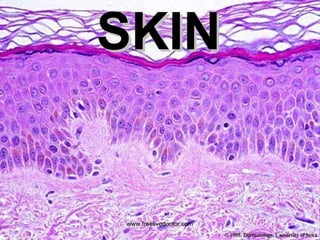
Skin is more than a fleshy surface for pimples, tattoos and wrinkles. Skin is the body's largest organ, and along with hair, nails, glands and nerves, is part of the integumentary system, according to Oregon State University. This system acts as a protective barrier between the outside and the inside of the body.
11. MACROSCOPIC TERMS Macule: Circumscribed lesion of up to 5 mm in diameter characterized by flatness and usually discolored (often red) Patch : Circumscribed lesion of more than 5 mm in diameter characterized by flatness and usually discolored (often red) Papule: Elevated dome-shaped or flat-topped lesion 5 mm or less across. Nodule: Elevated lesion with spherical contour greater than 5 mm across. Plaque: Elevated flat-topped lesion, usually greater than 5 mm across (may be caused by coalescent papules). Vesicle: Fluid-filled raised lesion 5 mm or less across. Bulla: Fluid-filled raised lesion greater than 5 mm across. Blister: Common term used for vesicle or bulla. Pustule: Discrete, pus-filled, raised lesion. Wheal: Itchy, transient, elevated lesion with variable blanching and erythema formed as the result of dermal edema. Scale: Dry, horny, platelike excrescence; usually the result of imperfect cornification (i.e., keratinization). Lichenification: Thickened and rough skin characterized by prominent skin markings; usually the result of repeated rubbing in susceptible persons. Excoriation: Traumatic lesion characterized by breakage of the epidermis, causing a raw linear area (i.e., a deep scratch) Onycholysis: Separation of nail plate from nail bed. www.freelivedoctor.com
12.
13. MICROSCOPIC TERMS Hyperkeratosis: Thickening of the stratum corneum, often associated with a qualitative abnormality of the keratin. Parakeratosis: Modes of keratinization characterized by the retention of the nuclei in the stratum corneum. On mucous membranes, parakeratosis is normal. Hypergranulosis: Hyperplasia of the stratum granulosum, often due to intense rubbing. Acanthosis: Diffuse epidermal hyperplasia. Papillomatosis: Surface elevation caused by hyperplasia and enlargement of contiguous dermal papillae. Dyskeratosis: Abnormal keratinization occurring prematurely within individual cells or groups of cells below the stratum granulosum. Generally the same as DYSPLASIA. Acantholysis: Loss of intercellular connections resulting in loss of cohesion between keratinocytes. Spongiosis: Intercellular edema of the epidermis. Hydropic swelling (ballooning): Intracellular edema of keratinocytes. Exocytosis: Infiltration of the epidermis by inflammatory or circulating blood cells. Erosion: Discontinuity of the skin exhibiting incomplete loss of the epidermis. Ulceration: Discontinuity of the skin exhibiting complete loss of the epidermis and often of portions of the dermis and even subcutaneous fat. Vacuolization: Formation of vacuoles within or adjacent to cells; often refers to basal cell-basement membrane zone area. Lentiginous: Referring to a linear pattern of melanocyte proliferation within the epidermal basal cell layer. Lentiginous melanocytic hyperplasia can occur as a reactive change or as part of a neoplasm of melanocytes. www.freelivedoctor.com