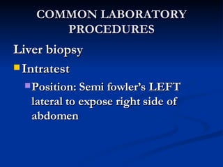
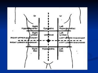
Medical Surgical Nursing The GASTRO-INTESTINAL System Nurse Licensure Examination Review
The Gastro-Intestinal System Review of the GIT Anatomy and Physiology Review of Common laboratory procedures Review of Common Symptoms and their nursing interventions Review of common disorders of the: Esophagus -gallbladder Stomach -exocrine pancreas Small intestine -liver Large Intestine
 
The GIT System: Anatomy and Physiology The GIT is composed of two general parts The main GIT starts from the mouth  Esophagus  Stomach  SI  LI The accessory organs are the  Salivary glands Liver Gallbladder Pancreas
The GIT ANATOMY The Mouth Contains the lips, cheeks, palate, tongue, teeth, salivary glands, masticatory/facial muscles and bones Anteriorly bounded by the lips Posteriorly bounded by the oropharynx
The GIT Physiology The Mouth Important for the mechanical digestion of food The saliva contains SALIVARY AMYLASE or PTYALIN that starts the INITIAL digestion of carbohydrates
The GIT ANATOMY The Esophagus A hollow collapsible tube Length- 10 inches Made up of stratified squamos epithelium
The GIT ANATOMY The Esophagus The upper third contains skeletal muscles The middle third contains mixed skeletal and smooth muscles The lower third contains smooth muscles and the esophago-gastric/ cardiac sphincter is found here
The GIT PHYSIOLOGY The Esophagus Functions to carry or propel foods from the oropharynx to the stomach Swallowing or deglutition is composed of three phases:
The GIT ANATOMY The stomach J-shaped organ in the epigastrium Contains four parts- the fundus, the cardia, the body and the pylorus The cardiac sphincter prevents the reflux of the contents into the esophagus The pyloric sphincter regulates the rate of gastric emptying into the duodenum Capacity is 1,500 ml!
The GIT PHYSIOLOGY The functions of the stomach are generally to digest the food (proteins) and to propel the digested materials into the SI for final digestion The Glands and cells in the stomach secrete digestive enzymes:
The GIT PHYSIOLOGY Stomach: 1. Parietal cells- HCl acid and Intrinsic factor 2. Chief cells- pepsin   digestion of PROTEINS! 3. Antral G-cells- gastrin 4. Argentaffin cells- serotonin 5. Mucus neck cells- mucus
The GIT ANATOMY The Small intestine Grossly divided into the Duodenum, Jejunum and Ileum The duodenum contains the two openings for the bile and pancreatic ducts The ileum is the longest part (about 12 feet)
The GIT physiology The intestinal glands secrete digestive enzymes that finalize the digestion of all foodstuff Enzymes for carbohydrates   disaccharidases Enzymes for proteins   dipeptidases and aminopeptidases Enzyme for lipids   intestinal lipase
The GIT ANATOMY The Large intestine Approximately 5 feet long, with parts: 1. The cecum   widest diameter, prone to rupture 2. The appendix 3. The ascending colon 4. The transverse colon 5. The descending colon 6. The sigmoid   most mobile, prone to twisting 7. The rectum
The GIT Physiology Absorbs water Eliminates wastes Bacteria in the colon synthesize Vitamin K Appendix participates in the immune system
The GIT Physiology SYMPATHETIC Generally  INHIBITORY! Decreased gastric secretions Decreased GIT motility But: Increased sphincteric tone and constriction of blood vessels PARASYMPATHETIC Generally  EXCITATORY! Increased gastric secretions Increased gastric motility But: Decreased sphincteric tone and dilation of blood vessels
The GIT ANATOMY The Liver The largest internal organ Located in the right upper quadrant Contains two lobes- the right and the left The hepatic ducts join together with the cystic duct to become the common bile duct
The GIT Physiology: LIVER Functions to store excess glucose, fats and amino acids Also stores the fat soluble vitamins- A, D and the water soluble- Vitamin B12 Produces the BILE for normal fat digestion The Von Kupffer cells remove bacteria in the portal blood Detoxifies ammonia into urea
The GIT anatomy The gallbladder Located below the liver  The cystic duct joins the hepatic duct to become the bile duct The common bile duct joins the pancreatic duct in the sphincter of Oddi in the first part of the duodenum
The GIT Physiology Stores and concentrates bile Contracts during the digestion of fats to deliver the bile Cholecystokinin  is released by the duodenal cells, causing the contraction of the gallbladder and relaxation of the sphincter of Oddi
The GIT anatomy The pancreas A retroperitoneal gland Functions as an endocrine and exocrine gland The pancreatic duct (major) joins the common bile duct in the sphincter of Oddi
The GIT Physiology The exocrine function of the pancreas is the secretion of digestive enzymes for carbohydrates, fats and proteins Pancreatic amylase   carbohydrates Pancreatic lipase (steapsin)   fats Trypsin, Chymotrypsin and Peptidases   proteins Bicarbonate   to neutralize the acidic chyme. Stimulated by SECRETIN!
Gastrointestinal Assessment Laboratory Procedures
COMMON LABORATORY PROCEDURES FECALYSIS Examination of stool consistency, color and the presence of occult blood. Special tests for fat, nitrogen, parasites, ova, pathogens and others
COMMON LABORATORY PROCEDURES FECALYSIS: Occult Blood Testing Instruct the patient to adhere to a 3-day meatless diet No intake of NSAIDS, aspirin and anti-coagulant Screening test for colonic cancer
COMMON LABORATORY PROCEDURES Upper GIT study: barium swallow Examines the upper GI tract Barium sulfate is usually used as contrast
COMMON LABORATORY PROCEDURES Upper GIT study: barium swallow Pre-test :  NPO post-midnight Post-test : Laxative is ordered, increase pt fluid intake, instruct that stools will turn white, monitor for obstruction
 
 
COMMON LABORATORY PROCEDURES Lower GIT study: barium enema Examines the lower GI tract Pre-test :  Clear liquid diet and laxatives, NPO post-midnight, cleansing enema prior to the test
COMMON LABORATORY PROCEDURES Lower GIT study: barium enema Post-test:  Laxative is ordered, increase patient fluid intake, instruct that stools will turn white, monitor for obstruction
 
COMMON LABORATORY PROCEDURES Gastric analysis Aspiration of gastric juice to measure pH, appearance, volume and contents Pre-test:  NPO 8 hours, avoidance of stimulants, drugs and smoking Post-test:  resume normal activities
COMMON LABORATORY PROCEDURES EGD (esophagogastroduodenoscopy) Visualization of the upper GIT by endoscope Pre-test : ensure consent, NPO 8 hours, pre-medications like atropine and anxiolytics
 
COMMON LABORATORY PROCEDURES EGD esophagogastroduodenoscopy Intra-test:  position :  LEFT  lateral to facilitate salivary drainage and easy access
COMMON LABORATORY PROCEDURES EGD (esophagogastroduodenoscopy) Post-test : NPO until gag reflex returns, place patient in  SIMS position until he awakens ,  monitor for complications, saline gargles for mild oral discomfort
COMMON LABORATORY PROCEDURES Lower GI- scopy Use of endoscope to visualize the anus, rectum, sigmoid and colon Pre-test:  consent, NPO 8 hours, cleansing enema until return is clear
 
COMMON LABORATORY PROCEDURES Lower GI- scopy Intra-test:  position is  LEFT  lateral, right leg is bent  and placed anteriorly Post-test:   bed rest, monitor for complications like bleeding and perforation
 
COMMON LABORATORY PROCEDURES Cholecystography Examination of the gallbladder to detect stones, its ability to concentrate, store and release the bile Pre-test:  ensure consent, ask allergies to iodine, seafood and dyes;  contrast medium is administered the night prior , NPO after contrast administration
COMMON LABORATORY PROCEDURES Cholecystography Post-test:  Advise that dysuria is common as the dye is excreted in the urine, resume normal activities
COMMON LABORATORY PROCEDURES Paracentesis Removal of peritoneal fluid for analysis
COMMON LABORATORY PROCEDURES Paracentesis Pre-test:  ensure consent, instruct to VOID and empty bladder, measure abdominal girth
COMMON LABORATORY PROCEDURES Paracentesis Intra-test:  Upright on the edge of the bed, back supported and feet resting on a foot stool
COMMON LABORATORY PROCEDURES Liver biopsy Pretest Consent NPO Check for the bleeding parameters
COMMON LABORATORY PROCEDURES Liver biopsy Intratest Position: Semi fowler’s LEFT lateral to expose right side of abdomen
COMMON LABORATORY PROCEDURES Liver biopsy Post-test : position on  RIGHT lateral with pillow underneath,  monitor VS and complications like bleeding, perforation. Instruct to avoid lifting objects for 1 week
The NURSING PROCESS in GIT Disorders Assessment Health history Nursing History PE Laboratory procedures
The ABDOMINAL examination The sequence to follow is: Inspection Auscultation Percussion Palpation
 
CONSTIPATION DIARRHEA DUMPING SYNDROME COMMON GIT SYMPTOMS AND MANAGEMENT
CONSTIPATION An abnormal infrequency and irregularity of defecation Multiple causations COMMON GIT SYMPTOMS AND MANAGEMENT
CONSTIPATION: Pathophysiology Interference with three functions of the colon 1. Mucosal transport 2. Myoelectric activity 3. Process of defecation COMMON GIT SYMPTOMS AND MANAGEMENT
COMMON GIT SYMPTOMS AND MANAGEMENT NURSING INTERVENTIONS 1. Assist physician in treating the underlying cause of constipation 2. Encourage to eat HIGH fiber diet to increase the bulk 3. Increase fluid intake 4. Administer prescribed laxatives, stool softeners 5. Assist in relieving stress
COMMON GIT SYMPTOMS AND MANAGEMENT Diarrhea Abnormal fluidity of the stool Multiple causes Gastrointestinal Diseases Hyperthyroidism Food poisoning
COMMON GIT SYMPTOMS AND MANAGEMENT Diarrhea Nursing Interventions 1. Increase fluid intake-  ORESOL is the most important treatment! 2. Determine and manage the cause 3. Anti-diarrheal drugs
COMMON GIT SYMPTOMS AND MANAGEMENT DUMPING SYNDROME A condition of rapid emptying of the gastric contents into the small intestine usually after a gastric surgery  Symptoms occur 30 minutes after eating
COMMON GIT SYMPTOMS AND MANAGEMENT PATHOPHYSIOLOGY Foods high in CHO and electrolytes must be diluted in the jejunum before absorption takes place.
COMMON GIT SYMPTOMS AND MANAGEMENT PATHOPHYSIOLOGY The rapid influx of stomach contents will cause distention of the jejunum early symptoms
COMMON GIT SYMPTOMS AND MANAGEMENT PATHOPHYSIOLOGY The hypertonic food bolus will draw fluid from the blood vessels to dilute the high concentrations of CHO and electrolytes in the food bolus
COMMON GIT SYMPTOMS AND MANAGEMENT Later, there is increased blood glucose stimulating the increased secretion of insulin
COMMON GIT SYMPTOMS AND MANAGEMENT Then, blood glucose will fall causing reactive hypoglycemia
COMMON GIT SYMPTOMS AND MANAGEMENT DUMPING SYNDROME ASSESSMENT FINDINGS: early symptoms 1. Nausea and Vomiting 2. Abdominal fullness 3. Abdominal cramping 4. Palpitation 5. Diaphoresis
COMMON GIT SYMPTOMS AND MANAGEMENT DUMPING SYNDROME ASSESSMENT FINDINGS: LATE symptoms:  6. Drowsiness  7. Weakness and Dizziness 8.  Hypoglycemia
COMMON GIT SYMPTOMS AND MANAGEMENT DS NURSING INTERVENTIONS 1. Advise patient to eat  LOW-carbohydrate  HIGH-fat and HIGH-protein diet 2. Instruct to eat  SMALL frequent  meals, include  MORE dry items . 3. Instruct to  AVOID consuming FLUIDS with meals
COMMON GIT SYMPTOMS AND MANAGEMENT DS NURSING INTERVENTIONS 4. Instruct to  LIE DOWN after meals 5. Administer anti-spasmodic medications to delay gastric emptying
GIT SYMPTOMS AND MANAGEMENT PERNICIOUS ANEMIA Results from Deficiency of vitamin B12 due to  autoimmune destruction of the parietal cells ,  lack of INTRINSIC FACTOR   or total removal of the stomach
GIT SYMPTOMS AND MANAGEMENT PERNICIOUS ANEMIA ASSESSMENT Severe pallor Fatigue Weight loss SMOOTH BEEFY-RED TONGUE Mild jaundice Paresthesia of extremities Balance disturbance
GIT SYMPTOMS AND MANAGEMENT NURSING INTERVENTION for Pernicious Anemia Lifetime injection of Vitamin B 12 weekly initially, then MONTHLY
Conditions of the GIT UPPER GI system
CONDITION OF THE ESOPHAGUS HIATAL HERNIA Protrusion of the esophagus into the diaphragm thru an opening Two types- Sliding hiatal hernia  ( most common) and Axial hiatal hernia
CONDITION OF THE ESOPHAGUS ASSESSMENT Findings in Hiatal hernia 1. Heartburn 2. Regurgitation 3. Dysphagia 4. 50%- without symptoms
CONDITION OF THE ESOPHAGUS DIAGNOSTIC TEST Barium swallow and fluoroscopy
CONDITION OF THE ESOPHAGUS NURSING INTERVENTIONS 1. Provide  small frequent feedings 2.  AVOID supine position  for 1 hour after eating 3.  Elevate the head  of the bed on 8-inch block 4. Provide pre-op and post-op care
CONDITION OF THE ESOPHAGUS Esophageal Varices Dilation and tortuosity of the submucosal veins in the distal esophagus  ETIOLOGY: commonly caused by PORTAL hypertension secondary to liver cirrhosis This is an Emergency condition!
CONDITION OF THE ESOPHAGUS ASSESSMENT findings for EV 1. Hematemesis 2. Melena 3. Ascites 4. jaundice 5. hepatomegaly/splenomegaly
CONDITION OF THE ESOPHAGUS ASSESSMENT findings for EV Signs of Shock- tachycardia, hypotension, tachypnea, cold clammy skin, narrowed pulse pressure
CONDITION OF THE ESOPHAGUS DIAGNOSTIC PROCEDURE Esophagoscopy
CONDITION OF THE ESOPHAGUS NURSING INTERVENTIONS FOR EV 1. Monitor VS strictly. Note for signs of shock 2. Monitor for LOC 3. Maintain NPO
CONDITION OF THE ESOPHAGUS NURSING INTERVENTIONS FOR EV 4. Monitor blood studies 5. Administer O2 6. prepare for blood transfusion
CONDITION OF THE ESOPHAGUS INTERVENTIONS FOR EV 7. prepare to administer Vasopressin and  Nitroglycerin 8. Assist in NGT and Sengstaken-Blakemore tube insertion for balloon tamponade
CONDITION OF THE ESOPHAGUS NURSING INTERVENTIONS FOR EV 9. Prepare to assist in surgical management: Endoscopic sclerotherapy Variceal ligation Shunt procedures
Conditions of the Stomach Gastro-esophageal reflux Backflow of gastric contents into the esophagus Usually due to incompetent lower esophageal sphincter , pyloric stenosis or motility disorder Symptoms may mimic ANGINA or MI
Conditions of the Stomach ASSESSMENT ( for GERD) Heartburn Dyspepsia Regurgitation Epigastric pain Difficulty swallowing Ptyalism
Conditions of the Stomach Diagnostic test Endoscopy or barium swallow Gastric ambulatory pH analysis Note for the pH of the esophagus, usually done for 24 hours The pH probe is located 5 inches above the lower esophageal sphincter The machine registers the different pH of the refluxed material into the esophagus
Conditions of the Stomach NURSING INTERVENTIONS 1. Instruct the patient to AVOID stimulus that increases stomach pressure and decreases GES pressure 2. Instruct to avoid spices, coffee, tobacco and carbonated drinks 3. Instruct to eat LOW-FAT, HIGH-FIBER diet
Conditions of the Stomach NURSING INTERVENTIONS 4. Avoid foods and drinks TWO hours before bedtime 5. Elevate the head of the bed with an approximately 8-inch block
Conditions of the Stomach NURSING INTERVENTIONS 6. Administer prescribed H2-blockers, PPI and prokinetic meds like cisapride, metochlopromide 7. Advise proper weight reduction
Conditions of the Stomach GASTRITIS Inflammation of the gastric mucosa May be Acute or Chronic Etiology: Acute- bacteria, irritating foods, NSAIDS, alcohol, bile and radiation Etiology: Chronic- Ulceration, bacteria, Autoimmune disease, diet, alcohol, smoking
Conditions of the Stomach PATHOPHYSIOLOGY OF Gastritis Insults   cause gastric mucosal damage   inflammation, hyperemia and edema   superficial erosions    decreased gastric secretions, ulcerations and bleeding
Conditions of the Stomach ASSESSMENT  (Acute) Dyspepsia Headache Anorexia Nausea/Vomiting ASSESSMENT (Chronic) Pyrosis Singultus Sour taste in the mouth Dyspepsia N/V/anorexia Pernicious anemia
Conditions of the Stomach DIAGNOSTIC PROCEDURE EGD- to visualize the gastric mucosa for inflammation Low levels of HCl Biopsy to establish correct diagnosis whether acute or chronic
Conditions of the Stomach NURSING INTERVENTIONS 1. Give  BLAND  diet 2. Monitor for signs of complications like bleeding, obstruction and pernicious anemia 3. Instruct to avoid spicy foods, irritating foods, alcohol and caffeine
Conditions of the Stomach NURSING INTERVENTIONS 4. Administer prescribed medications- H2 blockers, antibiotics, mucosal protectants 5. Inform the need for Vitamin B12 injection if deficiency is present
Conditions of the Stomach PEPTIC ULCER DISEASE An ulceration of the gastric and duodenal lining May be referred as to location as Gastric ulcer in the stomach, or Duodenal ulcer in the duodenum Most common Peptic ulceration: anterior part of the upper duodenum
Conditions of the Stomach PATHOPHYSIOLOGY of PUD Disturbance in acid secretion and mucosal protection Increased acidity or decreased mucosal resistance   erosion and ulceration
Conditions of the Stomach GASTRIC ULCER Ulceration  of the gastric mucosa, submucosa and rarely the muscularis
Conditions of the Stomach GASTRIC ULCER Risk factors: Stress, smoking, NSAIDS abuse, Alcohol, Helicobacter pylori infection, type A personality and History of gastritis  Incidence is high in older adults Acid secretion is NORMAL
Conditions of the Stomach ASSESSMENT (Gastric Ulcer) Epigastric pain Characteristic: Gnawing, sharp pain in the mid-epigastrium 1-2 hours  AFTER  eating, often  NOT RELIEVED  by food intake, sometimes AGGRAVATING the pain!
Conditions of the Stomach ASSESSMENT (Gastric Ulcer) Nausea  Vomiting is more common Hematemesis Weight loss
Conditions of the Stomach DIAGNOSTIC PROCEDURES 1. EGD to visualize the ulceration 2. Urea breath test for H. pylori infection 3. Biopsy- to rule out gastric cancer
Conditions of the Stomach NURSING INTERVENTIONS 1. Give BLAND diet, small frequent meals during the active phase of the disease 2. Administer prescribed medications- H2 blockers, PPI, mucosal barrier protectants and antacids
Conditions of the Stomach NURSING INTERVENTIONS 3. Monitor for complications of bleeding, perforation and intractable pain 4. provide teaching about stress reduction and relaxation techniques
Conditions of the Stomach NURSING INTERVENTIONS FOR BLEEDING 1. Maintain on NPO  2. Administer IVF and medications 3. Monitor hydration status, hematocrit and hemoglobin
Conditions of the Stomach NURSING INTERVENTIONS FOR BLEEDING 4. Assist with SALINE lavage 5. Insert  NGT for decompression and lavage
Conditions of the Stomach NURSING INTERVENTIONS FOR BLEEDING 6. Prepare to administer blood transfusion 7. Prepare to give VASOPRESSIN to induce vasoconstriction to reduce bleeding 8. Prepare patient for SURGERY if warranted
Conditions of the Stomach SURGICAL PROCEDURES FOR PUD Total gastrectomy, vagotomy, gastric resection, Billroth I and II, pyloroplasty
 
Conditions of the Stomach SURGICAL PROCEDURES FOR PUD Post-operative Nursing management 1. Monitor VS 2. Post-op position: FOWLER’S 3. NPO until peristalsis returns 4. Monitor for bowel sounds 5. Monitor for complications of surgery
Conditions of the Stomach Post-operative Nursing management 6. Monitor I and O, IVF 7. Maintain NGT 8. Diet progress: clear liquid   full liquid   six bland meals 9. Manage DUMPING SYNDROME
Condition of the Duodenum DUODENAL ULCER Ulceration of duodenal mucosa and submucosa Usually due to increased gastric acidity
Condition of the Duodenum DUODENAL ULCER ASSESSMENT PAIN characteristic: Burning pain in the mid-epigastrium 2-4 HOURS after eating or during the night,  RELIEVED  by food intake
 
Condition of the Duodenum DIAGNOSTIC TESTS EGD and Biopsy
 
 
Condition of the Duodenum NURSING INTERVENTIONS 1. Same as for gastric ulceration 2. Patient teaching-avoid alcohol,  smoking, caffeine and carbonated drinks Take NSAIDS with meals Adhere to medication regimen
Ulcers (-) cancer (+) cancer Less likely bleeding and vomiting Bleeding, weight loss and vomiting RELIEVES by food WORSENS by food, RELIEVED by VOMITING Pain late after eating (2-4 hours) Pain early after eating INCREASED acidity Normal Acidity Younger Older DUODENAL GASTRIC
Conditions of the Lower Tract Small and Large Intestine
CONDITIONS OF THE SMALL INTESTINE CROHN’S DISEASE Also called Regional Enteritis An inflammatory disease of the GIT affecting usually the small intestine
CONDITIONS OF THE SMALL INTESTINE CROHN’S DISEASE ETIOLOGY: unknown The terminal ileum thickens, with scarring, ulcerations, abscess formation and narrowing of the lumen
CONDITIONS OF THE SMALL INTESTINE ASSESSMENT findings for CD 1. Fever 2. Abdominal distention 3. Diarrhea 4. Colicky abdominal pain  5. Anorexia/N/V 6. Weight loss 7. Anemia
CONDITIONS OF THE LARGE INTESTINE ULCERATIVE COLITIS Ulcerative and inflammatory condition of the GIT usually affecting the large intestine The colon becomes edematous and develops bleeding ulcerations Scarring develops overtime with impaired water absorption and loss of elasticity
CONDITIONS OF THE LARGE INTESTINE ASSESSMENT findings for UC 1. Anorexia 2. Weight loss 3. Fever 4. SEVERE diarrhea with Rectal bleeding 5. Anemia 6. Dehydration 7. Abdominal pain and cramping
NURSING INTERVENTIONS for CD and UC 1. Maintain NPO during the active phase 2. Monitor for complications like severe bleeding, dehydration, electrolyte imbalance 3. Monitor bowel sounds, stool and blood studies 4. Restrict activities 5. Administer IVF, electrolytes and TPN if prescribed
NURSING INTERVENTIONS for CD and UC 6. Instruct the patient to AVOID gas-forming foods, MILK products and foods such as whole grains, nuts, RAW fruits and vegetables especially SPINACH, pepper, alcohol and caffeine 7. Diet progression- clear liquid   LOW residue, high protein diet 8. Administer drugs- anti-inflammatory, antibiotics, steroids, bulk-forming agents and vitamin/iron supplements
CONDITIONS OF THE LARGE INTESTINE APPENDICITIS Inflammation of the vermiform appendix
 
CONDITIONS OF THE LARGE INTESTINE APPENDICITIS ETIOLOGY: usually fecalith, lymphoid hyperplasia, foreign body and helminthic obstruction
CONDITIONS OF THE LARGE INTESTINE APPENDICITIS PATHOPHYSIOLOGY Obstruction of lumen   increased pressure   decreased blood supply   bacterial proliferation and mucosal inflammation   ischemia   necrosis   rupture
CONDITIONS OF THE LARGE INTESTINE ASSESSMENT FINDINGS for Appendicitis 1. Abdominal pain: begins in the umbilicus then localizes in the RLQ (Mc Burney’s point) 2. Anorexia 3. Nausea and Vomiting
CONDITIONS OF THE LARGE INTESTINE ASSESSMENT FINDINGS for Appendicitis 4. Fever 5. Rebound tenderness and abdominal rigidity (if perforated) 6. Constipation or diarrhea
CONDITIONS OF THE LARGE INTESTINE DIAGNOSTIC TESTS 1. CBC- reveals increased WBC count 2. Ultrasound 3. Abdominal X-ray
CONDITIONS OF THE LARGE INTESTINE NURSING INTERVENTIONS 1. Preoperative care NPO Consent Monitor for perforation and signs of shock
CONDITIONS OF THE LARGE INTESTINE NURSING INTERVENTIONS 1. Preoperative care Monitor bowel sounds, fever and hydration status POSITION of Comfort: RIGHT SIDELYING in a low FOWLER’S Avoid Laxatives, enemas & HEAT APPLICATION
CONDITIONS OF THE LARGE INTESTINE 2. Post-operative care Monitor VS and signs of surgical complications Maintain NPO until bowel function returns If rupture occurred, expect drains and IV antibiotics
CONDITIONS OF THE LARGE INTESTINE 2. Post-operative care POSITION post-op:  RIGHT side-lying, semi- fowler’s to decrease tension on incision, and  legs flexed to promote drainage Administer prescribed pain medications
CONDITIONS OF THE LARGE INTESTINE Hemorrhoids Abnormal dilation and weakness of the veins of the anal canal Variously classified as Internal or External, Prolapsed, Thrombosed and Reducible
CONDITIONS OF THE LARGE INTESTINE Hemorrhoids PATHOPHYSIOLOGY Increased pressure in the  hemorrhoidal tissue due to straining, pregnancy, etc   dilatation of veins
CONDITIONS OF THE LARGE INTESTINE Internal hemorrhoids These dilated veins lie above the internal anal sphincter Usually, the condition is PAINLESS
CONDITIONS OF THE LARGE INTESTINE External hemorrhoids These dilated veins lie below the internal anal sphincter Usually, the condition is PAINFUL
CONDITIONS OF THE LARGE INTESTINE ASSESSMENT findings for Hemorrhoids 1. Internal hemorrhoids- cannot be seen on the peri-anal area 2. External hemorrhoids- can be seen 3. Bright red bleeding with each defecation
CONDITIONS OF THE LARGE INTESTINE ASSESSMENT findings for Hemorrhoids 4. Rectal/ perianal pain 5. Rectal itching 6. Skin tags
CONDITIONS OF THE LARGE INTESTINE DIAGNOSTIC TEST 1. Anoscopy 2. Digital rectal examination
CONDITIONS OF THE LARGE INTESTINE NURSING INTERVENTIONS 1. Advise patient to apply cold packs to the anal/rectal area followed by a SITZ bath 2. Apply astringent like witch hazel soaks
CONDITIONS OF THE LARGE INTESTINE NURSING INTERVENTIONS 3. Encourage HIGH-fiber diet and fluids 4. Administer stool softener as prescribed
CONDITIONS OF THE LARGE INTESTINE Post-operative care for hemorrhoidectomy 1. Position:  Prone or Side-lying 2. Maintain dressing over the surgical site
CONDITIONS OF THE LARGE INTESTINE Post-operative care for hemorrhoidectomy 3. Monitor for bleeding 4. Administer analgesics and stool softeners 5. Advise  the use of SITZ bath 3-4 times a day
CONDITIONS OF THE LARGE INTESTINE DIVERTICULOSIS AND DIVERTICULITIS Diverticulosis Abnormal out-pouching of the intestinal mucosa occurring in any part of the LI most commonly in the sigmoid Diverticulitis Inflammation of the diverticulosis
CONDITIONS OF THE LARGE INTESTINE PATHOPHYSIOLOGY Increased intraluminal pressure, LOW volume in the lumen and Decreased muscle strength in the colon wall   herniation of the colonic mucosa
CONDITIONS OF THE LARGE INTESTINE ASSESSMENT findings for D/D 1. Left lower Quadrant pain 2. Flatulence 3. Bleeding per rectum 4. nausea and vomiting 5. Fever 6. Palpable, tender rectal mass
CONDITIONS OF THE LARGE INTESTINE DIAGNOSTIC STUDIES 1. If no active inflammation, COLONOSCOPY and Barium Enema 2. CT scan is the procedure of choice! 3. Abdominal X-ray
CONDITIONS OF THE LARGE INTESTINE NURSING INTERVENTIONS 1. Maintain NPO during acute phase 2. Provide bed rest 3. Administer antibiotics, analgesics like meperidine (morphine is not used) and anti-spasmodics 4. Monitor for potential complications like perforation, hemorrhage and fistula 5. Increase fluid intake
CONDITIONS OF THE LARGE INTESTINE NURSING INTERVENTIONS 6. Avoid gas-forming foods or HIGH-roughage foods containing seeds, nuts to avoid trapping 7. introduce soft, high fiber foods ONLY after the inflammation subsides 8. Instruct to avoid activities that increase intra-abdominal pressure
Conditions of the GIT accessory organs The liver
CONDITION OF THE LIVER Liver Cirrhosis A chronic, progressive disease characterized by a diffuse damage to the hepatic cells The liver heals with scarring, fibrosis and nodular regeneration
CONDITION OF THE LIVER Liver Cirrhosis ETIOLOGY:  Post-infection, Alcohol, Cardiac diseases, Schisostoma, Biliary obstruction
 
Liver physiology and Pathophysiology =  Gynecomastia, testes atrophy 8. Metabolizes estrogen = Deficiencies of Vit and min 7. Stores Vit and minerals =Hyperammonemia 6. Converts ammonia to urea = Jaundice and pruritus 5. Secreting bile = Bleeding tendencies 4. Synthesizes Clotting factors = Decreased Antibody formation 3. Synthesizes globulins = Hypoproteinemia 2. Synthesizes proteins = Hypoglycemia 1. Stores glycogen Abnormality in function Normal Function
CONDITION OF THE LIVER ASSESSMENT FINDINGS 1. Anorexia and weight loss 2. Jaundice 3. Fatigue
CONDITION OF THE LIVER ASSESSMENT FINDINGS 4. Early morning nausea and vomiting 5. RUQ abdominal pain 6. Ascites 7. Signs of Portal hypertension
 
CONDITION OF THE LIVER NURSING INTERVENTIONS 1. Monitor VS, I and O, Abdominal girth, weight, LOC and Bleeding 2. Promote rest.  Elevated the head of  the bed to minimize dyspnea
CONDITION OF THE LIVER NURSING INTERVENTIONS 3. Provide Moderate to LOW-protein (1 g/kg/day) and LOW-sodium diet 4. Provide supplemental vitamins (especially K) and minerals
CONDITION OF THE LIVER NURSING INTERVENTIONS 5. Administer prescribed Diuretics=  to reduce ascites and edema Lactulose=  to reduce NH4 in the bowel Antacids and  Neomycin =  to kill bacterial flora that cause NH production
CONDITION OF THE LIVER NURSING INTERVENTIONS 6. Avoid hepatotoxic drugs Paracetamol Anti-tubercular drugs
CONDITION OF THE LIVER NURSING INTERVENTIONS 7. Reduce the risk of injury  Side rails reorientation Assistance in ambulation Use of electric razor and soft-bristled toothbrush
CONDITION OF THE LIVER NURSING INTERVENTIONS 8. Keep equipments ready including Sengstaken-Blakemore tube, IV fluids, Medications to treat hemorrhage
CONDITION OF THE LIVER Albumin, Amino acid Vitamin K (menadione) Diuretics, Neomycin, Lactulose 6. Administer Medications: Done to relieve abdominal pressure 5. Assist in paracentesis To prevent bleeding 4. Pressure onto injection site To relieve pruritus 3. Benadryl and mild soap To reduce NH production 2. Low protein diet To reduce edema 1. Low sodium Diet Rationale Nursing Interventions
Conditions of the Accessory organs The Gallbladder
CONDITION OF THE GALLBLADDER Cholecystitis Inflammation of the gallbladder Can be acute or chronic
CONDITION OF THE GALLBLADDER Cholecystitis Acute cholecystitis usually is due to gallbladder stones
CONDITION OF THE GALLBLADDER Cholecystitis Chronic cholecystitis is usually due to long standing gall bladder inflammation
 
Cholelithiasis Formation of GALLSTONES in the biliary apparatus
Predisposing FACTORS “ F” Female Fat Forty Fertile Fair
 
Pathophysiology Supersaturated bile, Biliary stasis Stone formation Blockage of Gallbladder Inflammation, Mucosal Damage and WBC infiltration
Pathophysiology Less bile in the duodenum Impaired fat digestion and absorption Vitamin ADEK mal-absorption, STEATORHEA with increased gas formation Jaundice ACHOLIC stools
CONDITION OF THE GALLBLADDER ASSESSMENT findings for cholecystitis 1. Indigestion, belching and flatulence 2. Fatty food intolerance, steatorrhea
CONDITION OF THE GALLBLADDER ASSESSMENT findings for cholecystitis 3. Epigastric pain that radiates to the scapula or localized at the RUQ 4. Mass at the RUQ
CONDITION OF THE GALLBLADDER ASSESSMENT findings for cholecystitis 5. Murphy’s sign 6. Jaundice 7. dark orange and foamy urine
CONDITION OF THE GALLBLADDER DIAGNOSTIC PROCEDURES 1. Ultrasonography- can detect the stones  2. Abdominal X-ray 3. Cholecystography
CONDITION OF THE GALLBLADDER DIAGNOSTIC PROCEDURES 4. WBC count increased 5. Oral cholecystography cannot visualize the gallbladder 6. ERCP: revels inflamed gallbladder with gallstone
CONDITION OF THE GALLBLADDER NURSING INTERVENTIONS 1. Maintain NPO in the active phase 2. Maintain NGT decompression
CONDITION OF THE GALLBLADDER NURSING INTERVENTIONS 3. Administer prescribed medications to relieve pain.  Usually Demerol (MEPERIDINE) Codeine and Morphine may cause spasm of the Sphincter   increased pain. Morphine cause  MOREPAIN
CONDITION OF THE GALLBLADDER 4. Instruct patient to  AVOID HIGH- fat diet and GAS-forming foods 5. Assist in surgical and non-surgical measures 6. Surgical procedures- Cholecystectomy, Choledochotomy, laparoscopy
CONDITION OF THE GALLBLADDER PHARMACOLOGIC THERAPY Analgesic- Meperidine Chenodeoxycholic acid= to dissolve the gallstones Antacids Anti-emetics
 
 
CONDITION OF THE GALLBLADDER Post-operative nursing interventions 1. Monitor for surgical complications 2. Post-operative position after recovery from anesthesia-  LOW FOWLER’s
CONDITION OF THE GALLBLADDER Post-operative nursing interventions 3. Encourage early ambulation  4.  Administer medication before coughing and deep breathing exercises 5. Advise client to splint the abdomen to prevent discomfort during coughing
CONDITION OF THE GALLBLADDER Post-operative nursing interventions 6. Administer analgesics, antiemetics, antacids 7. Care of the biliary drainageor T-tube drainage 8. Fat restriction is only limited to 4-6 weeks. Normal diet is resumed
Conditions of the accessory organs The pancreas: Exocrine function
CONDITION OF THE PANCREAS Pancreatitis Inflammation of the pancreas Can be acute or chronic
CONDITION OF THE PANCREAS Pancreatitis Etiology and predisposing factors Alcoholism Hypercalcemia Trauma Hyperlipidemia
CONDITION OF THE PANCREAS Pancreatitis Etiology and predisposing factors Biliary tract disease - cholelithiasis Bacterial disease PUD Mumps
CONDITION OF THE PANCREAS PATHOPHYSIOLOGY of acute pancreatitis Self-digestion of the pancreas by its own digestive enzymes principally TRYPSIN
CONDITION OF THE PANCREAS PATHOPHYSIOLOGY of acute pancreatitis Spasm, edema or block in the Ampulla of Vater   reflux of proteolytic enzymes   auto digestion of the pancreas   inflammation
CONDITION OF THE PANCREAS PATHOPHYSIOLOGY of acute pancreatitis Autodigestion of pancreatic tissue Hemorrhage, Necrosis and Inflammation KININ ACTIVATION will result to increased permeability Loss of Protein-rich fluid into the peritoneum HYPOVOLEMIA
CONDITION OF THE PANCREAS ASSESSMENT findings 1. Abdominal pain- acute onset, occurring after a heavy meal or alcohol intake 2. Abdominal guarding
CONDITION OF THE PANCREAS ASSESSMENT findings 3. Bruising on the flanks and umbilicus 4. N/V, jaundice 5. Hypotension and hypovolemia 6.  HYPERGLYCEMIA, HYPOCALCEMIA 7. Signs of shock
CONDITION OF THE PANCREAS DIAGNOSTIC TESTS 1. Serum amylase and serum lipase 2. Ultrasound 3. WBC 4. Serum calcium 5. CT scan 6. Hemoglobin and hematocrit
CONDITION OF THE PANCREAS NURSING INTERVENTIONS 1. Assist in pain management. Usually,  Demerol is given . Morphine is AVOIDED 2. Assist in correction of Fluid and Blood loss
CONDITION OF THE PANCREAS NURSING INTERVENTIONS 3.  Place patient on NPO  to inhibit pancreatic stimulation 4.  NGT insertion to decompress distention and remove gastric secretions 5. Maintain on bed rest
CONDITION OF THE PANCREAS NURSING INTERVENTIONS 7. Position patient in  SEMI-FOWLER’s to decrease pressure on the diaphragm  8. Deep breathing and coughing exercises 9. Provide parenteral nutrition
CONDITION OF THE PANCREAS NURSING INTERVENTIONS 10. Introduce oral feedings gradually- HIGH carbo, LOW FAT 11. Maintain skin integrity 12. Manage shock and other complications
Quick Summary Peptic Ulcer Ulceration of mucosa; In the stomach or duodenum Outstanding Symptom: PAIN Nursing Goal: Allow ulcer to heal, prevent complication Rest: physical and Mental Eliminate certain foods Medications: antacid, H2 blockers, Proton Pump inhibitors, antibiotics, mucosal protectants Surgery: Vagotomy, Billroth 1 and 2
Quick Summary Liver Cirrhosis Destruction of liver with replacement by scars Common causes: alcoholism, post-hepatitic Manifestations related to liver derangements Jaundice, Ascites, splenomegaly, bleeding, enceph Nursing goal: Control manifestations and maximize liver function
Quick Summary Liver Cirrhosis Encourage rest Avoid hepatotoxic drugs Diet: HIGH calorie, Restricted protein, LOW Na Weight client and measure abdominal girth daily Provide skin care for jaundice and edema Assess for bleeding: esophageal, rectal, cutaneous DRUGS: Antacids, Diuretics, Albumin, Neomycin and Lactulose
Quick Summary Cholecystitis Inflammation of the gallbladder commonly caused by cholelithiasis (Female, Fat, Forty, Fertile, Fair) Manifestations: Fat intolerance, RUQ pain, Nausea and vomiting, Jaundice, Murphy’s sign Nursing Goal: Relieve symptoms and assist in stone removal
Quick Summary Cholecystitis Administer MEPERIDINE, avoid morphine Maintain Fluid and electrolyte balance Maintain a LOW fat diet Semi-fowler’s position Assist in surgery Care of the T-tube
Quick Summary Pancreatitis Inflammation of the pancreas brought about by the digestion of the organ by the enzyme it produces Common causes: Alcoholism, stone Manifestations: Extreme upper abdominal pain radiating into the back, vomiting, nausea, Abdominal distention, Steatorrhea and weight loss Laboratory: ELEVATED lipase and amylase
Quick Summary Pancreatitis Nursing Goal : relieve symptoms, maintain blood volume and GIT rest NPO Provide IVF and Parenteral nutrition Drugs: MEPERIDINE, never morphine, Antacids, anticholinergics After Acute phase: LOW fat diet, avoid alcohol, fat and vitamin replacements
End Of GIT SYSTEM

NurseReview.Org Gastrointestinal System

  • 1.
    Medical Surgical NursingThe GASTRO-INTESTINAL System Nurse Licensure Examination Review
  • 2.
    The Gastro-Intestinal SystemReview of the GIT Anatomy and Physiology Review of Common laboratory procedures Review of Common Symptoms and their nursing interventions Review of common disorders of the: Esophagus -gallbladder Stomach -exocrine pancreas Small intestine -liver Large Intestine
  • 3.
  • 4.
    The GIT System:Anatomy and Physiology The GIT is composed of two general parts The main GIT starts from the mouth  Esophagus  Stomach  SI  LI The accessory organs are the Salivary glands Liver Gallbladder Pancreas
  • 5.
    The GIT ANATOMYThe Mouth Contains the lips, cheeks, palate, tongue, teeth, salivary glands, masticatory/facial muscles and bones Anteriorly bounded by the lips Posteriorly bounded by the oropharynx
  • 6.
    The GIT PhysiologyThe Mouth Important for the mechanical digestion of food The saliva contains SALIVARY AMYLASE or PTYALIN that starts the INITIAL digestion of carbohydrates
  • 7.
    The GIT ANATOMYThe Esophagus A hollow collapsible tube Length- 10 inches Made up of stratified squamos epithelium
  • 8.
    The GIT ANATOMYThe Esophagus The upper third contains skeletal muscles The middle third contains mixed skeletal and smooth muscles The lower third contains smooth muscles and the esophago-gastric/ cardiac sphincter is found here
  • 9.
    The GIT PHYSIOLOGYThe Esophagus Functions to carry or propel foods from the oropharynx to the stomach Swallowing or deglutition is composed of three phases:
  • 10.
    The GIT ANATOMYThe stomach J-shaped organ in the epigastrium Contains four parts- the fundus, the cardia, the body and the pylorus The cardiac sphincter prevents the reflux of the contents into the esophagus The pyloric sphincter regulates the rate of gastric emptying into the duodenum Capacity is 1,500 ml!
  • 11.
    The GIT PHYSIOLOGYThe functions of the stomach are generally to digest the food (proteins) and to propel the digested materials into the SI for final digestion The Glands and cells in the stomach secrete digestive enzymes:
  • 12.
    The GIT PHYSIOLOGYStomach: 1. Parietal cells- HCl acid and Intrinsic factor 2. Chief cells- pepsin  digestion of PROTEINS! 3. Antral G-cells- gastrin 4. Argentaffin cells- serotonin 5. Mucus neck cells- mucus
  • 13.
    The GIT ANATOMYThe Small intestine Grossly divided into the Duodenum, Jejunum and Ileum The duodenum contains the two openings for the bile and pancreatic ducts The ileum is the longest part (about 12 feet)
  • 14.
    The GIT physiologyThe intestinal glands secrete digestive enzymes that finalize the digestion of all foodstuff Enzymes for carbohydrates  disaccharidases Enzymes for proteins  dipeptidases and aminopeptidases Enzyme for lipids  intestinal lipase
  • 15.
    The GIT ANATOMYThe Large intestine Approximately 5 feet long, with parts: 1. The cecum  widest diameter, prone to rupture 2. The appendix 3. The ascending colon 4. The transverse colon 5. The descending colon 6. The sigmoid  most mobile, prone to twisting 7. The rectum
  • 16.
    The GIT PhysiologyAbsorbs water Eliminates wastes Bacteria in the colon synthesize Vitamin K Appendix participates in the immune system
  • 17.
    The GIT PhysiologySYMPATHETIC Generally INHIBITORY! Decreased gastric secretions Decreased GIT motility But: Increased sphincteric tone and constriction of blood vessels PARASYMPATHETIC Generally EXCITATORY! Increased gastric secretions Increased gastric motility But: Decreased sphincteric tone and dilation of blood vessels
  • 18.
    The GIT ANATOMYThe Liver The largest internal organ Located in the right upper quadrant Contains two lobes- the right and the left The hepatic ducts join together with the cystic duct to become the common bile duct
  • 19.
    The GIT Physiology:LIVER Functions to store excess glucose, fats and amino acids Also stores the fat soluble vitamins- A, D and the water soluble- Vitamin B12 Produces the BILE for normal fat digestion The Von Kupffer cells remove bacteria in the portal blood Detoxifies ammonia into urea
  • 20.
    The GIT anatomyThe gallbladder Located below the liver The cystic duct joins the hepatic duct to become the bile duct The common bile duct joins the pancreatic duct in the sphincter of Oddi in the first part of the duodenum
  • 21.
    The GIT PhysiologyStores and concentrates bile Contracts during the digestion of fats to deliver the bile Cholecystokinin is released by the duodenal cells, causing the contraction of the gallbladder and relaxation of the sphincter of Oddi
  • 22.
    The GIT anatomyThe pancreas A retroperitoneal gland Functions as an endocrine and exocrine gland The pancreatic duct (major) joins the common bile duct in the sphincter of Oddi
  • 23.
    The GIT PhysiologyThe exocrine function of the pancreas is the secretion of digestive enzymes for carbohydrates, fats and proteins Pancreatic amylase  carbohydrates Pancreatic lipase (steapsin)  fats Trypsin, Chymotrypsin and Peptidases  proteins Bicarbonate  to neutralize the acidic chyme. Stimulated by SECRETIN!
  • 24.
  • 25.
    COMMON LABORATORY PROCEDURESFECALYSIS Examination of stool consistency, color and the presence of occult blood. Special tests for fat, nitrogen, parasites, ova, pathogens and others
  • 26.
    COMMON LABORATORY PROCEDURESFECALYSIS: Occult Blood Testing Instruct the patient to adhere to a 3-day meatless diet No intake of NSAIDS, aspirin and anti-coagulant Screening test for colonic cancer
  • 27.
    COMMON LABORATORY PROCEDURESUpper GIT study: barium swallow Examines the upper GI tract Barium sulfate is usually used as contrast
  • 28.
    COMMON LABORATORY PROCEDURESUpper GIT study: barium swallow Pre-test : NPO post-midnight Post-test : Laxative is ordered, increase pt fluid intake, instruct that stools will turn white, monitor for obstruction
  • 29.
  • 30.
  • 31.
    COMMON LABORATORY PROCEDURESLower GIT study: barium enema Examines the lower GI tract Pre-test : Clear liquid diet and laxatives, NPO post-midnight, cleansing enema prior to the test
  • 32.
    COMMON LABORATORY PROCEDURESLower GIT study: barium enema Post-test: Laxative is ordered, increase patient fluid intake, instruct that stools will turn white, monitor for obstruction
  • 33.
  • 34.
    COMMON LABORATORY PROCEDURESGastric analysis Aspiration of gastric juice to measure pH, appearance, volume and contents Pre-test: NPO 8 hours, avoidance of stimulants, drugs and smoking Post-test: resume normal activities
  • 35.
    COMMON LABORATORY PROCEDURESEGD (esophagogastroduodenoscopy) Visualization of the upper GIT by endoscope Pre-test : ensure consent, NPO 8 hours, pre-medications like atropine and anxiolytics
  • 36.
  • 37.
    COMMON LABORATORY PROCEDURESEGD esophagogastroduodenoscopy Intra-test: position : LEFT lateral to facilitate salivary drainage and easy access
  • 38.
    COMMON LABORATORY PROCEDURESEGD (esophagogastroduodenoscopy) Post-test : NPO until gag reflex returns, place patient in SIMS position until he awakens , monitor for complications, saline gargles for mild oral discomfort
  • 39.
    COMMON LABORATORY PROCEDURESLower GI- scopy Use of endoscope to visualize the anus, rectum, sigmoid and colon Pre-test: consent, NPO 8 hours, cleansing enema until return is clear
  • 40.
  • 41.
    COMMON LABORATORY PROCEDURESLower GI- scopy Intra-test: position is LEFT lateral, right leg is bent and placed anteriorly Post-test: bed rest, monitor for complications like bleeding and perforation
  • 42.
  • 43.
    COMMON LABORATORY PROCEDURESCholecystography Examination of the gallbladder to detect stones, its ability to concentrate, store and release the bile Pre-test: ensure consent, ask allergies to iodine, seafood and dyes; contrast medium is administered the night prior , NPO after contrast administration
  • 44.
    COMMON LABORATORY PROCEDURESCholecystography Post-test: Advise that dysuria is common as the dye is excreted in the urine, resume normal activities
  • 45.
    COMMON LABORATORY PROCEDURESParacentesis Removal of peritoneal fluid for analysis
  • 46.
    COMMON LABORATORY PROCEDURESParacentesis Pre-test: ensure consent, instruct to VOID and empty bladder, measure abdominal girth
  • 47.
    COMMON LABORATORY PROCEDURESParacentesis Intra-test: Upright on the edge of the bed, back supported and feet resting on a foot stool
  • 48.
    COMMON LABORATORY PROCEDURESLiver biopsy Pretest Consent NPO Check for the bleeding parameters
  • 49.
    COMMON LABORATORY PROCEDURESLiver biopsy Intratest Position: Semi fowler’s LEFT lateral to expose right side of abdomen
  • 50.
    COMMON LABORATORY PROCEDURESLiver biopsy Post-test : position on RIGHT lateral with pillow underneath, monitor VS and complications like bleeding, perforation. Instruct to avoid lifting objects for 1 week
  • 51.
    The NURSING PROCESSin GIT Disorders Assessment Health history Nursing History PE Laboratory procedures
  • 52.
    The ABDOMINAL examinationThe sequence to follow is: Inspection Auscultation Percussion Palpation
  • 53.
  • 54.
    CONSTIPATION DIARRHEA DUMPINGSYNDROME COMMON GIT SYMPTOMS AND MANAGEMENT
  • 55.
    CONSTIPATION An abnormalinfrequency and irregularity of defecation Multiple causations COMMON GIT SYMPTOMS AND MANAGEMENT
  • 56.
    CONSTIPATION: Pathophysiology Interferencewith three functions of the colon 1. Mucosal transport 2. Myoelectric activity 3. Process of defecation COMMON GIT SYMPTOMS AND MANAGEMENT
  • 57.
    COMMON GIT SYMPTOMSAND MANAGEMENT NURSING INTERVENTIONS 1. Assist physician in treating the underlying cause of constipation 2. Encourage to eat HIGH fiber diet to increase the bulk 3. Increase fluid intake 4. Administer prescribed laxatives, stool softeners 5. Assist in relieving stress
  • 58.
    COMMON GIT SYMPTOMSAND MANAGEMENT Diarrhea Abnormal fluidity of the stool Multiple causes Gastrointestinal Diseases Hyperthyroidism Food poisoning
  • 59.
    COMMON GIT SYMPTOMSAND MANAGEMENT Diarrhea Nursing Interventions 1. Increase fluid intake- ORESOL is the most important treatment! 2. Determine and manage the cause 3. Anti-diarrheal drugs
  • 60.
    COMMON GIT SYMPTOMSAND MANAGEMENT DUMPING SYNDROME A condition of rapid emptying of the gastric contents into the small intestine usually after a gastric surgery Symptoms occur 30 minutes after eating
  • 61.
    COMMON GIT SYMPTOMSAND MANAGEMENT PATHOPHYSIOLOGY Foods high in CHO and electrolytes must be diluted in the jejunum before absorption takes place.
  • 62.
    COMMON GIT SYMPTOMSAND MANAGEMENT PATHOPHYSIOLOGY The rapid influx of stomach contents will cause distention of the jejunum early symptoms
  • 63.
    COMMON GIT SYMPTOMSAND MANAGEMENT PATHOPHYSIOLOGY The hypertonic food bolus will draw fluid from the blood vessels to dilute the high concentrations of CHO and electrolytes in the food bolus
  • 64.
    COMMON GIT SYMPTOMSAND MANAGEMENT Later, there is increased blood glucose stimulating the increased secretion of insulin
  • 65.
    COMMON GIT SYMPTOMSAND MANAGEMENT Then, blood glucose will fall causing reactive hypoglycemia
  • 66.
    COMMON GIT SYMPTOMSAND MANAGEMENT DUMPING SYNDROME ASSESSMENT FINDINGS: early symptoms 1. Nausea and Vomiting 2. Abdominal fullness 3. Abdominal cramping 4. Palpitation 5. Diaphoresis
  • 67.
    COMMON GIT SYMPTOMSAND MANAGEMENT DUMPING SYNDROME ASSESSMENT FINDINGS: LATE symptoms: 6. Drowsiness 7. Weakness and Dizziness 8. Hypoglycemia
  • 68.
    COMMON GIT SYMPTOMSAND MANAGEMENT DS NURSING INTERVENTIONS 1. Advise patient to eat LOW-carbohydrate HIGH-fat and HIGH-protein diet 2. Instruct to eat SMALL frequent meals, include MORE dry items . 3. Instruct to AVOID consuming FLUIDS with meals
  • 69.
    COMMON GIT SYMPTOMSAND MANAGEMENT DS NURSING INTERVENTIONS 4. Instruct to LIE DOWN after meals 5. Administer anti-spasmodic medications to delay gastric emptying
  • 70.
    GIT SYMPTOMS ANDMANAGEMENT PERNICIOUS ANEMIA Results from Deficiency of vitamin B12 due to autoimmune destruction of the parietal cells , lack of INTRINSIC FACTOR or total removal of the stomach
  • 71.
    GIT SYMPTOMS ANDMANAGEMENT PERNICIOUS ANEMIA ASSESSMENT Severe pallor Fatigue Weight loss SMOOTH BEEFY-RED TONGUE Mild jaundice Paresthesia of extremities Balance disturbance
  • 72.
    GIT SYMPTOMS ANDMANAGEMENT NURSING INTERVENTION for Pernicious Anemia Lifetime injection of Vitamin B 12 weekly initially, then MONTHLY
  • 73.
    Conditions of theGIT UPPER GI system
  • 74.
    CONDITION OF THEESOPHAGUS HIATAL HERNIA Protrusion of the esophagus into the diaphragm thru an opening Two types- Sliding hiatal hernia ( most common) and Axial hiatal hernia
  • 75.
    CONDITION OF THEESOPHAGUS ASSESSMENT Findings in Hiatal hernia 1. Heartburn 2. Regurgitation 3. Dysphagia 4. 50%- without symptoms
  • 76.
    CONDITION OF THEESOPHAGUS DIAGNOSTIC TEST Barium swallow and fluoroscopy
  • 77.
    CONDITION OF THEESOPHAGUS NURSING INTERVENTIONS 1. Provide small frequent feedings 2. AVOID supine position for 1 hour after eating 3. Elevate the head of the bed on 8-inch block 4. Provide pre-op and post-op care
  • 78.
    CONDITION OF THEESOPHAGUS Esophageal Varices Dilation and tortuosity of the submucosal veins in the distal esophagus ETIOLOGY: commonly caused by PORTAL hypertension secondary to liver cirrhosis This is an Emergency condition!
  • 79.
    CONDITION OF THEESOPHAGUS ASSESSMENT findings for EV 1. Hematemesis 2. Melena 3. Ascites 4. jaundice 5. hepatomegaly/splenomegaly
  • 80.
    CONDITION OF THEESOPHAGUS ASSESSMENT findings for EV Signs of Shock- tachycardia, hypotension, tachypnea, cold clammy skin, narrowed pulse pressure
  • 81.
    CONDITION OF THEESOPHAGUS DIAGNOSTIC PROCEDURE Esophagoscopy
  • 82.
    CONDITION OF THEESOPHAGUS NURSING INTERVENTIONS FOR EV 1. Monitor VS strictly. Note for signs of shock 2. Monitor for LOC 3. Maintain NPO
  • 83.
    CONDITION OF THEESOPHAGUS NURSING INTERVENTIONS FOR EV 4. Monitor blood studies 5. Administer O2 6. prepare for blood transfusion
  • 84.
    CONDITION OF THEESOPHAGUS INTERVENTIONS FOR EV 7. prepare to administer Vasopressin and Nitroglycerin 8. Assist in NGT and Sengstaken-Blakemore tube insertion for balloon tamponade
  • 85.
    CONDITION OF THEESOPHAGUS NURSING INTERVENTIONS FOR EV 9. Prepare to assist in surgical management: Endoscopic sclerotherapy Variceal ligation Shunt procedures
  • 86.
    Conditions of theStomach Gastro-esophageal reflux Backflow of gastric contents into the esophagus Usually due to incompetent lower esophageal sphincter , pyloric stenosis or motility disorder Symptoms may mimic ANGINA or MI
  • 87.
    Conditions of theStomach ASSESSMENT ( for GERD) Heartburn Dyspepsia Regurgitation Epigastric pain Difficulty swallowing Ptyalism
  • 88.
    Conditions of theStomach Diagnostic test Endoscopy or barium swallow Gastric ambulatory pH analysis Note for the pH of the esophagus, usually done for 24 hours The pH probe is located 5 inches above the lower esophageal sphincter The machine registers the different pH of the refluxed material into the esophagus
  • 89.
    Conditions of theStomach NURSING INTERVENTIONS 1. Instruct the patient to AVOID stimulus that increases stomach pressure and decreases GES pressure 2. Instruct to avoid spices, coffee, tobacco and carbonated drinks 3. Instruct to eat LOW-FAT, HIGH-FIBER diet
  • 90.
    Conditions of theStomach NURSING INTERVENTIONS 4. Avoid foods and drinks TWO hours before bedtime 5. Elevate the head of the bed with an approximately 8-inch block
  • 91.
    Conditions of theStomach NURSING INTERVENTIONS 6. Administer prescribed H2-blockers, PPI and prokinetic meds like cisapride, metochlopromide 7. Advise proper weight reduction
  • 92.
    Conditions of theStomach GASTRITIS Inflammation of the gastric mucosa May be Acute or Chronic Etiology: Acute- bacteria, irritating foods, NSAIDS, alcohol, bile and radiation Etiology: Chronic- Ulceration, bacteria, Autoimmune disease, diet, alcohol, smoking
  • 93.
    Conditions of theStomach PATHOPHYSIOLOGY OF Gastritis Insults  cause gastric mucosal damage  inflammation, hyperemia and edema  superficial erosions  decreased gastric secretions, ulcerations and bleeding
  • 94.
    Conditions of theStomach ASSESSMENT (Acute) Dyspepsia Headache Anorexia Nausea/Vomiting ASSESSMENT (Chronic) Pyrosis Singultus Sour taste in the mouth Dyspepsia N/V/anorexia Pernicious anemia
  • 95.
    Conditions of theStomach DIAGNOSTIC PROCEDURE EGD- to visualize the gastric mucosa for inflammation Low levels of HCl Biopsy to establish correct diagnosis whether acute or chronic
  • 96.
    Conditions of theStomach NURSING INTERVENTIONS 1. Give BLAND diet 2. Monitor for signs of complications like bleeding, obstruction and pernicious anemia 3. Instruct to avoid spicy foods, irritating foods, alcohol and caffeine
  • 97.
    Conditions of theStomach NURSING INTERVENTIONS 4. Administer prescribed medications- H2 blockers, antibiotics, mucosal protectants 5. Inform the need for Vitamin B12 injection if deficiency is present
  • 98.
    Conditions of theStomach PEPTIC ULCER DISEASE An ulceration of the gastric and duodenal lining May be referred as to location as Gastric ulcer in the stomach, or Duodenal ulcer in the duodenum Most common Peptic ulceration: anterior part of the upper duodenum
  • 99.
    Conditions of theStomach PATHOPHYSIOLOGY of PUD Disturbance in acid secretion and mucosal protection Increased acidity or decreased mucosal resistance  erosion and ulceration
  • 100.
    Conditions of theStomach GASTRIC ULCER Ulceration of the gastric mucosa, submucosa and rarely the muscularis
  • 101.
    Conditions of theStomach GASTRIC ULCER Risk factors: Stress, smoking, NSAIDS abuse, Alcohol, Helicobacter pylori infection, type A personality and History of gastritis Incidence is high in older adults Acid secretion is NORMAL
  • 102.
    Conditions of theStomach ASSESSMENT (Gastric Ulcer) Epigastric pain Characteristic: Gnawing, sharp pain in the mid-epigastrium 1-2 hours AFTER eating, often NOT RELIEVED by food intake, sometimes AGGRAVATING the pain!
  • 103.
    Conditions of theStomach ASSESSMENT (Gastric Ulcer) Nausea Vomiting is more common Hematemesis Weight loss
  • 104.
    Conditions of theStomach DIAGNOSTIC PROCEDURES 1. EGD to visualize the ulceration 2. Urea breath test for H. pylori infection 3. Biopsy- to rule out gastric cancer
  • 105.
    Conditions of theStomach NURSING INTERVENTIONS 1. Give BLAND diet, small frequent meals during the active phase of the disease 2. Administer prescribed medications- H2 blockers, PPI, mucosal barrier protectants and antacids
  • 106.
    Conditions of theStomach NURSING INTERVENTIONS 3. Monitor for complications of bleeding, perforation and intractable pain 4. provide teaching about stress reduction and relaxation techniques
  • 107.
    Conditions of theStomach NURSING INTERVENTIONS FOR BLEEDING 1. Maintain on NPO 2. Administer IVF and medications 3. Monitor hydration status, hematocrit and hemoglobin
  • 108.
    Conditions of theStomach NURSING INTERVENTIONS FOR BLEEDING 4. Assist with SALINE lavage 5. Insert NGT for decompression and lavage
  • 109.
    Conditions of theStomach NURSING INTERVENTIONS FOR BLEEDING 6. Prepare to administer blood transfusion 7. Prepare to give VASOPRESSIN to induce vasoconstriction to reduce bleeding 8. Prepare patient for SURGERY if warranted
  • 110.
    Conditions of theStomach SURGICAL PROCEDURES FOR PUD Total gastrectomy, vagotomy, gastric resection, Billroth I and II, pyloroplasty
  • 111.
  • 112.
    Conditions of theStomach SURGICAL PROCEDURES FOR PUD Post-operative Nursing management 1. Monitor VS 2. Post-op position: FOWLER’S 3. NPO until peristalsis returns 4. Monitor for bowel sounds 5. Monitor for complications of surgery
  • 113.
    Conditions of theStomach Post-operative Nursing management 6. Monitor I and O, IVF 7. Maintain NGT 8. Diet progress: clear liquid  full liquid  six bland meals 9. Manage DUMPING SYNDROME
  • 114.
    Condition of theDuodenum DUODENAL ULCER Ulceration of duodenal mucosa and submucosa Usually due to increased gastric acidity
  • 115.
    Condition of theDuodenum DUODENAL ULCER ASSESSMENT PAIN characteristic: Burning pain in the mid-epigastrium 2-4 HOURS after eating or during the night, RELIEVED by food intake
  • 116.
  • 117.
    Condition of theDuodenum DIAGNOSTIC TESTS EGD and Biopsy
  • 118.
  • 119.
  • 120.
    Condition of theDuodenum NURSING INTERVENTIONS 1. Same as for gastric ulceration 2. Patient teaching-avoid alcohol, smoking, caffeine and carbonated drinks Take NSAIDS with meals Adhere to medication regimen
  • 121.
    Ulcers (-) cancer(+) cancer Less likely bleeding and vomiting Bleeding, weight loss and vomiting RELIEVES by food WORSENS by food, RELIEVED by VOMITING Pain late after eating (2-4 hours) Pain early after eating INCREASED acidity Normal Acidity Younger Older DUODENAL GASTRIC
  • 122.
    Conditions of theLower Tract Small and Large Intestine
  • 123.
    CONDITIONS OF THESMALL INTESTINE CROHN’S DISEASE Also called Regional Enteritis An inflammatory disease of the GIT affecting usually the small intestine
  • 124.
    CONDITIONS OF THESMALL INTESTINE CROHN’S DISEASE ETIOLOGY: unknown The terminal ileum thickens, with scarring, ulcerations, abscess formation and narrowing of the lumen
  • 125.
    CONDITIONS OF THESMALL INTESTINE ASSESSMENT findings for CD 1. Fever 2. Abdominal distention 3. Diarrhea 4. Colicky abdominal pain 5. Anorexia/N/V 6. Weight loss 7. Anemia
  • 126.
    CONDITIONS OF THELARGE INTESTINE ULCERATIVE COLITIS Ulcerative and inflammatory condition of the GIT usually affecting the large intestine The colon becomes edematous and develops bleeding ulcerations Scarring develops overtime with impaired water absorption and loss of elasticity
  • 127.
    CONDITIONS OF THELARGE INTESTINE ASSESSMENT findings for UC 1. Anorexia 2. Weight loss 3. Fever 4. SEVERE diarrhea with Rectal bleeding 5. Anemia 6. Dehydration 7. Abdominal pain and cramping
  • 128.
    NURSING INTERVENTIONS forCD and UC 1. Maintain NPO during the active phase 2. Monitor for complications like severe bleeding, dehydration, electrolyte imbalance 3. Monitor bowel sounds, stool and blood studies 4. Restrict activities 5. Administer IVF, electrolytes and TPN if prescribed
  • 129.
    NURSING INTERVENTIONS forCD and UC 6. Instruct the patient to AVOID gas-forming foods, MILK products and foods such as whole grains, nuts, RAW fruits and vegetables especially SPINACH, pepper, alcohol and caffeine 7. Diet progression- clear liquid  LOW residue, high protein diet 8. Administer drugs- anti-inflammatory, antibiotics, steroids, bulk-forming agents and vitamin/iron supplements
  • 130.
    CONDITIONS OF THELARGE INTESTINE APPENDICITIS Inflammation of the vermiform appendix
  • 131.
  • 132.
    CONDITIONS OF THELARGE INTESTINE APPENDICITIS ETIOLOGY: usually fecalith, lymphoid hyperplasia, foreign body and helminthic obstruction
  • 133.
    CONDITIONS OF THELARGE INTESTINE APPENDICITIS PATHOPHYSIOLOGY Obstruction of lumen  increased pressure  decreased blood supply  bacterial proliferation and mucosal inflammation  ischemia  necrosis  rupture
  • 134.
    CONDITIONS OF THELARGE INTESTINE ASSESSMENT FINDINGS for Appendicitis 1. Abdominal pain: begins in the umbilicus then localizes in the RLQ (Mc Burney’s point) 2. Anorexia 3. Nausea and Vomiting
  • 135.
    CONDITIONS OF THELARGE INTESTINE ASSESSMENT FINDINGS for Appendicitis 4. Fever 5. Rebound tenderness and abdominal rigidity (if perforated) 6. Constipation or diarrhea
  • 136.
    CONDITIONS OF THELARGE INTESTINE DIAGNOSTIC TESTS 1. CBC- reveals increased WBC count 2. Ultrasound 3. Abdominal X-ray
  • 137.
    CONDITIONS OF THELARGE INTESTINE NURSING INTERVENTIONS 1. Preoperative care NPO Consent Monitor for perforation and signs of shock
  • 138.
    CONDITIONS OF THELARGE INTESTINE NURSING INTERVENTIONS 1. Preoperative care Monitor bowel sounds, fever and hydration status POSITION of Comfort: RIGHT SIDELYING in a low FOWLER’S Avoid Laxatives, enemas & HEAT APPLICATION
  • 139.
    CONDITIONS OF THELARGE INTESTINE 2. Post-operative care Monitor VS and signs of surgical complications Maintain NPO until bowel function returns If rupture occurred, expect drains and IV antibiotics
  • 140.
    CONDITIONS OF THELARGE INTESTINE 2. Post-operative care POSITION post-op: RIGHT side-lying, semi- fowler’s to decrease tension on incision, and legs flexed to promote drainage Administer prescribed pain medications
  • 141.
    CONDITIONS OF THELARGE INTESTINE Hemorrhoids Abnormal dilation and weakness of the veins of the anal canal Variously classified as Internal or External, Prolapsed, Thrombosed and Reducible
  • 142.
    CONDITIONS OF THELARGE INTESTINE Hemorrhoids PATHOPHYSIOLOGY Increased pressure in the hemorrhoidal tissue due to straining, pregnancy, etc  dilatation of veins
  • 143.
    CONDITIONS OF THELARGE INTESTINE Internal hemorrhoids These dilated veins lie above the internal anal sphincter Usually, the condition is PAINLESS
  • 144.
    CONDITIONS OF THELARGE INTESTINE External hemorrhoids These dilated veins lie below the internal anal sphincter Usually, the condition is PAINFUL
  • 145.
    CONDITIONS OF THELARGE INTESTINE ASSESSMENT findings for Hemorrhoids 1. Internal hemorrhoids- cannot be seen on the peri-anal area 2. External hemorrhoids- can be seen 3. Bright red bleeding with each defecation
  • 146.
    CONDITIONS OF THELARGE INTESTINE ASSESSMENT findings for Hemorrhoids 4. Rectal/ perianal pain 5. Rectal itching 6. Skin tags
  • 147.
    CONDITIONS OF THELARGE INTESTINE DIAGNOSTIC TEST 1. Anoscopy 2. Digital rectal examination
  • 148.
    CONDITIONS OF THELARGE INTESTINE NURSING INTERVENTIONS 1. Advise patient to apply cold packs to the anal/rectal area followed by a SITZ bath 2. Apply astringent like witch hazel soaks
  • 149.
    CONDITIONS OF THELARGE INTESTINE NURSING INTERVENTIONS 3. Encourage HIGH-fiber diet and fluids 4. Administer stool softener as prescribed
  • 150.
    CONDITIONS OF THELARGE INTESTINE Post-operative care for hemorrhoidectomy 1. Position: Prone or Side-lying 2. Maintain dressing over the surgical site
  • 151.
    CONDITIONS OF THELARGE INTESTINE Post-operative care for hemorrhoidectomy 3. Monitor for bleeding 4. Administer analgesics and stool softeners 5. Advise the use of SITZ bath 3-4 times a day
  • 152.
    CONDITIONS OF THELARGE INTESTINE DIVERTICULOSIS AND DIVERTICULITIS Diverticulosis Abnormal out-pouching of the intestinal mucosa occurring in any part of the LI most commonly in the sigmoid Diverticulitis Inflammation of the diverticulosis
  • 153.
    CONDITIONS OF THELARGE INTESTINE PATHOPHYSIOLOGY Increased intraluminal pressure, LOW volume in the lumen and Decreased muscle strength in the colon wall  herniation of the colonic mucosa
  • 154.
    CONDITIONS OF THELARGE INTESTINE ASSESSMENT findings for D/D 1. Left lower Quadrant pain 2. Flatulence 3. Bleeding per rectum 4. nausea and vomiting 5. Fever 6. Palpable, tender rectal mass
  • 155.
    CONDITIONS OF THELARGE INTESTINE DIAGNOSTIC STUDIES 1. If no active inflammation, COLONOSCOPY and Barium Enema 2. CT scan is the procedure of choice! 3. Abdominal X-ray
  • 156.
    CONDITIONS OF THELARGE INTESTINE NURSING INTERVENTIONS 1. Maintain NPO during acute phase 2. Provide bed rest 3. Administer antibiotics, analgesics like meperidine (morphine is not used) and anti-spasmodics 4. Monitor for potential complications like perforation, hemorrhage and fistula 5. Increase fluid intake
  • 157.
    CONDITIONS OF THELARGE INTESTINE NURSING INTERVENTIONS 6. Avoid gas-forming foods or HIGH-roughage foods containing seeds, nuts to avoid trapping 7. introduce soft, high fiber foods ONLY after the inflammation subsides 8. Instruct to avoid activities that increase intra-abdominal pressure
  • 158.
    Conditions of theGIT accessory organs The liver
  • 159.
    CONDITION OF THELIVER Liver Cirrhosis A chronic, progressive disease characterized by a diffuse damage to the hepatic cells The liver heals with scarring, fibrosis and nodular regeneration
  • 160.
    CONDITION OF THELIVER Liver Cirrhosis ETIOLOGY: Post-infection, Alcohol, Cardiac diseases, Schisostoma, Biliary obstruction
  • 161.
  • 162.
    Liver physiology andPathophysiology = Gynecomastia, testes atrophy 8. Metabolizes estrogen = Deficiencies of Vit and min 7. Stores Vit and minerals =Hyperammonemia 6. Converts ammonia to urea = Jaundice and pruritus 5. Secreting bile = Bleeding tendencies 4. Synthesizes Clotting factors = Decreased Antibody formation 3. Synthesizes globulins = Hypoproteinemia 2. Synthesizes proteins = Hypoglycemia 1. Stores glycogen Abnormality in function Normal Function
  • 163.
    CONDITION OF THELIVER ASSESSMENT FINDINGS 1. Anorexia and weight loss 2. Jaundice 3. Fatigue
  • 164.
    CONDITION OF THELIVER ASSESSMENT FINDINGS 4. Early morning nausea and vomiting 5. RUQ abdominal pain 6. Ascites 7. Signs of Portal hypertension
  • 165.
  • 166.
    CONDITION OF THELIVER NURSING INTERVENTIONS 1. Monitor VS, I and O, Abdominal girth, weight, LOC and Bleeding 2. Promote rest. Elevated the head of the bed to minimize dyspnea
  • 167.
    CONDITION OF THELIVER NURSING INTERVENTIONS 3. Provide Moderate to LOW-protein (1 g/kg/day) and LOW-sodium diet 4. Provide supplemental vitamins (especially K) and minerals
  • 168.
    CONDITION OF THELIVER NURSING INTERVENTIONS 5. Administer prescribed Diuretics= to reduce ascites and edema Lactulose= to reduce NH4 in the bowel Antacids and Neomycin = to kill bacterial flora that cause NH production
  • 169.
    CONDITION OF THELIVER NURSING INTERVENTIONS 6. Avoid hepatotoxic drugs Paracetamol Anti-tubercular drugs
  • 170.
    CONDITION OF THELIVER NURSING INTERVENTIONS 7. Reduce the risk of injury Side rails reorientation Assistance in ambulation Use of electric razor and soft-bristled toothbrush
  • 171.
    CONDITION OF THELIVER NURSING INTERVENTIONS 8. Keep equipments ready including Sengstaken-Blakemore tube, IV fluids, Medications to treat hemorrhage
  • 172.
    CONDITION OF THELIVER Albumin, Amino acid Vitamin K (menadione) Diuretics, Neomycin, Lactulose 6. Administer Medications: Done to relieve abdominal pressure 5. Assist in paracentesis To prevent bleeding 4. Pressure onto injection site To relieve pruritus 3. Benadryl and mild soap To reduce NH production 2. Low protein diet To reduce edema 1. Low sodium Diet Rationale Nursing Interventions
  • 173.
    Conditions of theAccessory organs The Gallbladder
  • 174.
    CONDITION OF THEGALLBLADDER Cholecystitis Inflammation of the gallbladder Can be acute or chronic
  • 175.
    CONDITION OF THEGALLBLADDER Cholecystitis Acute cholecystitis usually is due to gallbladder stones
  • 176.
    CONDITION OF THEGALLBLADDER Cholecystitis Chronic cholecystitis is usually due to long standing gall bladder inflammation
  • 177.
  • 178.
    Cholelithiasis Formation ofGALLSTONES in the biliary apparatus
  • 179.
    Predisposing FACTORS “F” Female Fat Forty Fertile Fair
  • 180.
  • 181.
    Pathophysiology Supersaturated bile,Biliary stasis Stone formation Blockage of Gallbladder Inflammation, Mucosal Damage and WBC infiltration
  • 182.
    Pathophysiology Less bilein the duodenum Impaired fat digestion and absorption Vitamin ADEK mal-absorption, STEATORHEA with increased gas formation Jaundice ACHOLIC stools
  • 183.
    CONDITION OF THEGALLBLADDER ASSESSMENT findings for cholecystitis 1. Indigestion, belching and flatulence 2. Fatty food intolerance, steatorrhea
  • 184.
    CONDITION OF THEGALLBLADDER ASSESSMENT findings for cholecystitis 3. Epigastric pain that radiates to the scapula or localized at the RUQ 4. Mass at the RUQ
  • 185.
    CONDITION OF THEGALLBLADDER ASSESSMENT findings for cholecystitis 5. Murphy’s sign 6. Jaundice 7. dark orange and foamy urine
  • 186.
    CONDITION OF THEGALLBLADDER DIAGNOSTIC PROCEDURES 1. Ultrasonography- can detect the stones 2. Abdominal X-ray 3. Cholecystography
  • 187.
    CONDITION OF THEGALLBLADDER DIAGNOSTIC PROCEDURES 4. WBC count increased 5. Oral cholecystography cannot visualize the gallbladder 6. ERCP: revels inflamed gallbladder with gallstone
  • 188.
    CONDITION OF THEGALLBLADDER NURSING INTERVENTIONS 1. Maintain NPO in the active phase 2. Maintain NGT decompression
  • 189.
    CONDITION OF THEGALLBLADDER NURSING INTERVENTIONS 3. Administer prescribed medications to relieve pain. Usually Demerol (MEPERIDINE) Codeine and Morphine may cause spasm of the Sphincter  increased pain. Morphine cause MOREPAIN
  • 190.
    CONDITION OF THEGALLBLADDER 4. Instruct patient to AVOID HIGH- fat diet and GAS-forming foods 5. Assist in surgical and non-surgical measures 6. Surgical procedures- Cholecystectomy, Choledochotomy, laparoscopy
  • 191.
    CONDITION OF THEGALLBLADDER PHARMACOLOGIC THERAPY Analgesic- Meperidine Chenodeoxycholic acid= to dissolve the gallstones Antacids Anti-emetics
  • 192.
  • 193.
  • 194.
    CONDITION OF THEGALLBLADDER Post-operative nursing interventions 1. Monitor for surgical complications 2. Post-operative position after recovery from anesthesia- LOW FOWLER’s
  • 195.
    CONDITION OF THEGALLBLADDER Post-operative nursing interventions 3. Encourage early ambulation 4. Administer medication before coughing and deep breathing exercises 5. Advise client to splint the abdomen to prevent discomfort during coughing
  • 196.
    CONDITION OF THEGALLBLADDER Post-operative nursing interventions 6. Administer analgesics, antiemetics, antacids 7. Care of the biliary drainageor T-tube drainage 8. Fat restriction is only limited to 4-6 weeks. Normal diet is resumed
  • 197.
    Conditions of theaccessory organs The pancreas: Exocrine function
  • 198.
    CONDITION OF THEPANCREAS Pancreatitis Inflammation of the pancreas Can be acute or chronic
  • 199.
    CONDITION OF THEPANCREAS Pancreatitis Etiology and predisposing factors Alcoholism Hypercalcemia Trauma Hyperlipidemia
  • 200.
    CONDITION OF THEPANCREAS Pancreatitis Etiology and predisposing factors Biliary tract disease - cholelithiasis Bacterial disease PUD Mumps
  • 201.
    CONDITION OF THEPANCREAS PATHOPHYSIOLOGY of acute pancreatitis Self-digestion of the pancreas by its own digestive enzymes principally TRYPSIN
  • 202.
    CONDITION OF THEPANCREAS PATHOPHYSIOLOGY of acute pancreatitis Spasm, edema or block in the Ampulla of Vater  reflux of proteolytic enzymes  auto digestion of the pancreas  inflammation
  • 203.
    CONDITION OF THEPANCREAS PATHOPHYSIOLOGY of acute pancreatitis Autodigestion of pancreatic tissue Hemorrhage, Necrosis and Inflammation KININ ACTIVATION will result to increased permeability Loss of Protein-rich fluid into the peritoneum HYPOVOLEMIA
  • 204.
    CONDITION OF THEPANCREAS ASSESSMENT findings 1. Abdominal pain- acute onset, occurring after a heavy meal or alcohol intake 2. Abdominal guarding
  • 205.
    CONDITION OF THEPANCREAS ASSESSMENT findings 3. Bruising on the flanks and umbilicus 4. N/V, jaundice 5. Hypotension and hypovolemia 6. HYPERGLYCEMIA, HYPOCALCEMIA 7. Signs of shock
  • 206.
    CONDITION OF THEPANCREAS DIAGNOSTIC TESTS 1. Serum amylase and serum lipase 2. Ultrasound 3. WBC 4. Serum calcium 5. CT scan 6. Hemoglobin and hematocrit
  • 207.
    CONDITION OF THEPANCREAS NURSING INTERVENTIONS 1. Assist in pain management. Usually, Demerol is given . Morphine is AVOIDED 2. Assist in correction of Fluid and Blood loss
  • 208.
    CONDITION OF THEPANCREAS NURSING INTERVENTIONS 3. Place patient on NPO to inhibit pancreatic stimulation 4. NGT insertion to decompress distention and remove gastric secretions 5. Maintain on bed rest
  • 209.
    CONDITION OF THEPANCREAS NURSING INTERVENTIONS 7. Position patient in SEMI-FOWLER’s to decrease pressure on the diaphragm 8. Deep breathing and coughing exercises 9. Provide parenteral nutrition
  • 210.
    CONDITION OF THEPANCREAS NURSING INTERVENTIONS 10. Introduce oral feedings gradually- HIGH carbo, LOW FAT 11. Maintain skin integrity 12. Manage shock and other complications
  • 211.
    Quick Summary PepticUlcer Ulceration of mucosa; In the stomach or duodenum Outstanding Symptom: PAIN Nursing Goal: Allow ulcer to heal, prevent complication Rest: physical and Mental Eliminate certain foods Medications: antacid, H2 blockers, Proton Pump inhibitors, antibiotics, mucosal protectants Surgery: Vagotomy, Billroth 1 and 2
  • 212.
    Quick Summary LiverCirrhosis Destruction of liver with replacement by scars Common causes: alcoholism, post-hepatitic Manifestations related to liver derangements Jaundice, Ascites, splenomegaly, bleeding, enceph Nursing goal: Control manifestations and maximize liver function
  • 213.
    Quick Summary LiverCirrhosis Encourage rest Avoid hepatotoxic drugs Diet: HIGH calorie, Restricted protein, LOW Na Weight client and measure abdominal girth daily Provide skin care for jaundice and edema Assess for bleeding: esophageal, rectal, cutaneous DRUGS: Antacids, Diuretics, Albumin, Neomycin and Lactulose
  • 214.
    Quick Summary CholecystitisInflammation of the gallbladder commonly caused by cholelithiasis (Female, Fat, Forty, Fertile, Fair) Manifestations: Fat intolerance, RUQ pain, Nausea and vomiting, Jaundice, Murphy’s sign Nursing Goal: Relieve symptoms and assist in stone removal
  • 215.
    Quick Summary CholecystitisAdminister MEPERIDINE, avoid morphine Maintain Fluid and electrolyte balance Maintain a LOW fat diet Semi-fowler’s position Assist in surgery Care of the T-tube
  • 216.
    Quick Summary PancreatitisInflammation of the pancreas brought about by the digestion of the organ by the enzyme it produces Common causes: Alcoholism, stone Manifestations: Extreme upper abdominal pain radiating into the back, vomiting, nausea, Abdominal distention, Steatorrhea and weight loss Laboratory: ELEVATED lipase and amylase
  • 217.
    Quick Summary PancreatitisNursing Goal : relieve symptoms, maintain blood volume and GIT rest NPO Provide IVF and Parenteral nutrition Drugs: MEPERIDINE, never morphine, Antacids, anticholinergics After Acute phase: LOW fat diet, avoid alcohol, fat and vitamin replacements
  • 218.
    End Of GITSYSTEM