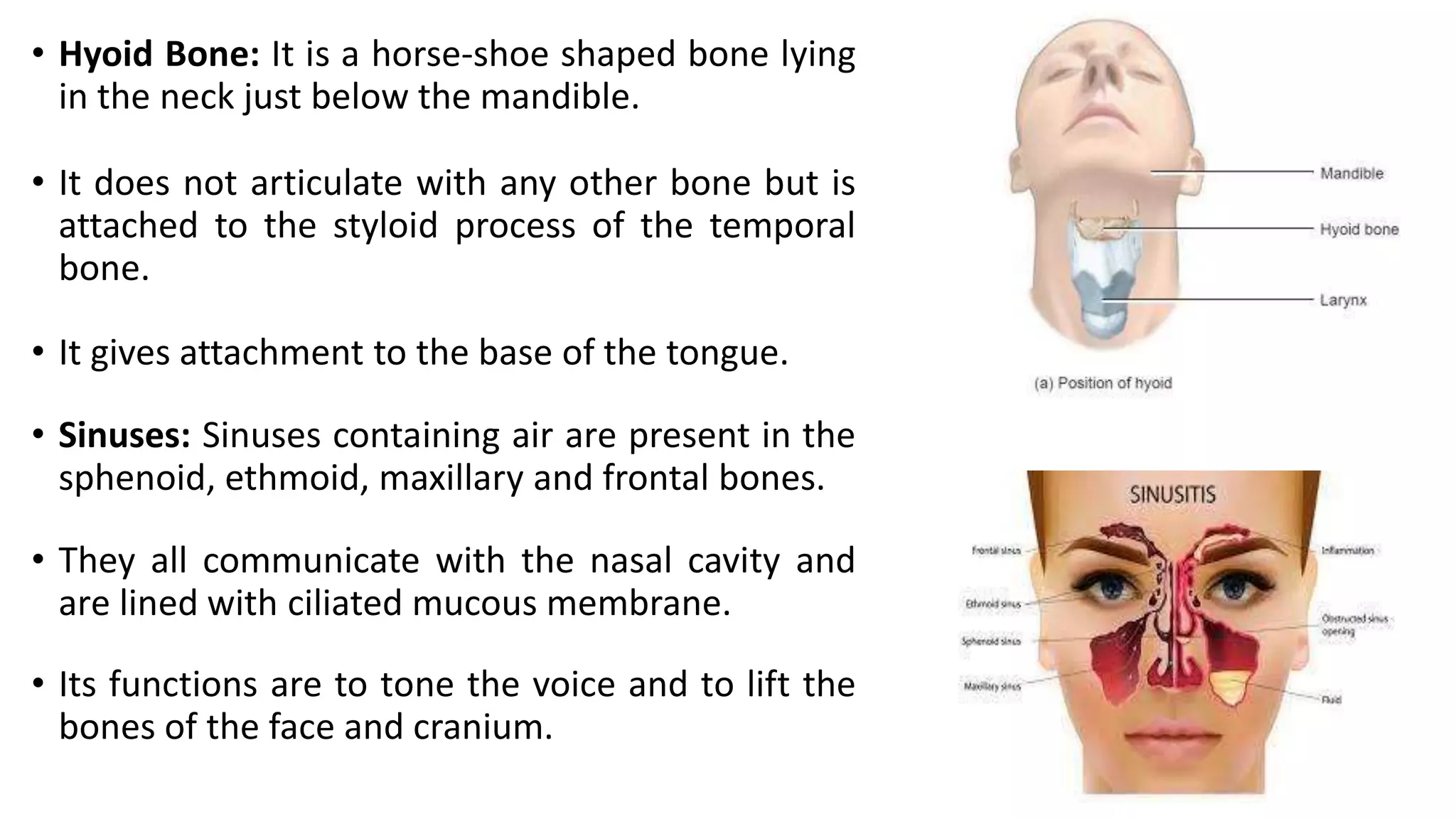
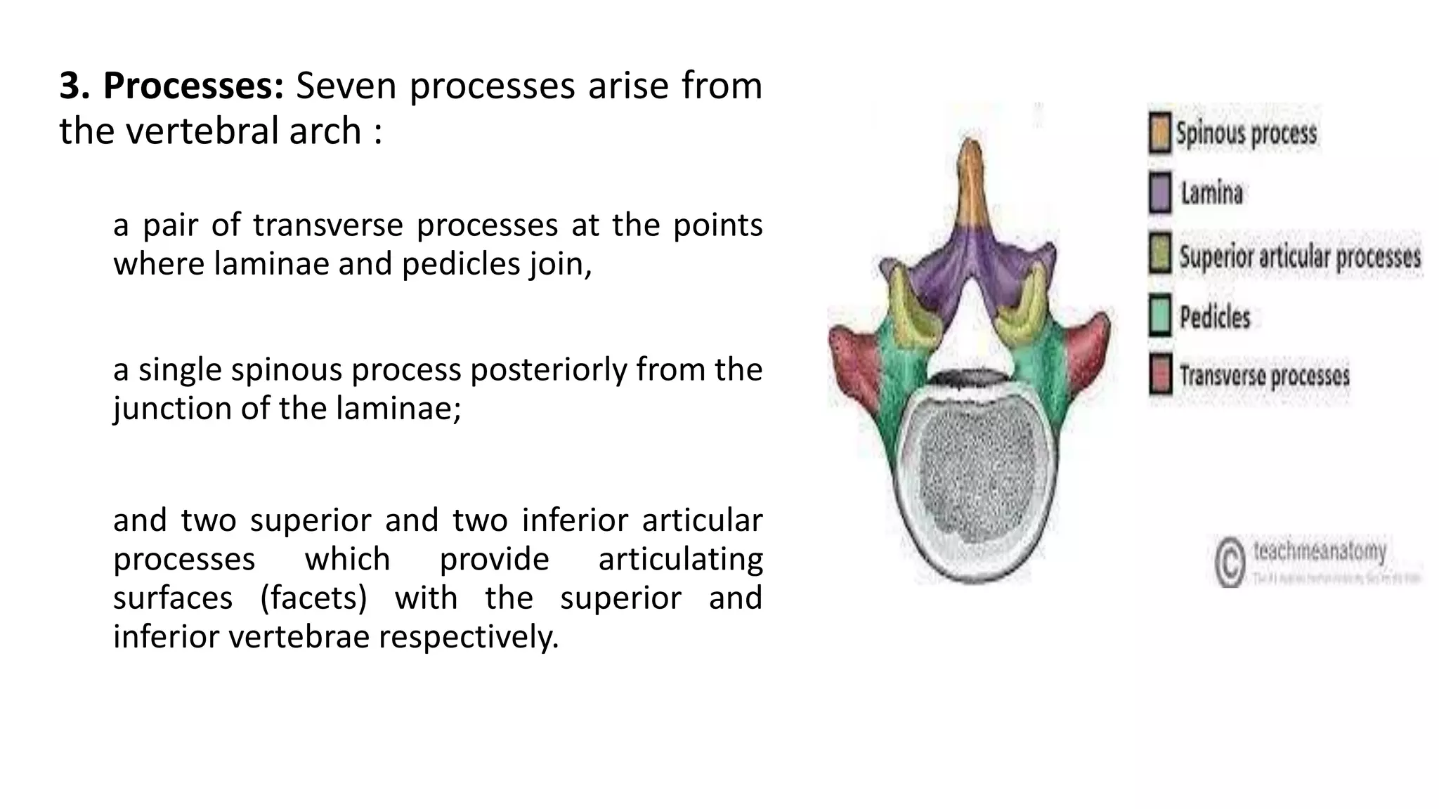
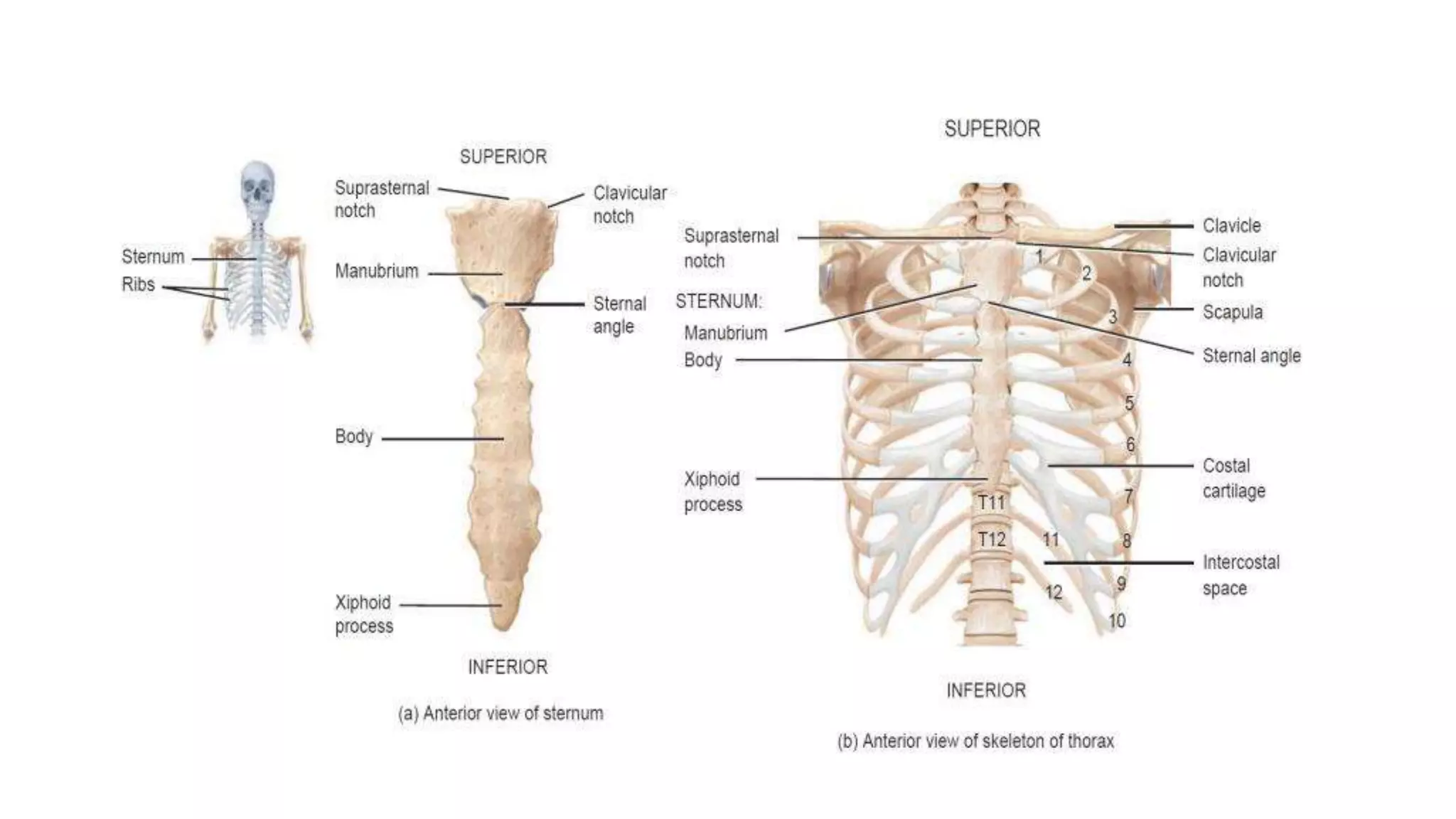
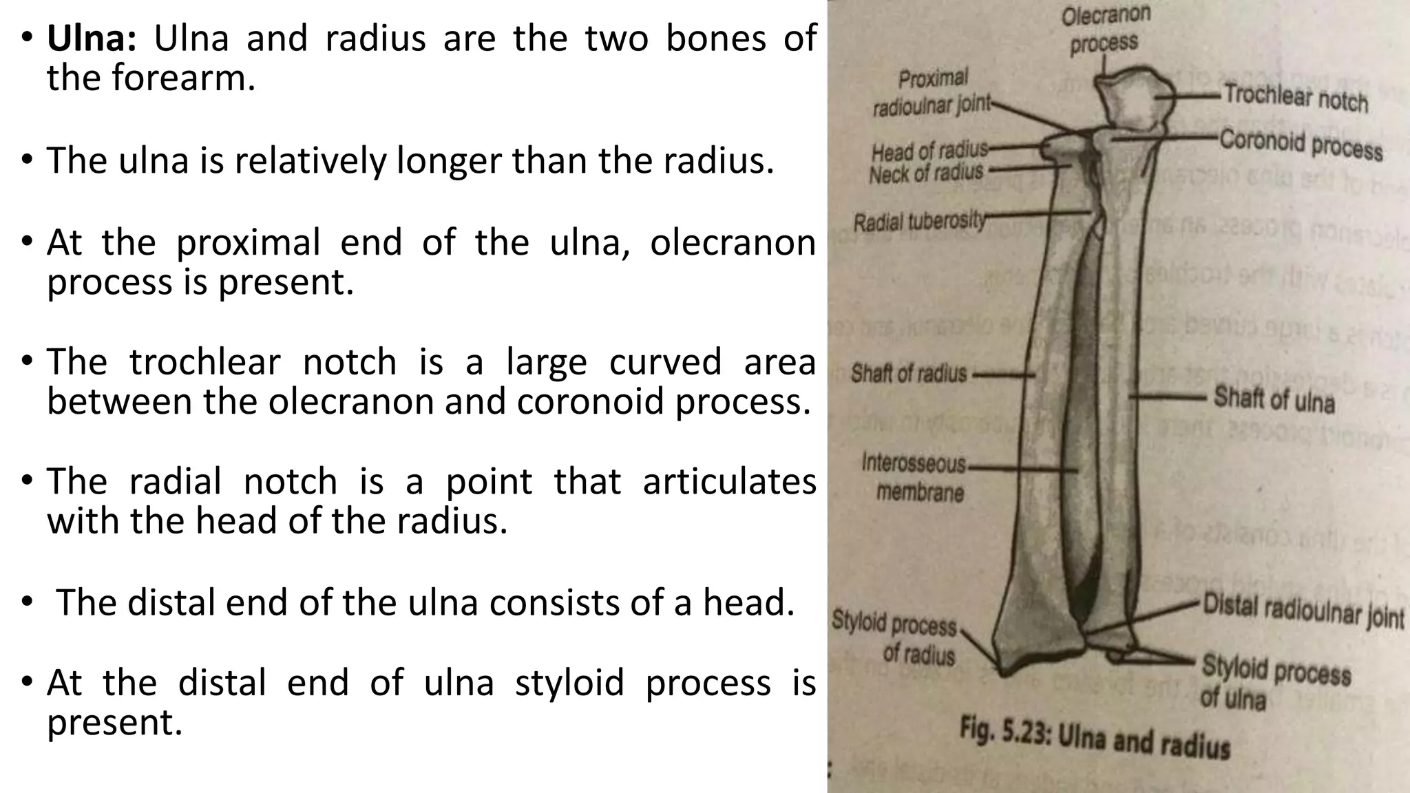
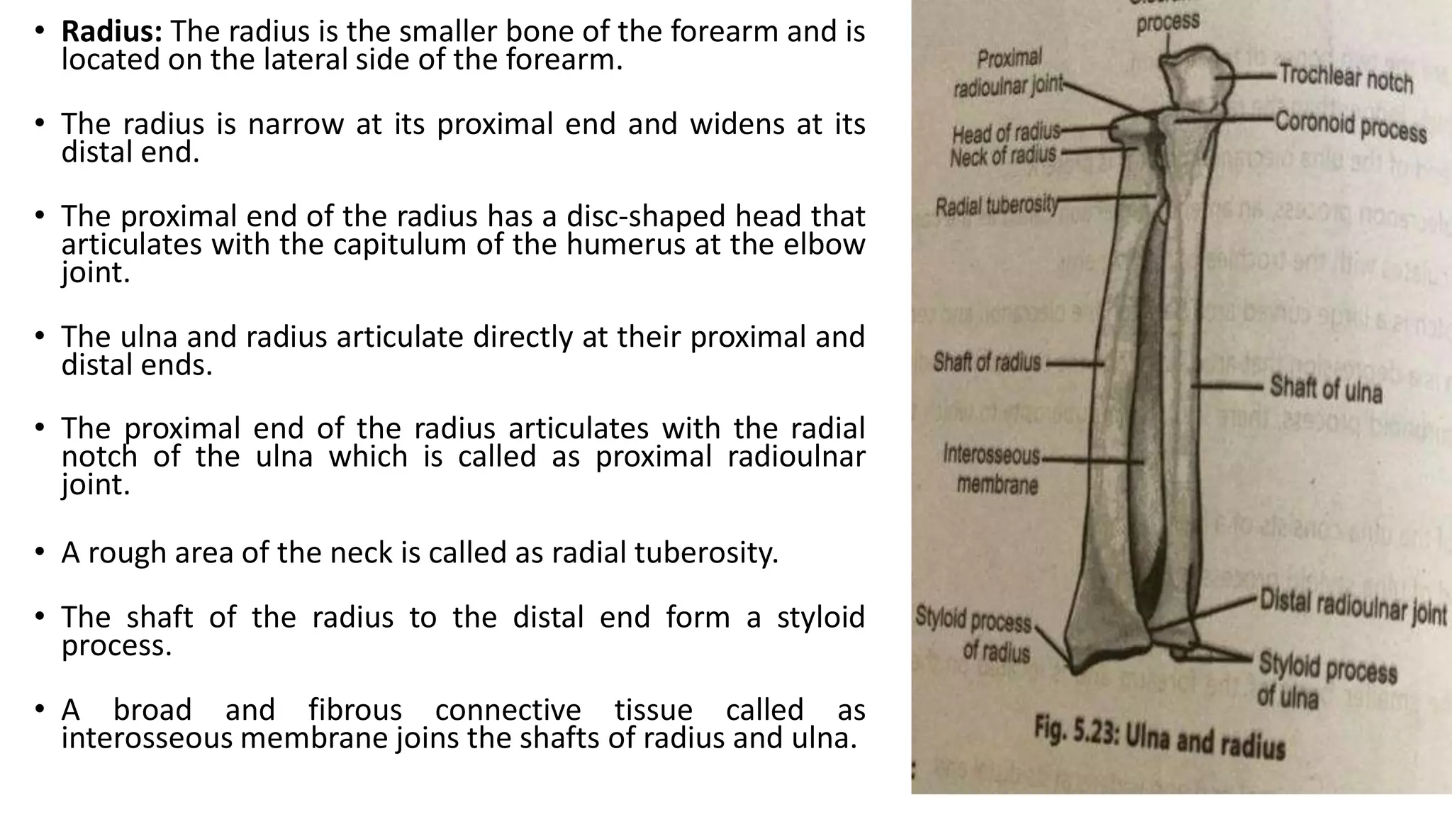
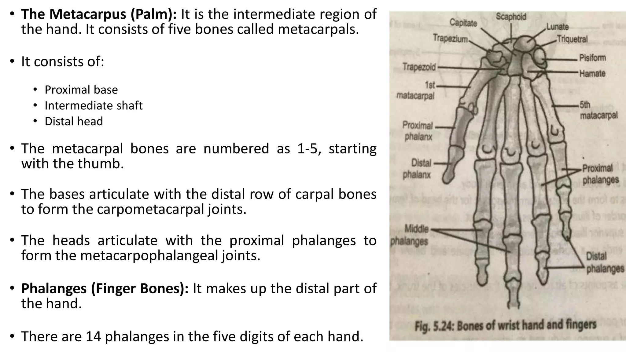
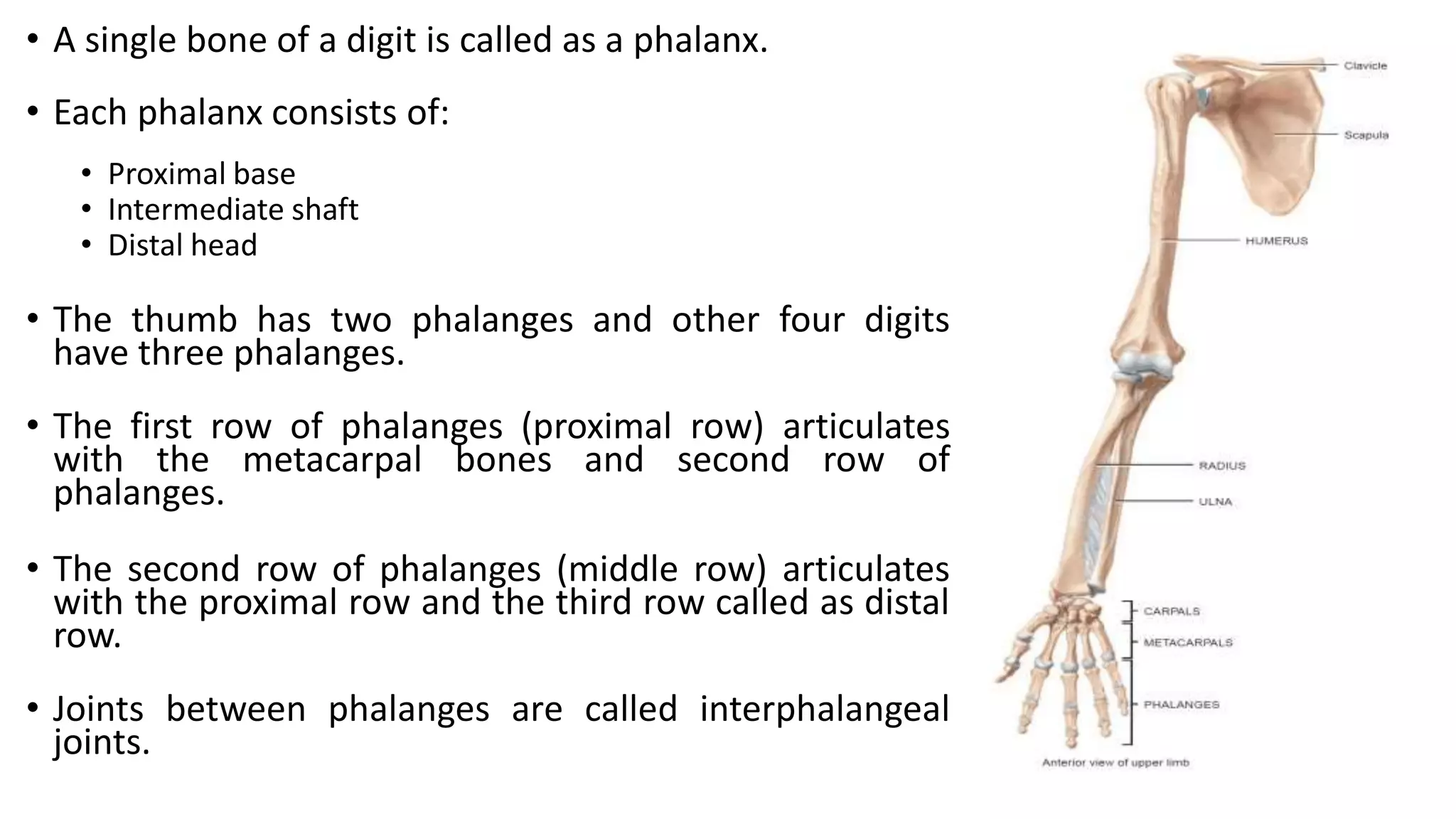
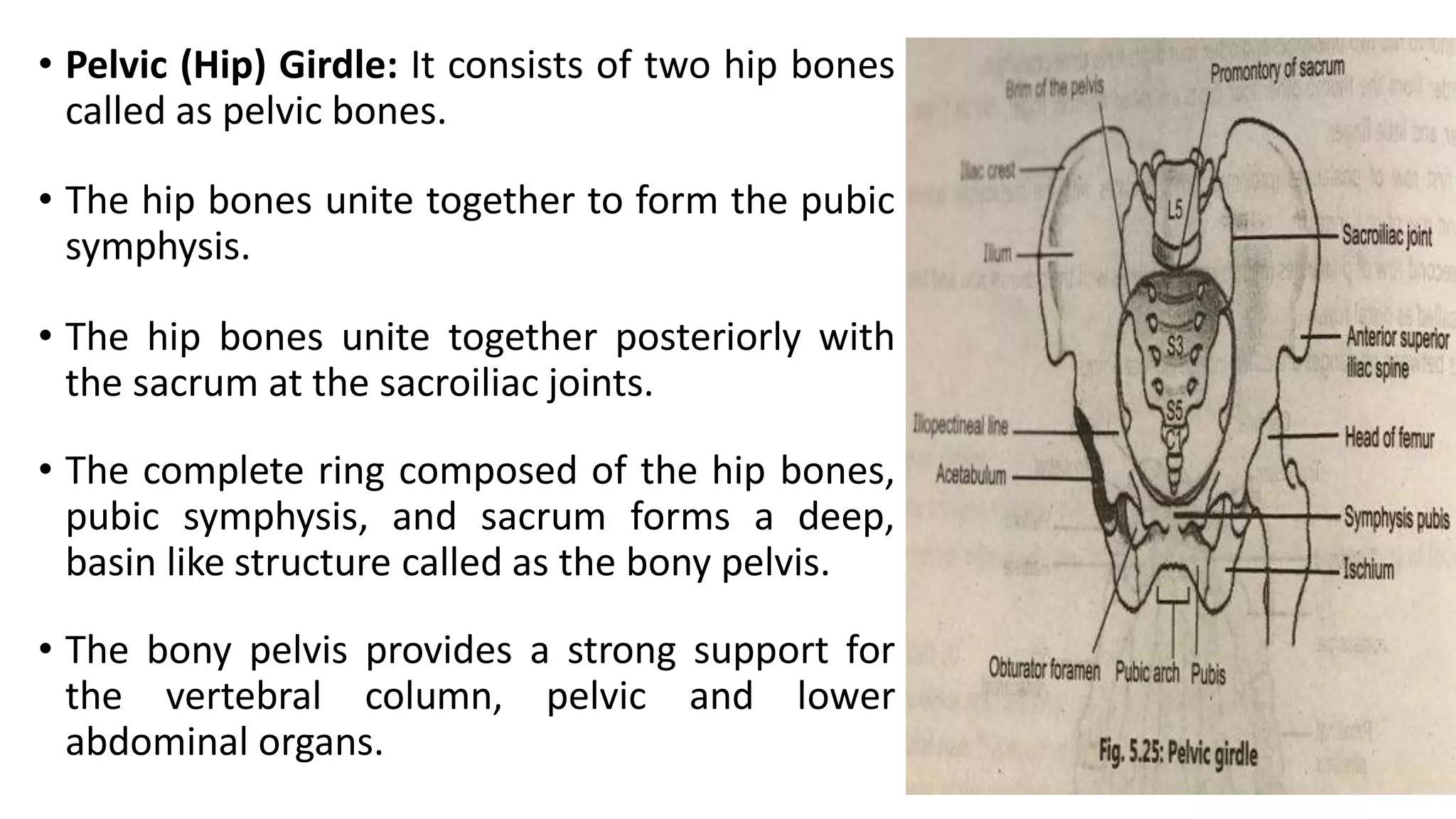
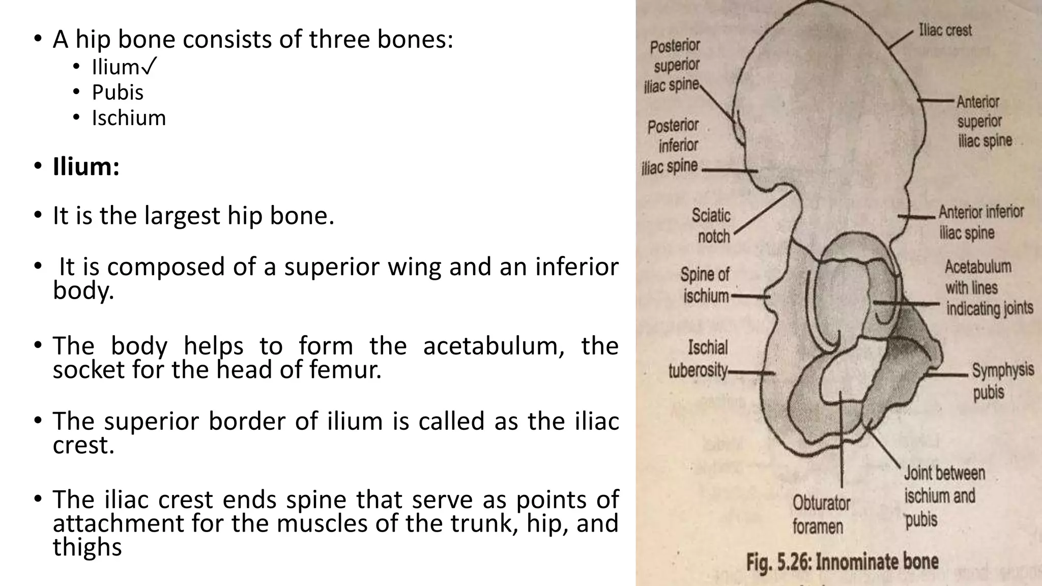
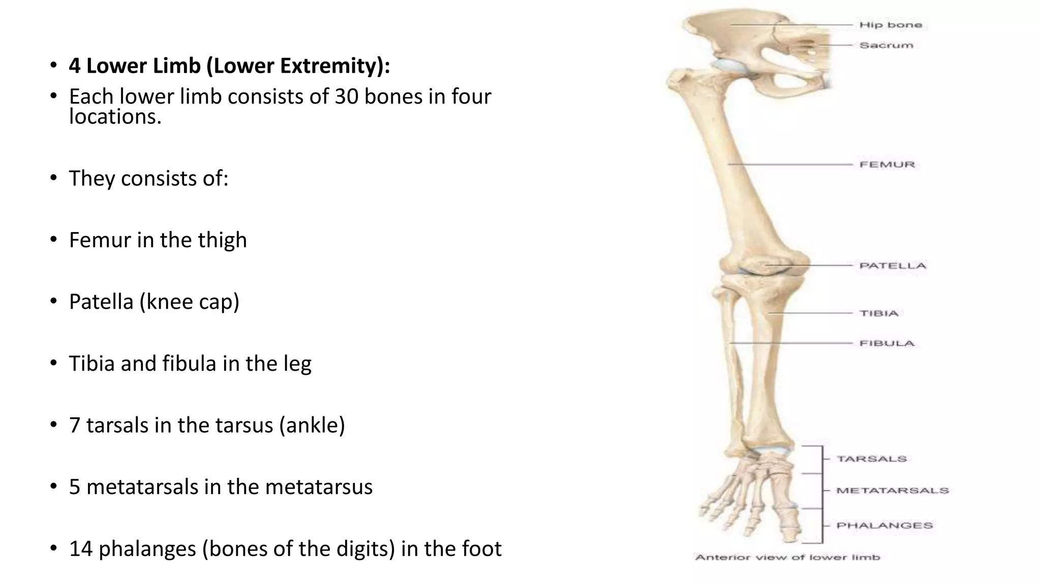
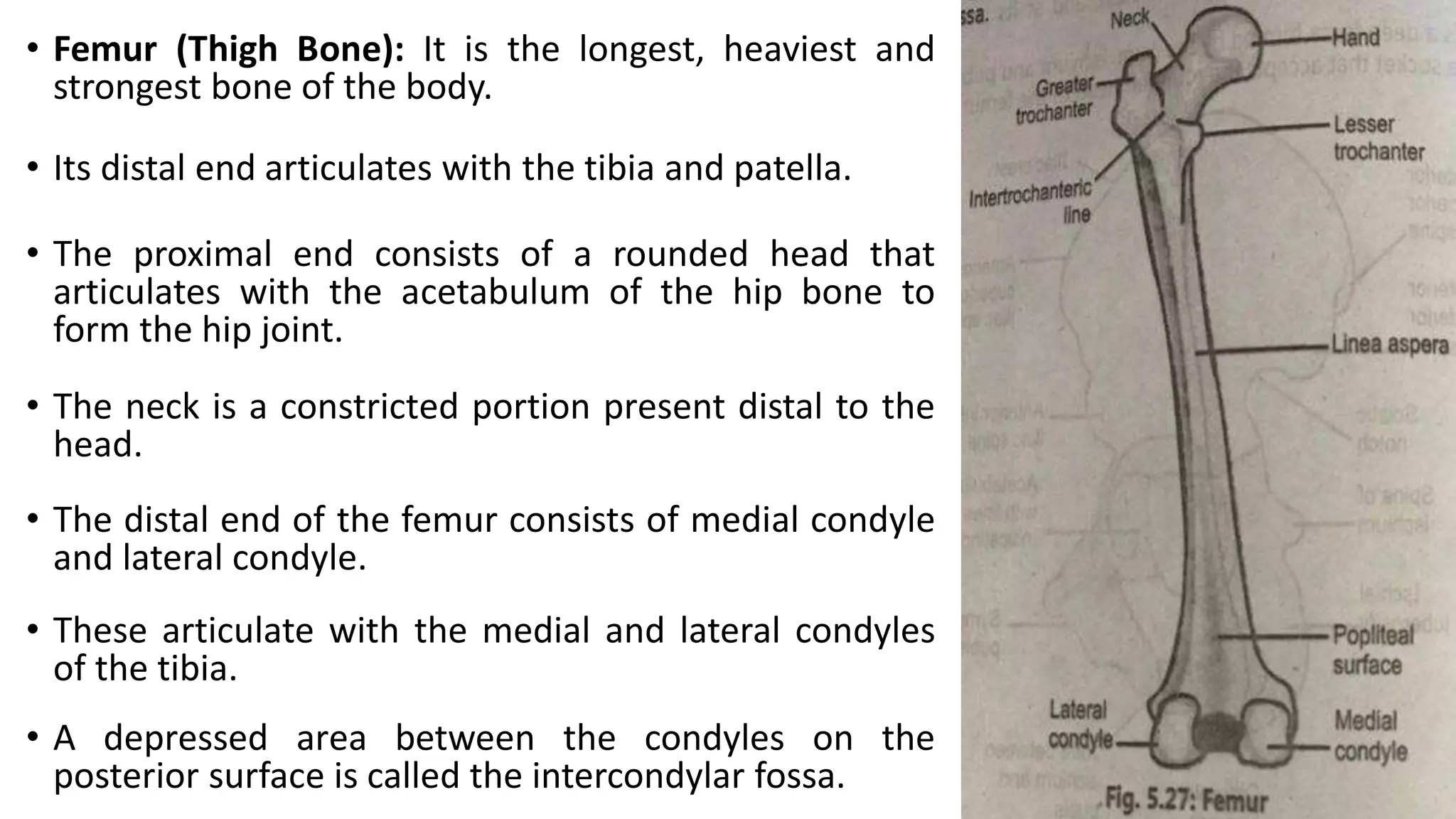
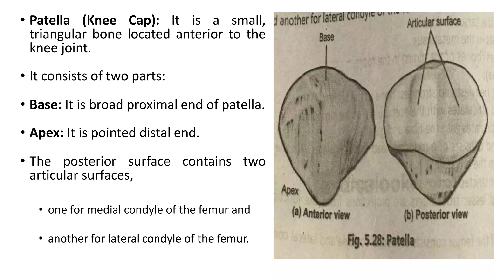
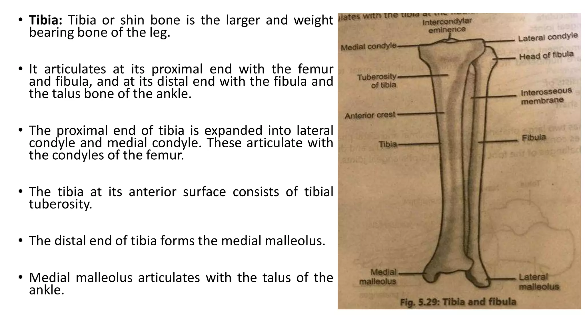
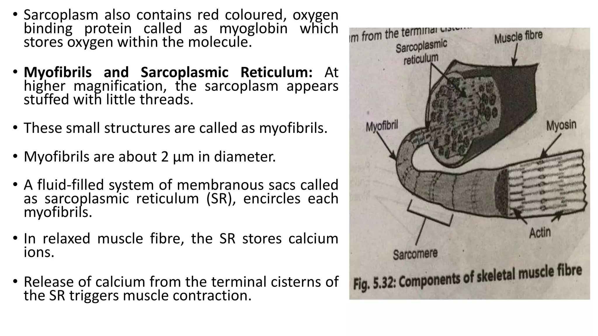
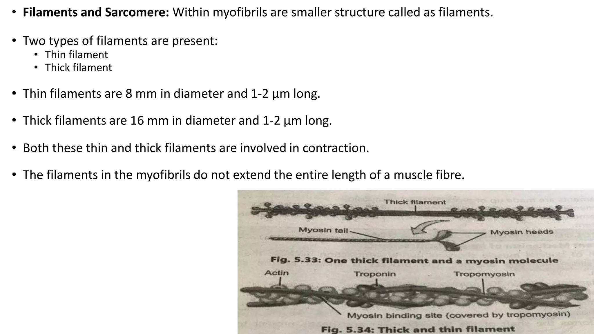
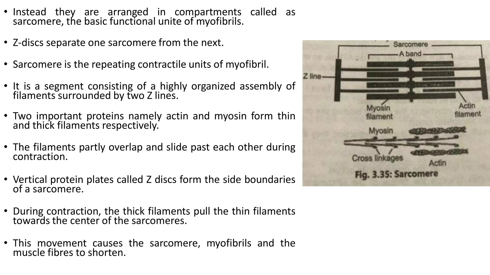
The document provides information about bones and the skeletal system. It discusses the following key points:
- Bones make up the skeletal system and provide structure, protection, movement, mineral storage, blood cell production, and fat storage.
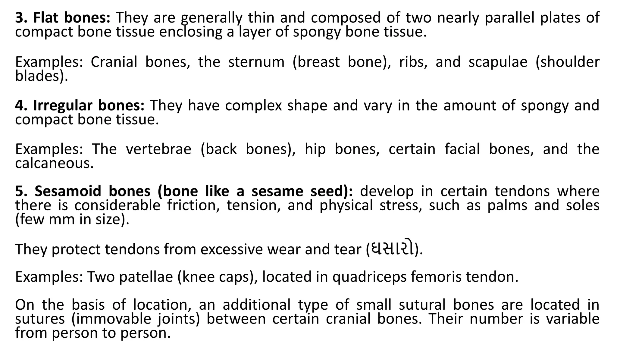
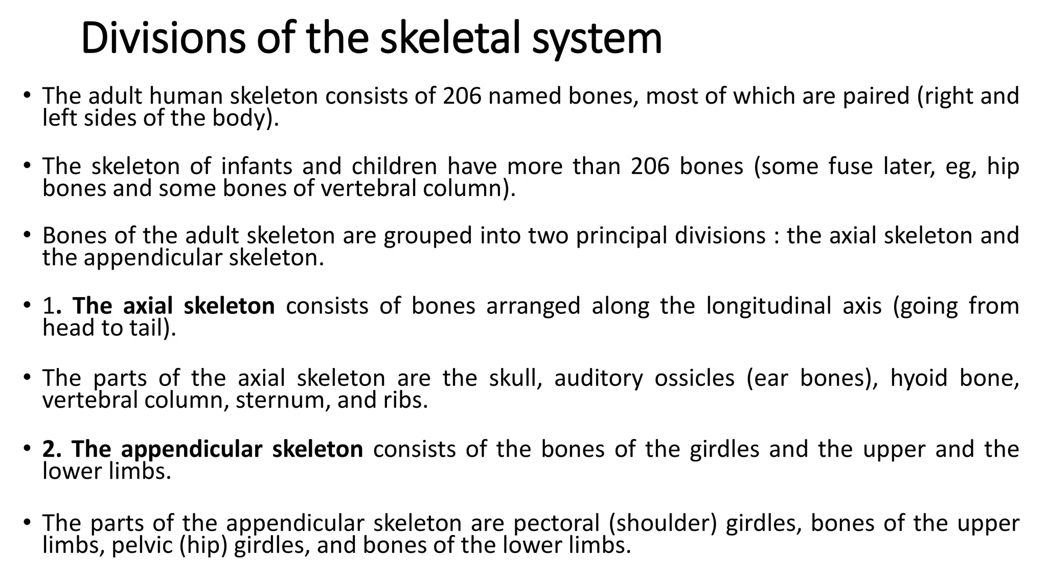
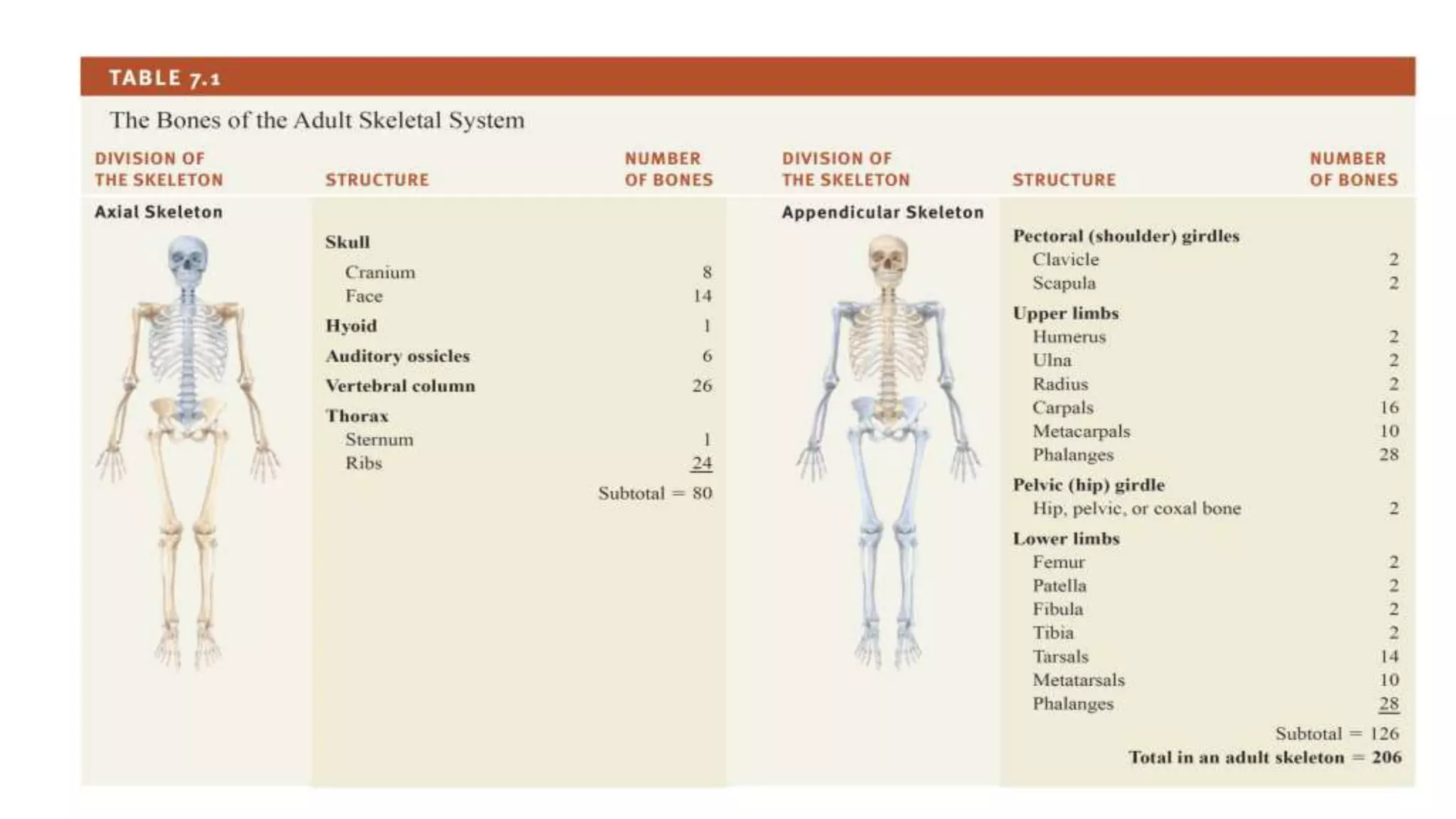
- The skeletal system includes long bones, short bones, flat bones, irregular bones, and sesamoid bones. It is divided into the axial skeleton and appendicular skeleton.
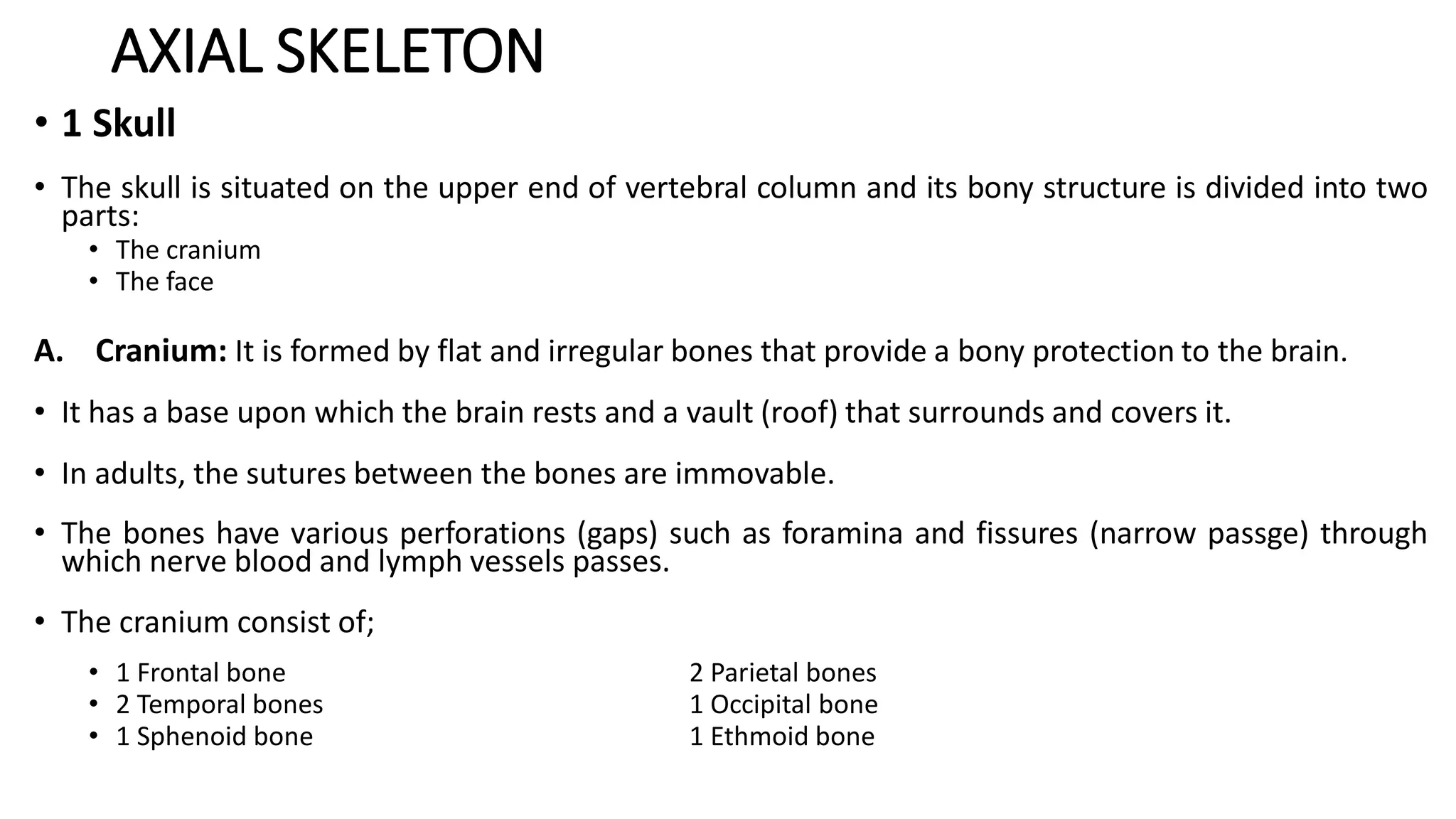
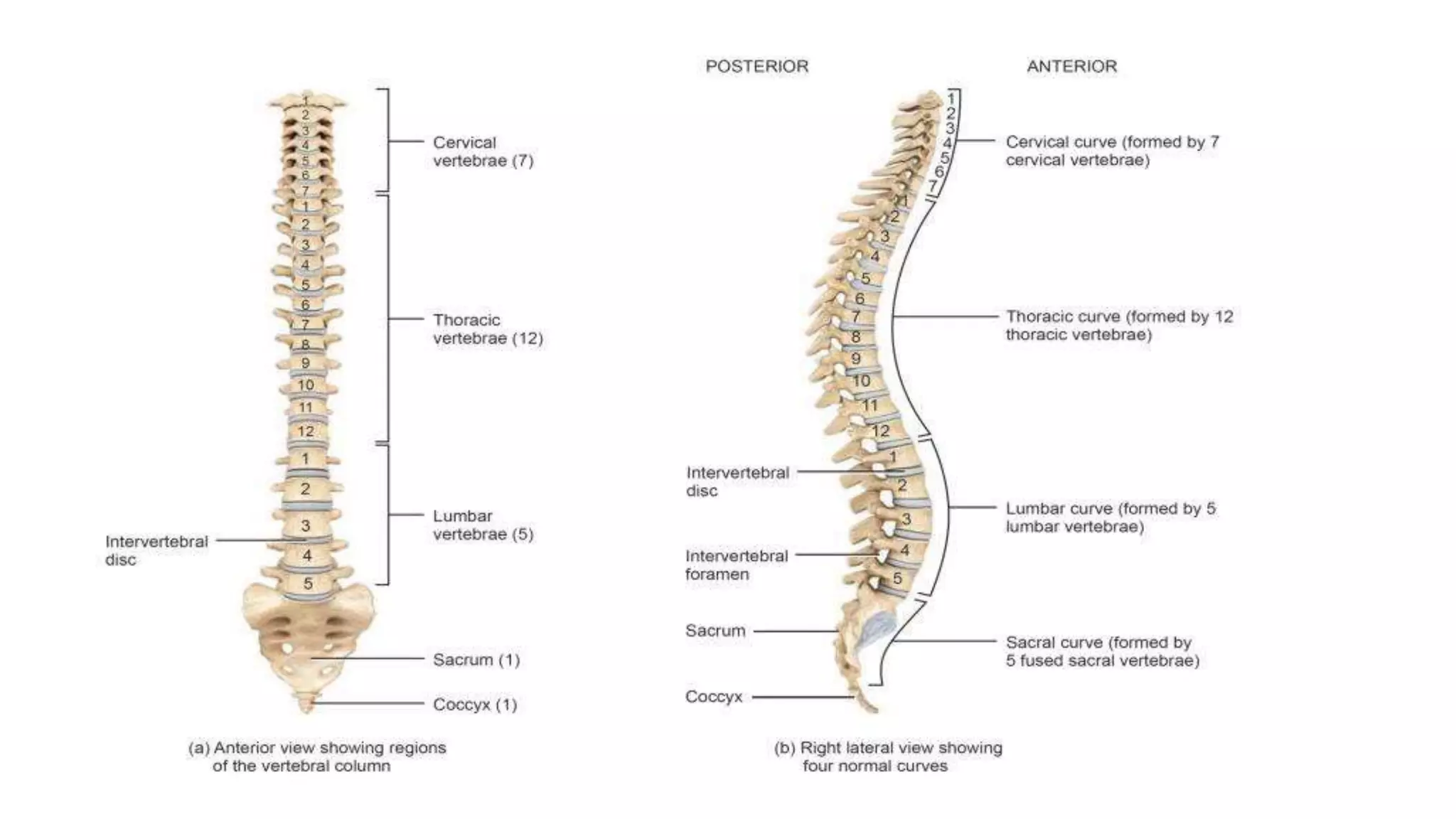
- The axial skeleton includes the skull, vertebral column, ribs, and sternum. It protects organs and allows movement.
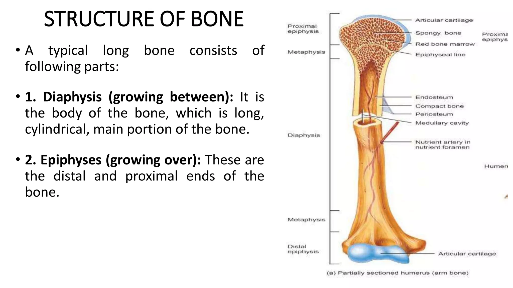
- Bones are made of compact bone, spongy bone, periosteum, and marrow. A typical long bone has a diaphysis, epiphyses
Rickets and osteomalacia are diseases caused by a lack of calcium and phosphorus that prevents bone mineralization. Rickets occurs in children before growth plate closure, while osteomalacia occurs in adults. The most common causes are vitamin D deficiency from inadequate intake or absorption and phosphate deficiency. Treatment involves replacing the deficient nutrient, usually with vitamin D, calcium, and phosphate supplements. Monitoring treatment involves checking serum and urine calcium levels until they normalize and x-rays show bone healing.
This document provides an overview of rickets and osteomalacia. It begins by defining rickets as a disease affecting the growth plates, causing deficient mineralization and impaired endochondral ossification due to failed apoptosis of hypertrophic chondrocytes. Osteomalacia is a similar disorder affecting mineralization of osteoid. Both result from insufficient calcium and phosphate levels. Vitamin D deficiency is a common cause and treatment involves vitamin D supplementation. Other causes discussed include hereditary disorders, prematurity, drugs, tumors, and renal osteodystrophy. Clinical features, radiographic findings, and management approaches are described for each condition.
This document discusses inflammatory diseases of the bones and joints, focusing on osteomyelitis. It defines osteomyelitis as inflammation of all bone anatomical structures that is typically caused by common bacteria like Staphylococcus aureus. It describes the pathogenesis of hematogenous osteomyelitis, how it spreads through the bones, and can lead to complications like sepsis and fractures. Diagnosis involves x-rays and CT scans to identify features like periosteal reaction and bone destruction. Treatment involves antibiotics, surgery to debride infected areas, and managing complications.
Osteoporosis is a progressive systemic skeletal disease characterized by low bone mass and microarchitecture deterioration of bone tissue, leading to enhanced bone fragility and a consequent increase in fracture risk.
Osteogenesis imperfecta (OI), or brittle bone disease, is a genetic disorder characterized by fragile bones that fracture easily. It is caused by a defect in the body's production of type 1 collagen, which is important for bone strength. The majority of OI cases are inherited in an autosomal dominant pattern, though some are the result of spontaneous genetic mutations or are inherited autosomally recessively. OI has been classified into eight types based on severity and symptoms, which can include multiple fractures, skeletal deformities, short stature, and in severe cases, lethality in infancy. While there is no cure for OI, treatment aims to increase bone strength and prevent fractures through medications, physical therapy,
Development,structure and organization of boneadityachakri
This document provides an overview of bone development, structure, and types. It discusses that bone is a mineralized connective tissue composed of cells and an intercellular matrix. There are two main types of bone tissue: compact bone, which makes up the hard outer surface and is made up of concentric lamellae; and spongy or cancellous bone, which is found in the interior and has a sponge-like appearance. Bone is also classified based on shape into long, short, flat, irregular, and sesamoid bones. The document outlines the development of bone through two processes: intramembranous ossification which forms flat bones; and endochondral ossification which replaces cartilage models with bone in
This document discusses osteoporosis and bone health. It begins by noting the impact of osteoporosis on older patients, including increased hospital admissions and length of stay. It then discusses definitions of osteoporosis from WHO and treatment gaps. The document covers bone structure, cells, mineralization, remodeling cycles, and factors influencing bone health like hormones and lifestyle. Diagnostic methods like DXA are summarized. Treatment options focused on prevention of fractures through calcium, vitamin D, bisphosphonates, PTH, and fall prevention are outlined.
This seminar consists of introduction, incidence, etiology, various classifications, history, clinical examination,sequelae of trauma of primary teeth followed by management
This document provides an introduction to common bone disorders. It begins by defining bone and its importance in providing structure, protection, and a site for blood cell production. Bone disorders are divided into congenital and acquired categories. Congenital disorders include Osteogenesis Imperfecta, Achondroplasia, and Osteopetrosis. Acquired or metabolic bone diseases are caused by abnormalities in minerals, vitamins, bone mass or structure and include Osteoporosis, Paget's Disease, Rickets/Osteomalacia, and Hyperparathyroidism. Each condition is briefly characterized in terms of symptoms and causes of abnormal bone structure or composition.
An avulsion fracture occurs when a tendon or ligament pulls off a piece of bone where it attaches. It is commonly seen in adolescent athletes, especially runners and hurdlers. Symptoms include severe pain in the affected area, swelling, bruising, and inability to move without pain. X-rays, ultrasounds, and MRIs can diagnose the fracture. Treatment focuses on RICE therapy as well as physical therapy exercises and modalities to regain strength and function.
Rickets and osteomalacia are diseases caused by inadequate bone mineralization due to vitamin D deficiency or impaired mineral metabolism. Rickets primarily affects children as it involves the growth plates, while osteomalacia affects adults. Symptoms include bone pain, deformities, and fractures. Diagnosis involves blood tests showing low calcium and phosphate levels and high alkaline phosphatase. X-rays show abnormalities in bone structure and density. Treatment focuses on correcting the underlying causes through vitamin D and calcium supplementation, with surgical intervention for severe deformities.
Osteogenesis imperfecta, or brittle bone disease, is caused by mutations in collagen genes COL1A1 and COL1A2 that result in abnormal bone formation and fragility. It is characterized by bones that break easily from minor trauma, along with other connective tissue problems like blue sclera and joint laxity. Treatment focuses on preventing fractures through braces and limiting activity, as well as bisphosphonates and surgery to insert rods for stabilization. The disease ranges in severity from mild cases with normal life expectancy to lethal prenatal forms.
Osteoporosis is a systemic skeletal disease characterized by low bone mass and deterioration of bone tissue, leading to fragile bones that are prone to fractures. It often presents without symptoms until a fracture occurs. Key risk factors include advanced age, female sex, small body frame, family history, smoking, excessive alcohol use, low calcium intake, physical inactivity, and certain medications. Screening high-risk individuals is important for diagnosis and treatment before fractures manifest.
This document provides information on osteoporosis from Dr. Ashutosh. It defines osteoporosis as a reduction in bone strength that increases fracture risk. The WHO defines it as a bone density 2.5 standard deviations below young adults of the same sex. Postmenopausal women with bone density between -1 and -2.5 SD also have increased fracture risk. Non-pharmacological prevention includes nutrition, lifestyle modifications, fall prevention, and hip protectors. Pharmacological management includes calcium and vitamin D supplementation, hormonal therapy, antiresorptive drugs, anabolic drugs, and drugs with dual actions.
Paget's disease, also known as osteitis deformans, is a chronic disease that involves abnormal bone remodeling. It results from increased and uncontrolled bone resorption by osteoclasts, followed by new bone formation that is poorly structured. Common symptoms include bone pain, deformities, and fractures. Diagnosis is based on x-rays showing abnormal bone structure and elevated alkaline phosphatase levels. Treatment focuses on controlling symptoms using bisphosphonates, calcitonin, or surgery. Complications can include fractures, deafness, and in rare cases cancer.
This document describes cherubism, a rare genetic condition characterized by bilateral swelling of the jaws causing a cherubic facial appearance. It results from mutations in the SH3BP2 gene in most cases, causing excess bone growth in the jaws during childhood that typically resolves by adulthood. Symptoms range from mild jaw enlargement to severe issues with vision, breathing, speech or swallowing depending on the severity.
Fracture Healing,Introduction,Pathology&Stages,Factors influencing osteogenesis,differences in healing of fractured bone by conservative&operative management.
This document provides an overview of diet, nutrition, and their importance in pediatric dentistry. It defines key terms like diet, food, and nutrition. It discusses the major nutrients including carbohydrates, proteins, lipids, vitamins, and minerals. It outlines the recommended dietary allowances and food sources for various nutrients. The document also discusses balanced diets, nutritional requirements by age, common nutritional disorders in pediatrics, and the role of dietary counseling.
A 3-year-old girl presented with a limp in her right leg and was diagnosed with coxa vara. Coxa vara is a decrease in the angle between the femoral neck and shaft to less than 120 degrees. It can be congenital, developmental, or secondary to other bone diseases. Surgical management involves subtrochanteric osteotomy to correct the angle, which is fixed internally with plates or screws. Postoperative care includes casting for 8-12 weeks until radiographic healing is seen. Complications can include recurrence, physeal injury, or leg length discrepancy.
The document discusses the mechanism of action of fluorides in preventing dental caries. It begins by providing background on fluorine and the structure of hydroxyapatite in enamel. It then discusses how fluoride is incorporated into enamel through different "pools" in the oral environment. The main proposed mechanisms of fluoride include increasing enamel resistance through formation of fluorapatite, enhancing remineralization, and interfering with plaque bacteria. Understanding fluoride's various modes of action helps develop more effective prevention products and programs.
Skeletal fluorosis is caused by excessive fluoride intake over long periods of time. The main sources of fluoride are drinking water, tea, and indoor air pollution from burning coal. Fluoride is deposited in bones and teeth. At low levels it strengthens teeth and bones, but at high levels it leads to skeletal and dental fluorosis. Skeletal fluorosis causes bone and joint pain and stiffness, and if severe, bone deformities and crippling. It is a major public health problem affecting millions in India, China, and other parts of Asia and Africa. Reducing fluoride intake and ensuring adequate calcium and vitamin D can help prevent and treat skeletal fluorosis.
This document summarizes flexible flatfoot, including its definition, symptoms, diagnosis, natural history, and treatment options. Flexible flatfoot is one of the most common foot deformities seen in children. The foot appears pronated and flattened during weight bearing but the arch reconstitutes when not bearing weight. Conservative treatment includes exercises, orthotics, and supportive shoes. For severe, symptomatic cases not improving with conservative care, surgical options like heel cord lengthening, lateral column lengthening, and talonavicular imbrication can provide pain relief and arch correction while preserving joint motion.
- Osteoarthritis is a degenerative joint disease affecting cartilage that commonly occurs in weight-bearing joints like the knee. It can be primary with no underlying cause or secondary to other joint issues.
- Symptoms include joint pain that worsens with use and improves with rest, morning stiffness, and crepitus. Conservative treatment focuses on lifestyle changes, physical therapy including exercises, bracing, and medications like acetaminophen, NSAIDs, or injections. Surgery is considered if conservative options provide insufficient relief.
The document discusses diseases of bone, including developmental disorders. It describes four developmental disorders:
1. Cherubism - A rare jaw condition seen in childhood characterized by bilateral swelling of the jaws due to fibrous dysplasia-like lesions. It is caused by a genetic mutation and lesions typically regress after puberty.
2. Osteopetrosis - A rare disease caused by a defect in bone remodeling resulting in abnormally dense bone that is fragile. It can cause anemia, fractures, and nerve compression.
3. Osteogenesis imperfecta - The most common inherited bone disorder caused by collagen defects, characterized by bone fragility and fractures that easily heal. Other features include blue sclera
This document provides information on physical therapy for osteoporosis. It begins with definitions and prevalence statistics on osteoporosis. It then describes the types of bone cells and bone remodeling process. The document outlines the goals of physical therapy for osteoporosis, which include maintaining posture, strengthening, balance training, flexibility exercises, and preventing further bone loss. Specific exercises recommended are extensions, chin tucks, and strengthening the lumbar spine and glutes. Physical therapy aims to improve function and reduce fractures for those with osteoporosis.
Skeletal System and division of axial and appendicularRupaSingh83
The skeletal system consists of the bones and joints of the body. The axial skeleton includes the skull, vertebral column, ribs and sternum, which form the core of the body. The appendicular skeleton includes the upper and lower limbs and their attachments. The skeletal system provides structure, protection, movement and mineral storage. It is divided into the axial and appendicular skeletons, with over 200 bones that can be classified by shape.
This document provides information about the skeletal system, specifically focusing on the axial skeleton. It defines the main types of bones and classifies them as long, short, flat, irregular, or sesamoid. It then describes the functions of bones and divides the skeletal system into the axial and appendicular skeleton. The majority of the document describes the bones that make up the axial skeleton, including the skull, vertebral column, ribs, and sternum. It provides details on the individual bones of the skull and vertebral column, including their features and locations.
ANATOMICAL FAETURES OF BONES FOR NURSING STUDENTS .pptxWINCY THIRUMURUGAN
A long bone has two parts: the diaphysis and the epiphysis.
The diaphysis is the tubular shaft that runs between the proximal and distal ends of the bone.
The hollow region in the diaphysis is called the medullary cavity, which is filled with yellow marrow.
The walls of the diaphysis are composed of dense and hard compact bone.
The wider section at each end of the bone is called the epiphysis (plural = epiphyses), which is filled with spongy bone.
The medullary cavity has a delicate membranous lining called the endosteum (end- = “inside”; oste- = “bone”), where bone growth, repair, and remodeling occur.The outer surface of the bone is covered with a fibrous membrane called the periosteum (peri- = “around” or “surrounding”). The periosteum contains blood vessels, nerves, and lymphatic vessels that nourish compact bone.Flat bones, like those of the cranium, consist of a layer of diploë (spongy bone), lined on either side by a layer of compact bone .Four types of cells are found within bone tissue: osteoblasts, osteocytes, osteogenic cells, and osteoclasts .Compact bone is the denser, stronger of the two types of bone tissue ,spongy bone, also known as cancellous bone, contains osteocytes housed in lacunae, but they are not arranged in concentric circles. Bones of the axial skeleton protect internal organs that includes skull (22), vertebral column (26), thoracic cage (25), ear bones (6) & Hyoid (1) TOTAL = 80
Bones of the appendicular skeleton facilitate movement with TOTAL 126 (64 in the upper & 62 in the lower) appendicular skeleton.
126+80=206.Skull bones:
The adult skull comprises 22 bones. These bones can be further classified by location:
Cranial bones: The 8 cranial bones form the bulk of your skull. They help to protect your brain.
Facial bones: There are 14 facial bones. They’re found on the front of the skull and make up the face.
Vertebral column:
The vertebral column is made up 33 bones.
Cervical vertebrae: These 7 bones are found in the head and neck.
Thoracic vertebrae: These 12 bones are found in the upper back.
Lumbar vertebrae: These 5 bones are found in the lower back.
The sacrum (5) and coccyx (4) are both made up of several fused vertebrae. Thoracic cage: The thoracic cage is made up of the sternum (breastbone) and 12 pairs of ribs.
These bones form a protective cage around the organs of the upper part, including the heart and lungs & gives attachment to muscles involved in respiration and upper limb movement.
The sternum consists of the manubrium, body of the sternum, and xiphoid process.
Ribs 1-7 are called true ribs because they attached directly to the sternum in front and vertebrae at back
Ribs 8-12 are known as false ribs.
the last two false ribs (11 & 12), have no anterior attachment, are called floating, fluctuating or vertebral ribs.
Ear bones (6):
Bones of the inner ear: Inside the temporal bone are the 3 smallest bones of the body:
Malleus
Incus
Stapes ( the smallest bone in the body) etc..
The musculoskeletal system consists of the skeletal and muscular systems. The skeletal system includes 206 bones that make up the axial skeleton (skull, vertebral column, rib cage) and appendicular skeleton (shoulder and pelvic girdles, upper and lower limbs). Bones develop through the processes of ossification and remodeling. The skeletal system works with muscles to allow movement and protect organs.
The skeletal system consists of 206 bones that form the framework of the body. There are two main parts - the axial skeleton along the body's central axis including the skull, vertebral column, and ribcage, and the appendicular skeleton of the limbs. The axial skeleton has 80 bones and protects internal organs. The appendicular skeleton has 126 bones and includes the shoulder and pelvic girdles and upper and lower limbs. Together the skeletal system provides structure, movement, protection, blood cell production, mineral storage, and triglyceride reserves for the body.
It is skeletal system of human body in detail description. In this ppt gives axial skeleton of body cranium thoracic cage and Vertibral coloumn . i gave structure and function of the bone , parts of axial skeleton with diagram
THE SKELETON SYSTEM ANATOMY AND PHYSIOLOGY SLIDESHARE Jitendra Bhargav
This document provides an overview of the skeletal system. It begins with classifying the different types of bones as long, short, flat, or irregular. It then describes the axial and appendicular skeleton in detail, identifying the specific bones that make up each part. Finally, it reviews bone tissue, the process of bone formation, growth and remodeling, and the three types of joints and movements they allow. The skeletal system has important functions of support, protection, movement, mineral storage, and blood cell formation.
The document provides details on the skeletal system, including:
1) There are 206 bones in the human body that make up the axial and appendicular skeleton. The axial skeleton includes the skull, vertebral column, and rib cage, providing protection, support, and carrying other body parts. The appendicular skeleton includes the bones of the upper and lower limbs.
2) Bones are composed of organic and inorganic materials and come in long, short, flat, and irregular shapes. They contain bone cells including osteoblasts, osteocytes, and osteoclasts that form and break down bone tissue.
3) The skeletal system functions to provide structure, protect organs, allow movement, store minerals, and produce blood cells.
The skeletal system includes all of the bones and joints in the body. Each bone is a complex living organ that is made up of many cells, protein fibers, and minerals. The skeleton acts as a scaffold by providing support and protection for the soft tissues that make up the rest of the body. The skeletal system also provides attachment points for muscles to allow movements at the joints. New blood cells are produced by the red bone marrow inside of our bones.
The document summarizes the skeletal system. It discusses that the skeletal system is composed of bones, cartilage, joints, and ligaments. It then describes the main components of the axial skeleton - the skull, vertebral column, and thoracic cage. The skull is made up of numerous flat and irregular bones that form the cranium and face. The vertebral column consists of 26 vertebrae and intervertebral discs. The thoracic cage is formed by the sternum, ribs, costal cartilages, and thoracic vertebrae.
The skeletal system has three main functions: providing structure and shape to the body, protecting vital organs, and allowing for bodily movement. It is made up of 206 bones that form the axial skeleton (skull, vertebrae, ribs, sternum) and appendicular skeleton (limbs and girdles). Bones are living tissues composed of compact bone, spongy bone, bone marrow, and various bone cells. They provide structure through their interaction with muscles, tendons, and ligaments at joints like the ball-and-socket hip. The skeletal system also plays roles in blood cell production and mineral storage.
The human skeleton is the internal framework of the body. It is composed of around 270 bones at birth – this total decreases to around 206 bones by adulthood after some bones have fused together.
The bone mass in the skeleton reaches maximum density around age 21. The human skeleton can be divided into the axial skeleton and the appendicular skeleton.
The axial skeleton is formed by the vertebral column, the rib cage, the skull and other associated bones. The appendicular skeleton, which is attached to the axial skeleton, is formed by the shoulder girdle, the pelvic girdle, and the bones of the upper and lower limbs.
he skeleton serves six major functions: support, movement, protection, production of blood cells, storage of minerals and endocrine regulation.
The skeleton provides the framework which supports the body and maintains its shape. The pelvis, associated ligaments and muscles provide a floor for the pelvic structures. Without the rib cages, costal cartilages, and intercostal muscles, the lungs would collapse.
ANATOMY OF THE HUMAN SKELETON POWERPOINTmhixgoodie
The document provides an outline for a presentation on the anatomy of the human skeleton. It describes the skeletal system as being divided into the axial skeleton, which includes the skull, vertebrae, ribs, and sternum, and the appendicular skeleton, which includes the bones of the extremities. It provides details on the types of bones, joints, microscopic structure of bones, and the individual bones that make up the skull, vertebrae, ribs, sternum, and extremities. The functions of the skeleton and common conditions that can affect it are also discussed.
The skeletal system consists of 206 bones that are divided into the axial skeleton (skull, vertebral column, ribs, sternum) and appendicular skeleton (limbs and their attaching girdles). Bones provide structure, protection, movement, mineral storage, blood cell formation, and are living tissues that undergo remodeling. The skeletal system includes various bone cell types and bone is composed of inorganic minerals and organic matrix. Common diseases include osteoporosis, rickets, osteomalacia, and Paget's disease.
The skeletal system consists of bones, cartilages, and ligaments that provide structure and protection. The axial skeleton forms the central axis and includes the skull, vertebral column, and thoracic cage. The skull has multiple bones that form the cranium and face. Bones of the skull include the parietal, temporal, frontal, occipital, maxilla, zygomatic, nasal, and mandible. The appendicular skeleton includes all bones of the upper and lower limbs attached to the axial skeleton.
The skeleton document describes the human skeletal system. It details that the skeleton is made up of 206 bones that form the axial skeleton (skull, vertebral column, rib cage) and appendicular skeleton (shoulder and pelvic girdles with attached upper and lower limbs). The skeleton enables movement, protects organs, produces blood cells, and stores minerals. It categorizes each bone and describes its location, structure, and relevant features.
This lecture help the students such as medical ,nursing , and any health care provider to understand the basic information about anatomy of skeletomuscular system.
The skeletal system comprises 206 bones that support the body and allow for movement. The axial skeleton includes the skull, vertebral column, and thoracic cage, while the appendicular skeleton includes the upper and lower limbs attached to the axial skeleton. The skull protects the brain and is divided into cranial and facial bones. The vertebral column is made up of individual vertebrae that protect the spinal cord and allow for flexibility. Bones are living tissues composed of minerals and connective tissues. The skeletal system provides structure, protects organs, allows body movement, stores minerals, and produces blood cells.
There are 206 bones in the human body grouped into the axial skeleton and appendicular skeleton. The axial skeleton includes 80 bones that make up the skull, vertebral column, ribs, and sternum. It forms the central core and foundation of the body. The appendicular skeleton includes 126 bones arranged in the upper and lower limbs, including their attaching girdles, forming the shoulders, arms, legs and allowing for movement.
Throughout history, the symbol of the skull and crossbones has served as a representation of mortality, likely owing to the fact that following death and decay, bones are the sole remnants. Many individuals perceive bones as inert, desiccated, and fragile. While these attributes accurately portray the bones of a preserved skeleton, the bones within a living human being are profoundly alive. Living bones exhibit strength and flexibility, serving as the primary components of the skeletal system.
This presentation was provided by Rebecca Benner, Ph.D., of the American Society of Anesthesiologists, for the second session of NISO's 2024 Training Series "DEIA in the Scholarly Landscape." Session Two: 'Expanding Pathways to Publishing Careers,' was held June 13, 2024.
How to Setup Warehouse & Location in Odoo 17 InventoryCeline George
In this slide, we'll explore how to set up warehouses and locations in Odoo 17 Inventory. This will help us manage our stock effectively, track inventory levels, and streamline warehouse operations.
This document provides an overview of wound healing, its functions, stages, mechanisms, factors affecting it, and complications.
A wound is a break in the integrity of the skin or tissues, which may be associated with disruption of the structure and function.
Healing is the body’s response to injury in an attempt to restore normal structure and functions.
Healing can occur in two ways: Regeneration and Repair
There are 4 phases of wound healing: hemostasis, inflammation, proliferation, and remodeling. This document also describes the mechanism of wound healing. Factors that affect healing include infection, uncontrolled diabetes, poor nutrition, age, anemia, the presence of foreign bodies, etc.
Complications of wound healing like infection, hyperpigmentation of scar, contractures, and keloid formation.
This presentation was provided by Racquel Jemison, Ph.D., Christina MacLaughlin, Ph.D., and Paulomi Majumder. Ph.D., all of the American Chemical Society, for the second session of NISO's 2024 Training Series "DEIA in the Scholarly Landscape." Session Two: 'Expanding Pathways to Publishing Careers,' was held June 13, 2024.
Philippine Edukasyong Pantahanan at Pangkabuhayan (EPP) CurriculumMJDuyan
(𝐓𝐋𝐄 𝟏𝟎𝟎) (𝐋𝐞𝐬𝐬𝐨𝐧 𝟏)-𝐏𝐫𝐞𝐥𝐢𝐦𝐬
𝐃𝐢𝐬𝐜𝐮𝐬𝐬 𝐭𝐡𝐞 𝐄𝐏𝐏 𝐂𝐮𝐫𝐫𝐢𝐜𝐮𝐥𝐮𝐦 𝐢𝐧 𝐭𝐡𝐞 𝐏𝐡𝐢𝐥𝐢𝐩𝐩𝐢𝐧𝐞𝐬:
- Understand the goals and objectives of the Edukasyong Pantahanan at Pangkabuhayan (EPP) curriculum, recognizing its importance in fostering practical life skills and values among students. Students will also be able to identify the key components and subjects covered, such as agriculture, home economics, industrial arts, and information and communication technology.
𝐄𝐱𝐩𝐥𝐚𝐢𝐧 𝐭𝐡𝐞 𝐍𝐚𝐭𝐮𝐫𝐞 𝐚𝐧𝐝 𝐒𝐜𝐨𝐩𝐞 𝐨𝐟 𝐚𝐧 𝐄𝐧𝐭𝐫𝐞𝐩𝐫𝐞𝐧𝐞𝐮𝐫:
-Define entrepreneurship, distinguishing it from general business activities by emphasizing its focus on innovation, risk-taking, and value creation. Students will describe the characteristics and traits of successful entrepreneurs, including their roles and responsibilities, and discuss the broader economic and social impacts of entrepreneurial activities on both local and global scales.
2. Introduction
• A bone is made up of several bone tissues bone, cartilage, dense connective
tissue, epithelium, adipose tissue, and nervous tissue.
• The entire framework of bones and their cartilages constitutes the skeletal
system.
• Movements such as walking require interactions between bones and muscles.
• The bones, muscles, and joints together form an integrated system called the
musculoskeletal system.
• The branch of medical science concerned with the prevention or correction of
disorders at the musculoskeletal system is called orthopedics (ortho correct;
pedi = child).
3. Functions of bone and the skeletal system
Bone tissue constitutes about 18% of total body weight and performs several
basic functions.
1. Support: It serves as the structural framework, supports soft tissues and
provides attachment for the tendons of most skeletal muscles.
2. Protection: The skeleton protects vital internal organs from injury, eg,
cranial bones protect the brain, vertebrae (backbones) protects the spinal
cord, and the rib cage protects the heart and lungs.
3. Assists Movement: Most skeletal muscles attach to bones and bring about
movements by pulling due to their contraction
4. Mineral Homeostasis: Bones stone several minerals, especially calcium and
phosphorus, which strengthen the bone.
4. On body demand, bone releases minerals into the blood to maintain critical
mineral balances (homeostasis) for distribution to other parts of the body.
5. Blood Cell Production: Red bone marrow (a connective tissue) within certain
bones produces RBCs, WBCs and platelets, a process called hemopoiesis.
• Red bone marrow consists of developing blood cells, fibroblasts, adipocytes,
and macrophages within network of reticular fibres.
• It occurs in developing bones of the foetus and in some adult bones like the
pelvis, ribs, breast bone, vertebrae, skull, and ends of the bones of the arm
and thigh.
• 6. Triglyceride storage: Yellow bone marrow consists mainly of adipose cells,
which store triglycerides (food reserve).
5. STRUCTURE OF BONE
• A typical long bone consists of
following parts:
• 1. Diaphysis (growing between): It is
the body of the bone, which is long,
cylindrical, main portion of the bone.
• 2. Epiphyses (growing over): These are
the distal and proximal ends of the
bone.
6. 3. Metaphyses (Between): These are the regions of mature bone where the diaphysis joins
the epiphyses.
In growing bone it represents the epiphyseal plate (a layer of hyaline cartilage that allows
the growth of bone in length).
At about ages 18-21 when a bone ceases to grow, cartilaginous epiphyseal plate is replaced
by bone, now called epiphyseal line.
4. Articular cartilage: It is a thin layer of hyaline cartilage covering that part of epiphysis
where the bone articulates with another bone.
It reduces friction and absorbs shock at freely movable joints.+
7. • 5. Periosteum: It is a tough sheath of dense irregular connective tissue that
surrounds the bone surface where there is no cartilage.
• Its bone forming cells enable bone to grow in thickness, but not in length.
• Periosteum also protects the bone, assists in fracture repair, helps to nourish
bone tissue, and serves as an attachment point for ligaments and tendons.
• 6. Medullary cavity (Marrow cavity): It is the space within the diaphysis that
contains fatty yellow bone marrow in adults.
• 7. Endosteum : It is a thin membrane that lines the medullary cavity. It contains
a single layer of bone forming cells and a small amount of connective tissue.
8. TYPES OF BONES
• Almost all bones of the body can be classified into five main types based on shape : long,
short, flat, irregular and sesamoid
1. Long bones: They have greater length than breadth, consist of a shaft and a variable
number of extremities (ends), and are slightly curved for strength.
• Long bones consist mostly of compact bone tissue in their diaphysis but have considerable
amounts of spongy bone tissue in their epiphyses.
• Examples: Femur (thigh bone), tibia and fibula (leg bones), humerus (arm bone), ulna and
radius (forearm bones), and phalanges (fingers and toes).
2. Short bones: These are somewhat cube shaped and nearly equal in length and width.
• They consist of spongy bone tissue surrounded by a thin layer of compact bone tissue.
• Examples: Carpal bones (except pisiform; sesamoid) and the tarsal (ankle) bones (except
calcaneous or heal bone; irregular).
9. 3. Flat bones: They are generally thin and composed of two nearly parallel plates of
compact bone tissue enclosing a layer of spongy bone tissue.
Examples: Cranial bones, the sternum (breast bone), ribs, and scapulae (shoulder
blades).
4. Irregular bones: They have complex shape and vary in the amount of spongy and
compact bone tissue.
Examples: The vertebrae (back bones), hip bones, certain facial bones, and the
calcaneous.
5. Sesamoid bones (bone like a sesame seed): develop in certain tendons where
there is considerable friction, tension, and physical stress, such as palms and soles
(few mm in size).
They protect tendons from excessive wear and tear (ઘસારો).
Examples: Two patellae (knee caps), located in quadriceps femoris tendon.
On the basis of location, an additional type of small sutural bones are located in
sutures (immovable joints) between certain cranial bones. Their number is variable
from person to person.
10. Divisions of the skeletal system
• The adult human skeleton consists of 206 named bones, most of which are paired (right and
left sides of the body).
• The skeleton of infants and children have more than 206 bones (some fuse later, eg, hip
bones and some bones of vertebral column).
• Bones of the adult skeleton are grouped into two principal divisions : the axial skeleton and
the appendicular skeleton.
• 1. The axial skeleton consists of bones arranged along the longitudinal axis (going from
head to tail).
• The parts of the axial skeleton are the skull, auditory ossicles (ear bones), hyoid bone,
vertebral column, sternum, and ribs.
• 2. The appendicular skeleton consists of the bones of the girdles and the upper and the
lower limbs.
• The parts of the appendicular skeleton are pectoral (shoulder) girdles, bones of the upper
limbs, pelvic (hip) girdles, and bones of the lower limbs.
11.
12. AXIAL SKELETON
• 1 Skull
• The skull is situated on the upper end of vertebral column and its bony structure is divided into two
parts:
• The cranium
• The face
A. Cranium: It is formed by flat and irregular bones that provide a bony protection to the brain.
• It has a base upon which the brain rests and a vault (roof) that surrounds and covers it.
• In adults, the sutures between the bones are immovable.
• The bones have various perforations (gaps) such as foramina and fissures (narrow passge) through
which nerve blood and lymph vessels passes.
• The cranium consist of;
• 1 Frontal bone 2 Parietal bones
• 2 Temporal bones 1 Occipital bone
• 1 Sphenoid bone 1 Ethmoid bone
13.
14. • Frontal bone: It is the forehead bone.
• It forms part of eye sockets and the noticeable points
above the eyes, the supraorbital margins.
• Just above the supraorbital margins within the bone,
there are two air-filled cavities which have opening in to
the nasal cavity.
• The coronal suture joins the frontal and parietal bones.
• Parietal bones: These bones form the sides and roof of
the skull.
• They articulate with each other at the sagittal suture,
with the frontal bone at the coronal suture, with the
occipital bone at the lambdoidal suture and with the
temporal bones at the squamous sutures.
15. • Temporal bones: These bones lie one on each side of the head and
form immovable joints with the parietal, occipital, sphenoid and
zygomatic bones.
• The squamous part articulates with the parietal bone.
• The zygomatic process articulates with the zygomatic bone to form the
zygomatic arch.
• The mastoid part contains the mastoid process, a thickened portion
behind the ear.
• The temporal bone articulates with the mandible at the temporo-
mandibular joint.
• Occipital bone: It forms the back of head and base of skull.
• It has immovable joints with the parietal, temporal and sphenoid
bones.
• Sphenoid bone: It occupies the middle portion of base of skull and it
articulates with the occipital, temporal, parietal and frontal bones.
16. • Ethmoid bone: It occupies the anterior part of
base of the skull and helps to form the orbital
cavity, the nasal septum and the lateral walls of
the nasal cavity.
• It is very delicate bone containing many air
sinuses that opens in to the nasal cavity.
• (B) Face:
• The face is formed by 13 bones in addition to
the frontal bone.
• This includes;
• 2 Cheek bones 1 Maxilla
• 2 Nasal bones 2Lacrimal bones
• 1 Vomer 2Palatine bones
• 2 Inferior conchae 1 mandible
17. • Cheek bones: It forms the prominences of the
cheeks, part of the floor and lateral walls of orbital
cavities.
• Maxilla (Upper jaw bone):It forms the upper jaw,
the anterior part of roof of the mouth, the lateral
walls of the nasal cavity and part of the floor of
orbital cavities.
• The alveolar process projects downwards and
carries the upper teeth.
• Nasal bones: These are two small flat bones which
form the part of the lateral and superior surfaces
of the nose.
• Lacrimal bones: These are small bones present
posterior and lateral to the nasal bones and form
part of the medial walls of the orbital cavities.
18. • Vomer: It is a thin flat bone which extends upwards
from the hard palate to form the nasal septum.
• Palatine bones: These are two L-shaped bones.
• The horizontal parts of bones unite to form posterior
part of hard palate and to form part of the nasal
cavity.
• Inferior conchae: It is a scroll-shaped bone which
forms part of nasal cavity.
• The superior and middle conchae are parts of the
ethmoid bone.
• Mandible: It is the only movable bone of the skull.
• Each half consists of two main parts: a curved body
with the alveolar ridge containing lower teeth and a
ramus projects upwards to the posterior end of the
body.
19. • Hyoid Bone: It is a horse-shoe shaped bone lying
in the neck just below the mandible.
• It does not articulate with any other bone but is
attached to the styloid process of the temporal
bone.
• It gives attachment to the base of the tongue.
• Sinuses: Sinuses containing air are present in the
sphenoid, ethmoid, maxillary and frontal bones.
• They all communicate with the nasal cavity and
are lined with ciliated mucous membrane.
• Its functions are to tone the voice and to lift the
bones of the face and cranium.
20. Vertebral column
• Structures
• The vertebral column (spine or backbone) is composed of a series of bones called
vertebrae.
• The vertebral column, the sternum, and the ribs form the skeleton of the trunk of the
body.
• The vertebral column consists of bone and connective tissue; the spinal cord that it
surrounds and protects consists of nervous and connective tissue.
• The average length of vertebral column is about 71 cm in adult male and about 61 cm in
adult female.
• The total number of vertebrae during early development is 33.
• As a child grows, several vertebrae in the sacral and coccygeal regions fuse.
• The vertebral column in the adult typically contains 26 vertebrae.
21. • These are distributed as follows:
• 7 cervical vertebrae (cervic- = neck) are in the neck region.
• 12 thoracic vertebrae (thorax = chest) are posterior to the thoracic cavity.
• 5 lumbar vertebrae (lumb- = loin) support the lower back.
• 1 sacrum (= sacred bone) consists of five fused sacral vertebrae.
• 1 coccyx (= cuckoo; resembles the bill of cuckoo bird) usually consists of four fused
coccygeal vertebrae.
• The cervical, thoracic, and lumbar vertebrae are movable, but the sacrum and
coccyx are not.
22.
23. • Functions of Vertebral Column
• It functions as a strong, flexible rod with elements that can move forward, backward, and
sideways, and can rotate.
• It forms the axis on which body weight is received.
• All the movable vertebrae have a canal in them and all are arranged in such a way that
they make one continuous passage or neural canal through which the spinal cord runs
from head to tail.
• It provides protection to the spinal cord.
• The first vertebra (atlas) articulates with the skull and it provides support to head as well
as its up-down and sideway movements.
• It serves as a point of attachment for the ribs, and together they protect the vital organs
like heart and lungs.
• It also provides a point of attachment to pelvic girdle and muscles of the back.
• Many internal organs get support or suspension with it
24. • Intervertebral Discs
• Intervertebral discs are found between the
bodies of adjacent vertebrae from the second
cervical (C₂) vertebra to the sacrum.
• Each disc has an outer fibrocartilaginous ring
(annulus fibrosus) and an inner soft, pulpy,
highly elastic substance called nucleus pulposus.
• The discs form strong joints, permit various
movements of the vertebral column, and absorb
vertical shock.
• Under compression, they flatten and broaden
with age, nucleus pulposus hardens and
becomes less elastic.
25. • Parts of a Typical Vertebra:
• Vertebrae typically consist of a body, a vertebral arch, and
several processes.
1. Body: It is thick, disc shaped, weight bearing anterior
part of a vertebra.
• Its superior and inferior portions are roughned for the
attachment of discs.
• The anterior and lateral surfaces contain nutrient foramina
openings through which blood vessels deliver nutrients
and oxygen and remove carbon dioxide and wastes from
bone tissue.
2. Vertebral arch: Two short, thick processes (pedicles: a
small stalk like structure connecting an organ or other part
to the body) project posteriorly from the vertebral body to
unite with the flat lamina to form the vertebral arch.
26. • Together with the body it forms vertebral
(spinal) foramen for the passage of spinal
cord and connective tissue.
• The pedicles exhibit superior and inferior
indentations called vertebral notches.
• When the vertebral notches are stacked on
top of one another, they form an opening
between adjoining vertebrae on both sides of
the column.
• Each opening, called an intervertebral
foramen, permits the passage of a single
spinal nerve that passes to a specific region
of the body.
27. 3. Processes: Seven processes arise from
the vertebral arch :
a pair of transverse processes at the points
where laminae and pedicles join,
a single spinous process posteriorly from the
junction of the laminae;
and two superior and two inferior articular
processes which provide articulating
surfaces (facets) with the superior and
inferior vertebrae respectively.
28. Bones of the thorax
• The skeletal part of the thorax (chest), the thoracic cage, consists of sternum, costal
cartilages, ribs and the bodies of the thoracic vertebrae.
• The thoracic cage is narrower at the superior end and broader at its inferior end
and is flattened from front to back.
• Functions: It encloses and protects the organs in the thoracic and superior
abdominal cavities.
• It provides support for the bones of the shoulder (pectoral) girdle and upper limbs.
• 1 Sternum : The sternum (breast bone, about 15 cm long) is a flat, narrow bone
located in the center of the anterior thoracic wall and consists of 3 parts:
• the manubrium (handle like superior part),
• the body (the middle and largest part), and
• the xiphoid process (sword shaped inferior, smallest part).
29.
30. 2. Ribs:
• Twelve pairs of ribs give structural support to the sides of thoracic cavity.
• They increase in length from 1 to 7, and then decrease in length from 8 to 12.
• Each rib articulates posteriorly with the corresponding thoracic vertebra.
• The first 1-7 pairs of ribs have a direct anterior attachment to the sternum by a strip of
hyaline cartilage (costal cartilage), providing elasticity to the thoracic cage.
• These ribs are called true (vertebrosternal) ribs forming sternocostal joints with the
sternum.
• The remaining 5 pairs (8-12) are called false ribs (either attach indirectly to the sternum or
do not attach to sternum).
• The cartilages of 8-10 pairs of ribs attach to one another and then to the cartilages of 7th
pair of ribs; these are called vertebrochondral ribs.
31. • The 11th and 12th pairs are called floating (vertebral) ribs and do not attach to the
sternum at all.
• Costochondritis (inflammation of one or more costal cartilages) causes local
inflammation and pain and mimics the chest pain associated with a heart attack
(angina pectoris).
• The posterior portion of the rib connects to a thoracic vertebra by its head
(vertebrocostal joint) and the articular part of a tubercle (a bony eminence on the
temporal bone in the skull).
• Intercostal spaces (between ribs) are occupied by intercostal muscles, blood vessels,
and nerves.
• The costal cartilages are sufficiently elastic in younger individuals to permit
considerable bending without breaking.
• Special rib refractors are used to create a wide separation between ribs to gain
access for surgery of lungs or other structures.
32. Appendicular skeleton
• The appendicular skeleton consists of the pectoral girdle
with the upper limbs and the pelvic girdle with the
lower limbs.
• 1 Pectoral (Shoulder Girdle): The human body has two
pectoral girdles that attach the bones of the upper limbs
to the axial skeleton.
• The pectoral girdles consists of: 1 Clavicle and 1 Scapula
a. Clavicle (Collar Bone): It is 'S' shaped long bone which
has a double curve.
• The medial end is rounded and articulates with the
manubrium of the sternum to form the sternoclavicular
joint.
33. • The broad, fat, lateral end the acromial end, articulates
with the acromion of the scapula form the
acromioclavicular joint.
• The clavicle provides the bony link between the upper limb
and the axial skeleton.
b. Scapula: It is a large, flat, triangular shaped bone, lying on
the posterior chest wall superficial to the ribs.
• At the lateral angle there is a thin articular surface called as
glenoid cavity in which the head of the humerus fits and
forms the shoulder joint.
• On the posterior surface there is a spinous process called
as acromion process.
• Acromion process articulates with the clavicle at the
acromioclavicular joint.
• The coracoid process, a projection from the upper border
of the bone, gives attachment to muscles that move the
shoulder joint.
34. • 2 Upper Limbs (Upper Extremity): Each upper limb has 30
bones in three locations.
• It consists of:
• The 1 Humerus in the arm
• The 1 Ulna and 1 Radius in the forearm
• The 8 Carpals in the carpus (wrist)
• The 5 Metacarpals in the metacarpus (palm)
• The 14 Phalanges (bones of the digits) in the hand
• Humerus: It is the longest and largest bone of the upper
limb.
• It articulates proximally with the scapula and distally to the
elbow with two bones the ulna and the radius.
• The proximal end of the humerus has rounded head that
articulates with the glenoid cavity of the scapula.
• Neck is present distal to the head of humerus.
• The greater tubercle is a lateral projection present distal to
the neck.
35. • The lesser tubercle projects anteriorly.
• Between the two tubercles there is a groove called as intertubercular sulcus.
• The shaft of the humerus is roughly cylindrical at its proximal end, but it
becomes flattened and broad at its distal end.
• At the middle of the shaft, there is a roughened V-shaped area called as
deltoid tuberosity.
• The capitulum is a rounded knob that articulates with the head of the radius.
• The trochlea located medial to the capitulum articulates with the ulna.
• The medial epicondyle and lateral epicondyle are rough projections present
on either side of distal end of the humerus.
36. • Ulna: Ulna and radius are the two bones of
the forearm.
• The ulna is relatively longer than the radius.
• At the proximal end of the ulna, olecranon
process is present.
• The trochlear notch is a large curved area
between the olecranon and coronoid process.
• The radial notch is a point that articulates
with the head of the radius.
• The distal end of the ulna consists of a head.
• At the distal end of ulna styloid process is
present.
37. • Radius: The radius is the smaller bone of the forearm and is
located on the lateral side of the forearm.
• The radius is narrow at its proximal end and widens at its
distal end.
• The proximal end of the radius has a disc-shaped head that
articulates with the capitulum of the humerus at the elbow
joint.
• The ulna and radius articulate directly at their proximal and
distal ends.
• The proximal end of the radius articulates with the radial
notch of the ulna which is called as proximal radioulnar
joint.
• A rough area of the neck is called as radial tuberosity.
• The shaft of the radius to the distal end form a styloid
process.
• A broad and fibrous connective tissue called as
interosseous membrane joins the shafts of radius and ulna.
38. • The distal end of the radius articulates with three
bones of the wrist (the lunate, the scaphoid, and
the triquetrum) to form the radiocarpal joint.
• The Carpels (Wrist): It is the proximal region of
the hand. It consists of eight small bones
arranged in two transverse rows consisting of
four bones each.
• It consists of:
• Proximal row: Scaphoid, lunate, triquetrum, pisiform.
• Distal row: Trapezium, trapezoid, capitate, hamate.
• The bones of the proximal row are associated
with the wrist joint.
• The bones of the distal row form joints with the
metacarpal bones.
39. • The Metacarpus (Palm): It is the intermediate region of
the hand. It consists of five bones called metacarpals.
• It consists of:
• Proximal base
• Intermediate shaft
• Distal head
• The metacarpal bones are numbered as 1-5, starting
with the thumb.
• The bases articulate with the distal row of carpal bones
to form the carpometacarpal joints.
• The heads articulate with the proximal phalanges to
form the metacarpophalangeal joints.
• Phalanges (Finger Bones): It makes up the distal part of
the hand.
• There are 14 phalanges in the five digits of each hand.
40. • A single bone of a digit is called as a phalanx.
• Each phalanx consists of:
• Proximal base
• Intermediate shaft
• Distal head
• The thumb has two phalanges and other four digits
have three phalanges.
• The first row of phalanges (proximal row) articulates
with the metacarpal bones and second row of
phalanges.
• The second row of phalanges (middle row) articulates
with the proximal row and the third row called as distal
row.
• Joints between phalanges are called interphalangeal
joints.
41. • Pelvic (Hip) Girdle: It consists of two hip bones
called as pelvic bones.
• The hip bones unite together to form the pubic
symphysis.
• The hip bones unite together posteriorly with
the sacrum at the sacroiliac joints.
• The complete ring composed of the hip bones,
pubic symphysis, and sacrum forms a deep,
basin like structure called as the bony pelvis.
• The bony pelvis provides a strong support for
the vertebral column, pelvic and lower
abdominal organs.
42. • A hip bone consists of three bones:
• Ilium✓
• Pubis
• Ischium
• Ilium:
• It is the largest hip bone.
• It is composed of a superior wing and an inferior
body.
• The body helps to form the acetabulum, the
socket for the head of femur.
• The superior border of ilium is called as the iliac
crest.
• The iliac crest ends spine that serve as points of
attachment for the muscles of the trunk, hip, and
thighs
43. • Ischium: It is the posterior portion of the hip bone.
• It is composed of a superior body and an inferior ramus.
• The ramus is the portion of ischium that fuses with the pubis.
• Together the ramus and the pubis surround the obturator foramen, the
largest foramen in the skeleton.
• Pubic: It is the inferior part of hip bone.
• The acetabulum is a deep fossa formed by the ilium, ischium and pubis.
• It functions as the socket that accepts the rounded head of the femur.
44. • 4 Lower Limb (Lower Extremity):
• Each lower limb consists of 30 bones in four
locations.
• They consists of:
• Femur in the thigh
• Patella (knee cap)
• Tibia and fibula in the leg
• 7 tarsals in the tarsus (ankle)
• 5 metatarsals in the metatarsus
• 14 phalanges (bones of the digits) in the foot
45. • Femur (Thigh Bone): It is the longest, heaviest and
strongest bone of the body.
• Its distal end articulates with the tibia and patella.
• The proximal end consists of a rounded head that
articulates with the acetabulum of the hip bone to
form the hip joint.
• The neck is a constricted portion present distal to the
head.
• The distal end of the femur consists of medial condyle
and lateral condyle.
• These articulate with the medial and lateral condyles
of the tibia.
• A depressed area between the condyles on the
posterior surface is called the intercondylar fossa.
46. • Patella (Knee Cap): It is a small,
triangular bone located anterior to the
knee joint.
• It consists of two parts:
• Base: It is broad proximal end of patella.
• Apex: It is pointed distal end.
• The posterior surface contains two
articular surfaces,
• one for medial condyle of the femur and
• another for lateral condyle of the femur.
47. • Tibia: Tibia or shin bone is the larger and weight
bearing bone of the leg.
• It articulates at its proximal end with the femur
and fibula, and at its distal end with the fibula and
the talus bone of the ankle.
• The proximal end of tibia is expanded into lateral
condyle and medial condyle. These articulate with
the condyles of the femur.
• The tibia at its anterior surface consists of tibial
tuberosity.
• The distal end of tibia forms the medial malleolus.
• Medial malleolus articulates with the talus of the
ankle.
48. • Fibula: The fibula is parallel and lateral to the tibia, but it is smaller.
• The proximal head of the fibula articulates with the lateral condyle of the
tibia to form the proximal tibiofibular joint.
• The distal end is arrowhead shaped and consists of a projection called as
lateral malleolus that articulates with the talus of the ankle.
• The tibia and fibula are connected by an interosseous membrane.
• The fibula articulates with the tibia at the fibular notch to form the distal
tibiofibular joint.
49. • Tarsals, Metatarsals and
Phalanges:
• Tarsals (Ankle): It is the proximal region of
the foot. It consists of seven tarsal bones.
• They consist of talus and calcaneus located in
the posterior part of the foot.
• The talus is the most superior tarsal bone.
• The calcaneus is the largest and strongest
tarsal bone.
• The anterior tarsal bones are the navicular,
three cuneiform bones, and the cuboid.
50. • Joints between tarsal bones are called as intertarsal joints.
• Metatarsus: It is the intermediate region of the foot, consists of five
metatarsal bones numbered 1-5 from the medial to lateral position.
• Each metatarsal consists of:
• Proximal base
• Intermediate shaft
• Distal head
• The metatarsals articulate proximally with the first, second, and third
cuneiform bone and with the cuboid to form the tarsometatarsal joints.
• Distally they articulate with the proximal row of phalanges to form
metatarsophalangeal joints.
51. • Phalanges: It is the distal component of the foot.
• The phalanges are numbered from 1-5 beginning with the great toe, from medial
to lateral.
• Each phalanx consists of:
• Proximal base
• Intermediate shaft
• Distal head
• The toe has two large phalanges called proximal and distal phalanges.
• The other four toes consist of three phalanges proximal, middle and distal.
• Joints between phalanges of the foot are called interphalangeal joints.
52. Organization of skeletal muscle
• 1 Skeletal Muscle Tissue. Skeletal
muscles are made up of hundreds to
thousands of cells which are called as
muscle fibres.
• The muscle fibres are of elongated
shapes.
• The outermost layer encircling the
entire muscle called as epimysium.
• Perimysium surrounds a group of 10 to
100 or more muscle fibres separating
them into bundles called as fascicles.
• Endomysium is a sheath of areolar
connective tissue that penetrates the
interior of each fascicle and separates
individual muscle fibres from one
another.
53. • The epimysium, perimysium and endomysium binds the fibres into highly organized
structure and blends together at the end of muscles to form tendons which are rope
shaped but sometimes it takes sheet like structure called as aponeurosis.
• The tendon attaches the muscle to bones.
• Skeletal Muscle Fibre: It is roughly cylindrical in shape.
• They lie parallel to one another with alternate dark and light strips.
• Individual fibre may be very long, up to 25 cm in the longest muscle.
• Each cell has several nuclei which are situated just under the cell membrane called
as sarcolemma.
• The cytoplasm of muscle cells is called as sarcoplasm.
• Skeletal muscle fibres contain many mitochondria which are used for production of
ATP from glucose and oxygen.
54. • Sarcoplasm also contains red coloured, oxygen
binding protein called as myoglobin which
stores oxygen within the molecule.
• Myofibrils and Sarcoplasmic Reticulum: At
higher magnification, the sarcoplasm appears
stuffed with little threads.
• These small structures are called as myofibrils.
• Myofibrils are about 2 µm in diameter.
• A fluid-filled system of membranous sacs called
as sarcoplasmic reticulum (SR), encircles each
myofibrils.
• In relaxed muscle fibre, the SR stores calcium
ions.
• Release of calcium from the terminal cisterns of
the SR triggers muscle contraction.
55. • Filaments and Sarcomere: Within myofibrils are smaller structure called as filaments.
• Two types of filaments are present:
• Thin filament
• Thick filament
• Thin filaments are 8 mm in diameter and 1-2 µm long.
• Thick filaments are 16 mm in diameter and 1-2 μm long.
• Both these thin and thick filaments are involved in contraction.
• The filaments in the myofibrils do not extend the entire length of a muscle fibre.
56. • Instead they are arranged in compartments called as
sarcomere, the basic functional unite of myofibrils.
• Z-discs separate one sarcomere from the next.
• Sarcomere is the repeating contractile units of myofibril.
• It is a segment consisting of a highly organized assembly of
filaments surrounded by two Z lines.
• Two important proteins namely actin and myosin form thin
and thick filaments respectively.
• The filaments partly overlap and slide past each other during
contraction.
• Vertical protein plates called Z discs form the side boundaries
of a sarcomere.
• During contraction, the thick filaments pull the thin filaments
towards the center of the sarcomeres.
• This movement causes the sarcomere, myofibrils and the
muscle fibres to shorten.
57. • Muscle protein: Myofibrils are made up of three types of proteins.
1. Contractile protein: It helps in contraction process.
2. Regulatory protein: It regulates the contraction process by switching on or shutting the
process.
3. Structural protein: It keeps thick and thin filaments in proper alignment and responsible for
myofibril elasticity and extensibility.
• Thick filaments are made up of protein called as myosin.
• Thin filaments are made up of protein called as actin.
• Smaller amount of two regulatory proteins, tropomyosin and troponin are also
part of thin filament.
58. Physiology of muscle contraction
• Sliding Filament Mechanism of Muscle Contraction:
• The length of skeletal muscle shortens during contraction because the thick and thin filaments slide
over one another.
• The process is known as the sliding filament mechanism.
• The thick filaments contain 300 myosin molecules.
• It consists of two parts:
• Myosin tail
• Myosin heads
• Myosin tail forms the shaft (tube) of the thick filament and heads project towards the thin filament.
• Thin filaments contain actin, troponin and tropomyosin. At the onset of contraction, the sarcoplasmic
reticulum releases calcium ions (Ca+2) in to the cytosol.
• There, they bind to troponin and cause the troponin-tropomyosin complexes to move away from
myosin binding site on actin
59. • Once the binding sites are free, the repeating sequence of events of the
contraction cycle occurs that causes the filaments to slide on each other.
• The contraction cycle consists of four steps:
1. ATP Hydrolysis: The myosin head includes an ATP-binding site and an ATPase, an
enzyme that hydrolyses ATP into ADP (adenosine diphosphate) and a phosphate
group.
• This hydrolysis gives energy to myosin head.
• ADP and a phosphate group remain attached to the myosin head.
2. Attachment of myosin to actin to form cross-bridges: The energized myosin head
attaches to the myosin binding site on actin and releases the previously hydrolysed
phosphate group.
60. When the myosin heads attach to actin during contraction, they are referred to as
crossbridges.
3. Power stroke: Once the cross-bridges are formed, the power stroke occurs.
• The cross-bridge rotates towards the centre of the sarcomere and releases the
ADP molecule.
• The cross-bridge generates a force which slides the thin filament over the thick
filament.
4. Detachment of myosin from actin: At the end of the power stroke, the cross-
bridge remains firmly attached to actin until it binds another molecule of ATP.
As ATP binds to the ATP binding site on the myosin head, the myosin head detaches
from actin
61.
62. Neuromuscular Junction (NMJ)
• The neurons that stimulate the skeletal muscle
fibres to contract are called somatic motor
neurons.
• Neuromuscular junction is the synapse between a
somatic motor neuron and a skeletal muscle fibre.
• A synapseis a region between two neurons, or
between a neuron and a target cells (between
somatic motor neuron and muscle fibre).
• Synapse contains a small gap, called as synaptic
cleft which separates the two cells.
• The first cell communicates with the second cell by
releasing a chemical called as neurotransmitter.
• At the NMJ, the end of the motor neuron called as
axon terminal, divides into a cluster of synaptic
end bulbs.
63. • In the cytosol within each synaptic end bulb contains hundreds of membrane
enclosed sacs called synaptic vesicles.
• Inside each synaptic vesicle are thousands of molecules of acetylcholine (Ach),
the neurotransmitter, released at the NMJ.
• The region of the sarcolemma opposite the synaptic end bulbs, called the
motor end plate is the muscle fibre part of the NMJ.
• Within each motor end plate are 30 to 40 million acetylcholine receptors are
present and provides a large surface area for Ach binding.
• A neuromuscular junction includes all the synaptic end bulbs on one side of
the synaptic cleft, plus the motor end plate of the muscle fibre on the other
side.