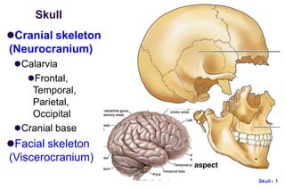
Skull
Cranial skeleton
(Neurocranium)
Calarvia
Frontal,
Temporal,
Parietal,
Occipital
Cranial base
Facial skeleton
(Viscerocranium)
Skull - 1
Neurocranium: cranial vault
 Frontal, Parietal, Temporal
 Mainly membranous bone formation
Skull - 2
Neurocranium:
cranial base
Midline
Ethmoid
Sphenoid
Occipital
Bilateral
Temporal
 Foramen
magnum
Skull - 3
Viscerocranium:
anterior view
Viscerocranium
Ethmoid,
Vomer,
Mandible
Maxilla, Zygoma,
Nasal, Lacrimal,
Inferior nasal
chonae, Palatine
Skull - 4
nasal cavities: septum
 nasal septum: perpendicular plate of ethmoid + vomer
Skull - 5
Lateral wall of nasal cavity
 Inferior nasal chonae
Ethmoid bone
Skull - 6
Viscerocranium: inferior view
 Palatine
 Maxilla
 Zygoma
Skull - 7
Skull - 8
Sutures and Fontanelles
 Coronal suture
 Sagittal suture
 Bregma
 Lambdoid suture
 Metopic suture
coronal
Skull: posterior
view
 external occipital
protuberance
(inion)
 external occipital
crest
 superior nuchal
line
 inferior nuchal line
Skull - 9
Superior nuchal line
ckwards
 Attachment of back muscles; e.g. Splenius capitis (“bandage”)
from spinous process of C7/T1-3 to superior nuchal line; draw
head ba
Superior nuchal line
Splenius capitis
Skull - 10
Skull - 11
Skull: lateral view
 Frankfurt plane
(anatomical position,
OrbitoMeatal line):
upper margin of ext.
acoustic meatus - orbit
floor  horizontal
 superior temporal line;
inferior temporal line
 external acoustic
meatus; mastoid
process
 level of ant., mid., post.
cranial fossae
OrbitoMeatal line (OM line) in
radiology
 from lateral canthus to
external acoustic meatus
Skull - 12
Pterion
Skull - 13
Superior temporal line and
Temporalis muscle
Posterior view
Skull - 14
Skull from front
 Supercillary arch
 Zygomatic proc.
of frontal bone
 Glabella
 Zygomatic bone
 Frontal proc. of
maxilla
 Frontal proc. of
zygoma
Skull - 15
Internal surface of the skull: The roof (vault)
e
 sagittal fissure
 coronal fissure
 lambdoid fissur
 grooves for
middle
meningeal vein,
artery
Skull - 16
Sk
Grooves for middle mengigeal arteries
Foramen
spinosum
ull - 17
Skull -
Sagittal fissure: superior sagittal sinus
 sup. sagittal sinus (SSS)
sagittal suture
18
Sagittal fissure: falx cerebri
 Dura extending from skull
Skull - 19
Dural sinuses in Posterior cranial fossa
 groove for transverse sinus; confluence of the sinus - internal
occipital protuberance
 tentorium cerebelli: separating occipital lobe from cerebellum
 internal occipital crest [- falx cerebelli]
Skull - 20
Inner surface of Anterior cranial fossa
 Frontal bone: Orbital plate: thin except near superciliary arch; frontal
air sinus
 Lesser wing of sphenoid: ant. clinoid processes
 Ethmoid bone: crista galli (attachment of falx cerebri)
 Cribriform plate
Skull - 21
Skul
Inner surface of Middle cranial fossa (1/2)
 Temporal bone: petrous part: thick, contains inner ear
 Hypophyseal fossa = sella turcica (Turk‘s saddle); tuberculum sellae,
dorsum sellae
 Ant. clinoid process (clinoid in Latin: bed-side); Post. clinoid
process; Diaphragma sellae
Anterior clinoid process
l - 22
Sella turcica
 Tuberculum sella
 Hypophyseal fossa:
pituitary gland
 Dorsum sella
Skull - 23
Sella turcica in
sphenoid bone
Skull - 24
Inner surface of Middle cranial fossa (2/2)
Superior orbital fissure (V1), foramen Rotundum (V2), foramen Ovale (V3)
foramen spinosum; opening of Int. Carodtid Art – Cavernous sinus
Skull - 25
Inner surface of Posterior fossa
 petrous portion of temporal (inner ear)
 int. acoustic meatus (N. VII and N. VIII.)
 to and from neck: foramen magnum; jugular foramen; hypoglossal canal
Skull - 26
Inner surface of Occipital bone, Temporal bone
 Occipital bone: Basilar part, [clivus]; Lateral parts;
Squamous part (squama occipitalis)
 Temporal bone: Squamous part; Petrous part; (Mastoid
part; Sytloid process)
Skull - 27
Floor (Outer surface) of middle cranial fossa-1
 foramen ovale (V3); foramen spinosum: (spine of
sphenoid bone close to foramen) for middle meningeal a.
 foramen lacerum: cartilage (internal carotid artery)
Skull - 28
Floor (Outer surface) of middle cranial fossa-2
 mandibular fossa, articular tubercle
Skull - 29
Outer surface of
Post. cranial fossa
 sphenoid and
occipital fused in the
midline
 opening of carotid
canal (ICA)
 stylomastoid
foramen (CN VII)
 pharyngeal tubercle
Skull - 30
carotid canal for
internal carotid artery
http://dc311.4shared.com/doc/WfGJhex5/preview.html Skull - 31
Skull - 32
internal carotid artery (ICA)
http://dermatologic.com.ar/7.htm
Outer surface of
Post. cranial fossa
 jugular foramen
 occipital condyle
 hypoglossal canal
(medial opening hidden
under condyle)
 styloid process:
stylohyoid lig.
 mastoid process: air
cell  middle ear
sternocleidomastoid
Skull - 33
Occoital-Atas joint
 Occipital bone:
(condyle)
 C1 Atlas: sup. Facet
(lateral mass)
Skull - 34
Skull (cranial skeleton): review
 Neurocranium
External surface
Interior (cranial fossa)
Anterior
Middle
Posterior
 Openings and contents
through them
Skull - 35
Facial skeleton
Cranial skeleton (Neurocranium)
Facial skeleton (Viscerocranium)
Ear
Orbit
Nasal cavity / Nasopharynx
Oral cavity / Palate and Jaw
Mandible
Bones
Temporal bone
Sphenoid bone
Skull - 36
Ear  External ear
External auditory meatus
 Middle ear (tympanic cavity)
Mastoid antrum
Pharyngotympanic tube
Auditroy ossicles
 Inner ear (petrous part)
Skull - 37
external auditory meatus
Skull - 38
3 ossicles
 malleus (handle
on tympanic
membrane)
 incus
 stapes (rest on
fenestra vestibuli)
Skull - 39
Inner ear: petrous part of temporal bone
 bony labyrinth
Cochlea
Vestibula, Semicircular canels
 internal accoustic meatus:
vestibulocochlear nerve (8th)
and facial nerve (7th)
Skull - 40
Facial canal: internal acoustic meatus;
stylomastoid foramen
Skull - 41
Temporal bone
 External surface
 Squamous part
 Zygomatic process
 Mastoid part
 Styloid process
 Internal surface
 Petrous part
Skull - 42
3
Skull - 4
Bony orbits: roof
 superciliary arch of frontal bone: thickened
 supraorbital notch (foramen): supraorbital n. (V1), vessels
 floor of ant. cranial fossa: thin
 lesser wing of sphenoid
 Lacrimal gland
Lesser wing,
Sphenoid
Sphenoid bone
 Greater wing
 Lesser wing
 Sphenoid body
 Medial pterygoid plate
 Lateral pterygoid plate
 Superior orbital fissure
Skull - 44
Bony orbits: lateral wall-1
 frontal proc. of
zygomatic bone
 zygomatic proc. of
frontal bone
Skull - 45
Zygomatic bone
 Frontal process
 Maxillary process
 Temporal process
F
Skull - 46
M
T
Bony orbits: lateral
wall-2
 Greater wing of
sphenoid
 superior orbital
fissure (where roof
and lateral wall meet)
 inferior orbital
fissure (where lateral
wall and floor meet)
Skull - 47
Skull - 48
Bony orbits: floor-1
 thin orbital floor
 maxillary process of zygomatic bone
zygomaticofacial foramen on malar surface
Maxillary proc.,
Zygoma
Skull - 49
Bony orbits: floor-2
 Maxilla
maxillary sinus
Skull - 50
Bony orbits: floor-3
 infraorbital n.: enters
from pterygopalatine
fossa through inf.
orbital fissure 
infraorbital groove 
infraorbital canal 
infraorbital foramen
Infraorbital
foramen
medial
lateral
Right orbit
Skull - 5
Bony orbits:
medial wall-1
 frontal process of maxilla
 frontal bone
 lacrimal bone
 fossa for lacrimal sac 
nasolacrimal canal 
inferior nasal meatus
 orbital plate of ethmoid:
(ethmoidal air cells medial
to this)
Frontal
process
of
maxilla
Frontal bone
1
medial
lateral
Right orbit
Skull - 5
Bony orbits:
medial wall-2
 orbital plate of
palatine bone
 body of sphenoid:
completes the lower
part of optic canal
 optic canal (where
roof and medial wall
meet)
Body of
sphenoid
2
Right orbit
lateral
medial
Skull - 5
Bony orbits:
medial wall-3
ant. & post. ethmoidal
foramina
transmitting
corresponding vessels
(br. of ophthalmic a.) to
supply nasal cavity
and ethmoidal air cells
Frontal
process
of
maxilla
Frontal bone
3
Skull -
Frontal bone
Openings of orbit
54
 1. optic foramen (canal)
 2. sup. orbital fissure
 3. inf. orbital fissure
 4. nasolacrimal canal
 5. ant. & post.
ethmoidal foramina
Frontal
process
of
maxilla
Skull - 55
Contents of orbital openings
III, IV, V1, VI
V2 branch
Nasal cavity-1
 piriform aperture; cartilage
plates; maxilla; nasal bone
Skull - 56
nasal cavities and nasopharynx-2
 nasal septum: perpendicular plate of ethmoid + vomer
Skull - 57
Roof of nasal
cavity
 cribriform plate
of ethmoid;
frontal & nasal
bone anteriorly;
sphenoid
posteriorly
Skull - 58
Floor of nasal cavity
 ant. 2/3: palatine
process of maxilla
 post. 1/3: horizontal
plate of palatine
Skull - 59
S
Lateral wall of nasal cavity-1
 Superior: ethmoid (ethmoidal air cells); inferior: maxilla
(maxillary sinus); posterior: perpendicular plate of palatine
 perpendicular plate separates the nasal cavity from
pterygopalatine fossa
Ethmoid bone
kull - 60
Pterygopalatine fossa
Skull - 61
Skull - 62
Lateral wall of nasal cavity-2
 sphenopalatine foramen
opens from nasal cavity to
pterygopalatine fossa.
 further posteriorly, the
medial surface of medial
pterygoid plate completes
the lateral wall
Medial pterygoid
plate, palatine bone
Extension of bones into Nasal cavity-1
 Shells of bones
extending into the
nasal cavities:
 1) superior concha:
part of ethmoid
 2) middle concha: part
of ethmoid
 3) inferior concha: a
separate bone
Skull - 63
Extension of bones into Nasal cavity-2
 4 channels in nasal cavity:
 1) sphenoethmoidal recess: above superior concha, 2)
sup. 3) mid. and 4) inf. nasal meatus
Skull - 64
Ethmoid bone
 Cristal galli
 Perpendicular plate
 Cribriform plate
 Lateral and inferior
extension
 Superior concha
 Middle concha
Skull - 65
Paranasal sinuses
communicate with nasal cavities and nasopharynx
frontal sinus, ethomidal air cells, maxillary & sphenoidal sinus
Skull - 66
Nasopharynx
Skull - 67
Bony walls of nasopharynx
 pharynx (raphe) attaches to pharyngeal tubercle; medial
pterygoid plate; auditory tube (cartilaginous part)
Skull - 68
Oral cavity: upper jaw
 maxilla; hard palate; alveolar process
Skull - 69
Bony palate-1
 incisor fossa/foramen: septal br. of sphenopalatine a. +
nasopalatine n. (connection between oral and nasal cavity)
Skull - 70
Skull - 71
Bony palate-2
 midline suture; transverse suture: palatine/ maxilla
 greater palatine foramen:  pterygopalatine fossa
 pterygoid hamulus of medial pterygoid plate
Transverse suture
Pterygoid of sphenoid bone
 Lateral pterygoid plate (lamina)
 Medial pterygoid plate (lamina)
 Pterygoid hamulus
Skull - 72
lower jaw (mandible)
 body; alveolar process; angle; ramus: head of mandible
(condylar proc.); coronoid proc.
Coronoid process
Skull - 73
Medial surface of mandible-1
mandibular foramen
inf. alveolar n. (branch
of V3), vessels
Entrance into
mandibular canal
Lingula: triangular, for
sphenomandibular lig.
mylohyoid groove:
mylohyoid n., vessels
mylohyoid line:
attachment of mylohyoid
muscle
Mylohyoid groove
Skull - 74
Medial surface of mandible-2
 rough area at angle and post. margin of ramus:
med. pterygoid muscle attachment
Skull - 75
Lateral surface of mandible
 masseter muscle attachment;
mental foramen
Skull - 76
Mandibular foramen; Mental foramen
Skull - 77
Temporomandibular
joint (TM joint, TMJ)
 head of mandible 
mandibular fossa +
articular tubercle
Skull - 78
Hyoid bone
 Body
 greater cornu
 lesser cornu
(stylohyoid lig.)
Skull - 79
Skull: review
 Neurocranium
External surface
Interior: Anterior, Middle,
Posterior cranial fossa
 Facial skeleton
Ear, Orbit
Nasal and Oral cavities
Mandible
 Openings and contents
through them
Skull - 80

Bone_Skull.pptx

  • 1.
  • 2.
    Neurocranium: cranial vault Frontal, Parietal, Temporal  Mainly membranous bone formation Skull - 2
  • 3.
  • 4.
  • 5.
    nasal cavities: septum nasal septum: perpendicular plate of ethmoid + vomer Skull - 5
  • 6.
    Lateral wall ofnasal cavity  Inferior nasal chonae Ethmoid bone Skull - 6
  • 7.
    Viscerocranium: inferior view Palatine  Maxilla  Zygoma Skull - 7
  • 8.
    Skull - 8 Suturesand Fontanelles  Coronal suture  Sagittal suture  Bregma  Lambdoid suture  Metopic suture coronal
  • 9.
    Skull: posterior view  externaloccipital protuberance (inion)  external occipital crest  superior nuchal line  inferior nuchal line Skull - 9
  • 10.
    Superior nuchal line ckwards Attachment of back muscles; e.g. Splenius capitis (“bandage”) from spinous process of C7/T1-3 to superior nuchal line; draw head ba Superior nuchal line Splenius capitis Skull - 10
  • 11.
    Skull - 11 Skull:lateral view  Frankfurt plane (anatomical position, OrbitoMeatal line): upper margin of ext. acoustic meatus - orbit floor  horizontal  superior temporal line; inferior temporal line  external acoustic meatus; mastoid process  level of ant., mid., post. cranial fossae
  • 12.
    OrbitoMeatal line (OMline) in radiology  from lateral canthus to external acoustic meatus Skull - 12
  • 13.
  • 14.
    Superior temporal lineand Temporalis muscle Posterior view Skull - 14
  • 15.
    Skull from front Supercillary arch  Zygomatic proc. of frontal bone  Glabella  Zygomatic bone  Frontal proc. of maxilla  Frontal proc. of zygoma Skull - 15
  • 16.
    Internal surface ofthe skull: The roof (vault) e  sagittal fissure  coronal fissure  lambdoid fissur  grooves for middle meningeal vein, artery Skull - 16
  • 17.
    Sk Grooves for middlemengigeal arteries Foramen spinosum ull - 17
  • 18.
    Skull - Sagittal fissure:superior sagittal sinus  sup. sagittal sinus (SSS) sagittal suture 18
  • 19.
    Sagittal fissure: falxcerebri  Dura extending from skull Skull - 19
  • 20.
    Dural sinuses inPosterior cranial fossa  groove for transverse sinus; confluence of the sinus - internal occipital protuberance  tentorium cerebelli: separating occipital lobe from cerebellum  internal occipital crest [- falx cerebelli] Skull - 20
  • 21.
    Inner surface ofAnterior cranial fossa  Frontal bone: Orbital plate: thin except near superciliary arch; frontal air sinus  Lesser wing of sphenoid: ant. clinoid processes  Ethmoid bone: crista galli (attachment of falx cerebri)  Cribriform plate Skull - 21
  • 22.
    Skul Inner surface ofMiddle cranial fossa (1/2)  Temporal bone: petrous part: thick, contains inner ear  Hypophyseal fossa = sella turcica (Turk‘s saddle); tuberculum sellae, dorsum sellae  Ant. clinoid process (clinoid in Latin: bed-side); Post. clinoid process; Diaphragma sellae Anterior clinoid process l - 22
  • 23.
    Sella turcica  Tuberculumsella  Hypophyseal fossa: pituitary gland  Dorsum sella Skull - 23
  • 24.
  • 25.
    Inner surface ofMiddle cranial fossa (2/2) Superior orbital fissure (V1), foramen Rotundum (V2), foramen Ovale (V3) foramen spinosum; opening of Int. Carodtid Art – Cavernous sinus Skull - 25
  • 26.
    Inner surface ofPosterior fossa  petrous portion of temporal (inner ear)  int. acoustic meatus (N. VII and N. VIII.)  to and from neck: foramen magnum; jugular foramen; hypoglossal canal Skull - 26
  • 27.
    Inner surface ofOccipital bone, Temporal bone  Occipital bone: Basilar part, [clivus]; Lateral parts; Squamous part (squama occipitalis)  Temporal bone: Squamous part; Petrous part; (Mastoid part; Sytloid process) Skull - 27
  • 28.
    Floor (Outer surface)of middle cranial fossa-1  foramen ovale (V3); foramen spinosum: (spine of sphenoid bone close to foramen) for middle meningeal a.  foramen lacerum: cartilage (internal carotid artery) Skull - 28
  • 29.
    Floor (Outer surface)of middle cranial fossa-2  mandibular fossa, articular tubercle Skull - 29
  • 30.
    Outer surface of Post.cranial fossa  sphenoid and occipital fused in the midline  opening of carotid canal (ICA)  stylomastoid foramen (CN VII)  pharyngeal tubercle Skull - 30
  • 31.
    carotid canal for internalcarotid artery http://dc311.4shared.com/doc/WfGJhex5/preview.html Skull - 31
  • 32.
    Skull - 32 internalcarotid artery (ICA) http://dermatologic.com.ar/7.htm
  • 33.
    Outer surface of Post.cranial fossa  jugular foramen  occipital condyle  hypoglossal canal (medial opening hidden under condyle)  styloid process: stylohyoid lig.  mastoid process: air cell  middle ear sternocleidomastoid Skull - 33
  • 34.
    Occoital-Atas joint  Occipitalbone: (condyle)  C1 Atlas: sup. Facet (lateral mass) Skull - 34
  • 35.
    Skull (cranial skeleton):review  Neurocranium External surface Interior (cranial fossa) Anterior Middle Posterior  Openings and contents through them Skull - 35
  • 36.
    Facial skeleton Cranial skeleton(Neurocranium) Facial skeleton (Viscerocranium) Ear Orbit Nasal cavity / Nasopharynx Oral cavity / Palate and Jaw Mandible Bones Temporal bone Sphenoid bone Skull - 36
  • 37.
    Ear  Externalear External auditory meatus  Middle ear (tympanic cavity) Mastoid antrum Pharyngotympanic tube Auditroy ossicles  Inner ear (petrous part) Skull - 37
  • 38.
  • 39.
    3 ossicles  malleus(handle on tympanic membrane)  incus  stapes (rest on fenestra vestibuli) Skull - 39
  • 40.
    Inner ear: petrouspart of temporal bone  bony labyrinth Cochlea Vestibula, Semicircular canels  internal accoustic meatus: vestibulocochlear nerve (8th) and facial nerve (7th) Skull - 40
  • 41.
    Facial canal: internalacoustic meatus; stylomastoid foramen Skull - 41
  • 42.
    Temporal bone  Externalsurface  Squamous part  Zygomatic process  Mastoid part  Styloid process  Internal surface  Petrous part Skull - 42
  • 43.
    3 Skull - 4 Bonyorbits: roof  superciliary arch of frontal bone: thickened  supraorbital notch (foramen): supraorbital n. (V1), vessels  floor of ant. cranial fossa: thin  lesser wing of sphenoid  Lacrimal gland Lesser wing, Sphenoid
  • 44.
    Sphenoid bone  Greaterwing  Lesser wing  Sphenoid body  Medial pterygoid plate  Lateral pterygoid plate  Superior orbital fissure Skull - 44
  • 45.
    Bony orbits: lateralwall-1  frontal proc. of zygomatic bone  zygomatic proc. of frontal bone Skull - 45
  • 46.
    Zygomatic bone  Frontalprocess  Maxillary process  Temporal process F Skull - 46 M T
  • 47.
    Bony orbits: lateral wall-2 Greater wing of sphenoid  superior orbital fissure (where roof and lateral wall meet)  inferior orbital fissure (where lateral wall and floor meet) Skull - 47
  • 48.
    Skull - 48 Bonyorbits: floor-1  thin orbital floor  maxillary process of zygomatic bone zygomaticofacial foramen on malar surface Maxillary proc., Zygoma
  • 49.
    Skull - 49 Bonyorbits: floor-2  Maxilla maxillary sinus
  • 50.
    Skull - 50 Bonyorbits: floor-3  infraorbital n.: enters from pterygopalatine fossa through inf. orbital fissure  infraorbital groove  infraorbital canal  infraorbital foramen Infraorbital foramen medial lateral Right orbit
  • 51.
    Skull - 5 Bonyorbits: medial wall-1  frontal process of maxilla  frontal bone  lacrimal bone  fossa for lacrimal sac  nasolacrimal canal  inferior nasal meatus  orbital plate of ethmoid: (ethmoidal air cells medial to this) Frontal process of maxilla Frontal bone 1 medial lateral Right orbit
  • 52.
    Skull - 5 Bonyorbits: medial wall-2  orbital plate of palatine bone  body of sphenoid: completes the lower part of optic canal  optic canal (where roof and medial wall meet) Body of sphenoid 2 Right orbit lateral medial
  • 53.
    Skull - 5 Bonyorbits: medial wall-3 ant. & post. ethmoidal foramina transmitting corresponding vessels (br. of ophthalmic a.) to supply nasal cavity and ethmoidal air cells Frontal process of maxilla Frontal bone 3
  • 54.
    Skull - Frontal bone Openingsof orbit 54  1. optic foramen (canal)  2. sup. orbital fissure  3. inf. orbital fissure  4. nasolacrimal canal  5. ant. & post. ethmoidal foramina Frontal process of maxilla
  • 55.
    Skull - 55 Contentsof orbital openings III, IV, V1, VI V2 branch
  • 56.
    Nasal cavity-1  piriformaperture; cartilage plates; maxilla; nasal bone Skull - 56
  • 57.
    nasal cavities andnasopharynx-2  nasal septum: perpendicular plate of ethmoid + vomer Skull - 57
  • 58.
    Roof of nasal cavity cribriform plate of ethmoid; frontal & nasal bone anteriorly; sphenoid posteriorly Skull - 58
  • 59.
    Floor of nasalcavity  ant. 2/3: palatine process of maxilla  post. 1/3: horizontal plate of palatine Skull - 59
  • 60.
    S Lateral wall ofnasal cavity-1  Superior: ethmoid (ethmoidal air cells); inferior: maxilla (maxillary sinus); posterior: perpendicular plate of palatine  perpendicular plate separates the nasal cavity from pterygopalatine fossa Ethmoid bone kull - 60
  • 61.
  • 62.
    Skull - 62 Lateralwall of nasal cavity-2  sphenopalatine foramen opens from nasal cavity to pterygopalatine fossa.  further posteriorly, the medial surface of medial pterygoid plate completes the lateral wall Medial pterygoid plate, palatine bone
  • 63.
    Extension of bonesinto Nasal cavity-1  Shells of bones extending into the nasal cavities:  1) superior concha: part of ethmoid  2) middle concha: part of ethmoid  3) inferior concha: a separate bone Skull - 63
  • 64.
    Extension of bonesinto Nasal cavity-2  4 channels in nasal cavity:  1) sphenoethmoidal recess: above superior concha, 2) sup. 3) mid. and 4) inf. nasal meatus Skull - 64
  • 65.
    Ethmoid bone  Cristalgalli  Perpendicular plate  Cribriform plate  Lateral and inferior extension  Superior concha  Middle concha Skull - 65
  • 66.
    Paranasal sinuses communicate withnasal cavities and nasopharynx frontal sinus, ethomidal air cells, maxillary & sphenoidal sinus Skull - 66
  • 67.
  • 68.
    Bony walls ofnasopharynx  pharynx (raphe) attaches to pharyngeal tubercle; medial pterygoid plate; auditory tube (cartilaginous part) Skull - 68
  • 69.
    Oral cavity: upperjaw  maxilla; hard palate; alveolar process Skull - 69
  • 70.
    Bony palate-1  incisorfossa/foramen: septal br. of sphenopalatine a. + nasopalatine n. (connection between oral and nasal cavity) Skull - 70
  • 71.
    Skull - 71 Bonypalate-2  midline suture; transverse suture: palatine/ maxilla  greater palatine foramen:  pterygopalatine fossa  pterygoid hamulus of medial pterygoid plate Transverse suture
  • 72.
    Pterygoid of sphenoidbone  Lateral pterygoid plate (lamina)  Medial pterygoid plate (lamina)  Pterygoid hamulus Skull - 72
  • 73.
    lower jaw (mandible) body; alveolar process; angle; ramus: head of mandible (condylar proc.); coronoid proc. Coronoid process Skull - 73
  • 74.
    Medial surface ofmandible-1 mandibular foramen inf. alveolar n. (branch of V3), vessels Entrance into mandibular canal Lingula: triangular, for sphenomandibular lig. mylohyoid groove: mylohyoid n., vessels mylohyoid line: attachment of mylohyoid muscle Mylohyoid groove Skull - 74
  • 75.
    Medial surface ofmandible-2  rough area at angle and post. margin of ramus: med. pterygoid muscle attachment Skull - 75
  • 76.
    Lateral surface ofmandible  masseter muscle attachment; mental foramen Skull - 76
  • 77.
    Mandibular foramen; Mentalforamen Skull - 77
  • 78.
    Temporomandibular joint (TM joint,TMJ)  head of mandible  mandibular fossa + articular tubercle Skull - 78
  • 79.
    Hyoid bone  Body greater cornu  lesser cornu (stylohyoid lig.) Skull - 79
  • 80.
    Skull: review  Neurocranium Externalsurface Interior: Anterior, Middle, Posterior cranial fossa  Facial skeleton Ear, Orbit Nasal and Oral cavities Mandible  Openings and contents through them Skull - 80