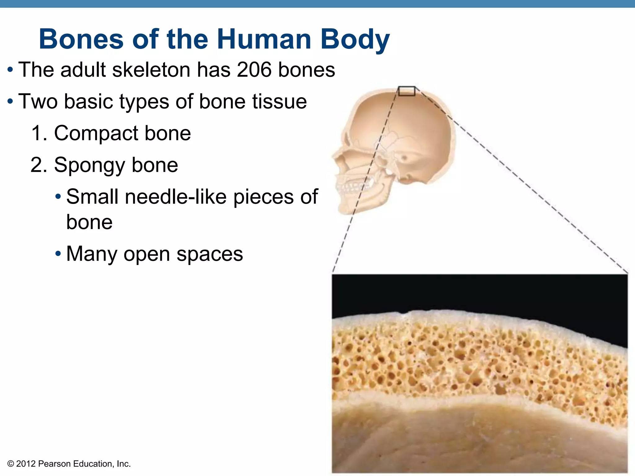
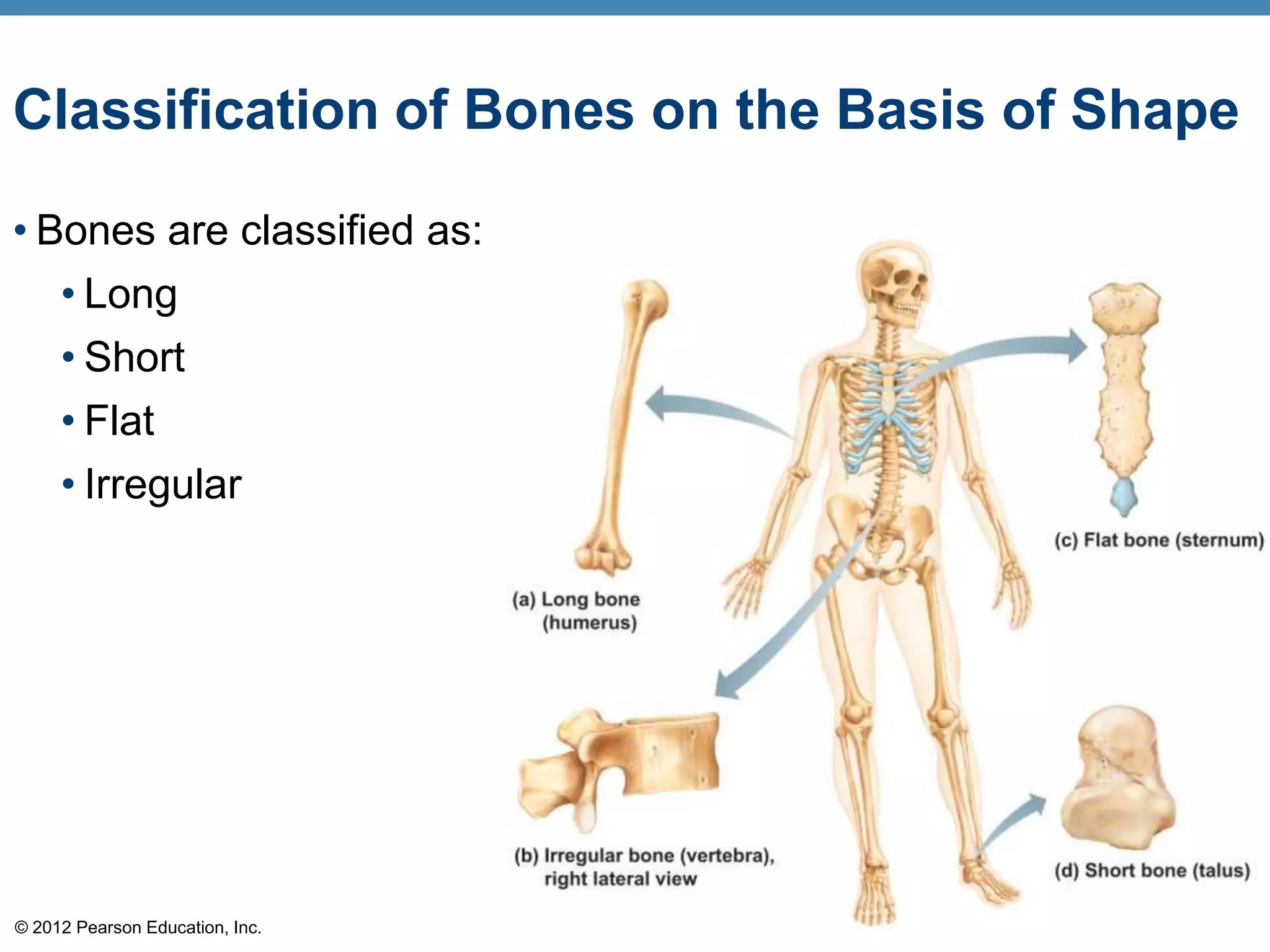
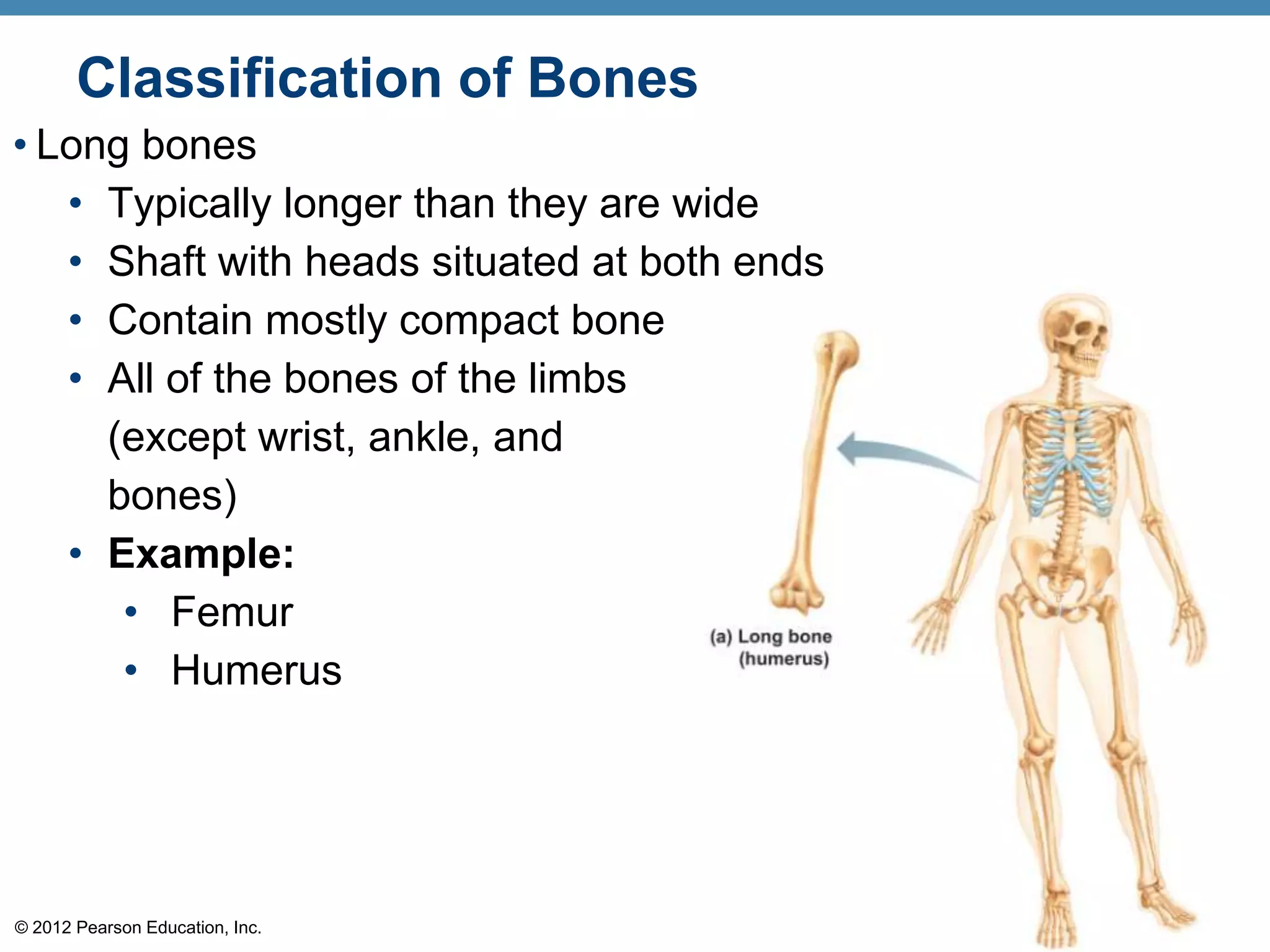
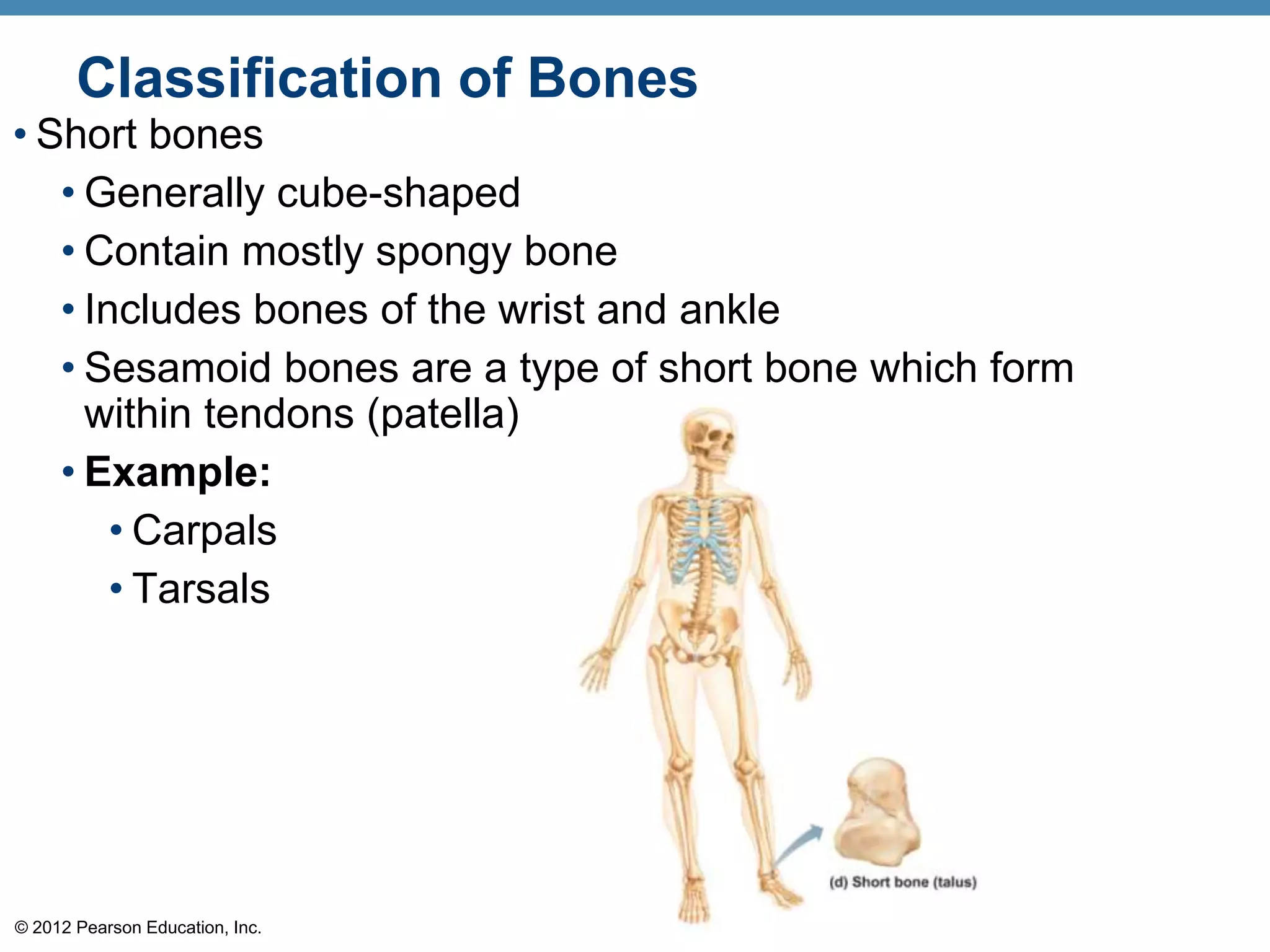
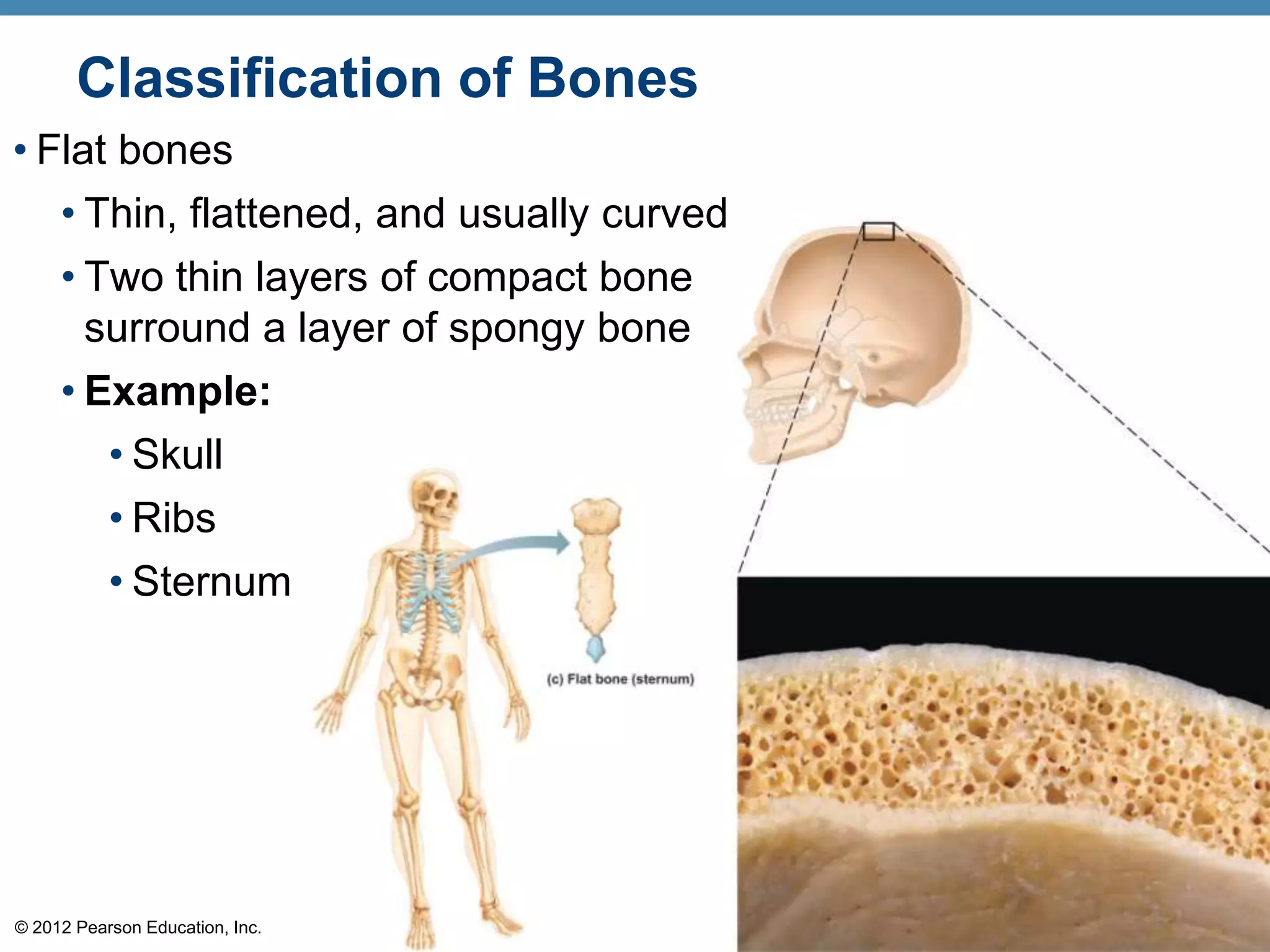
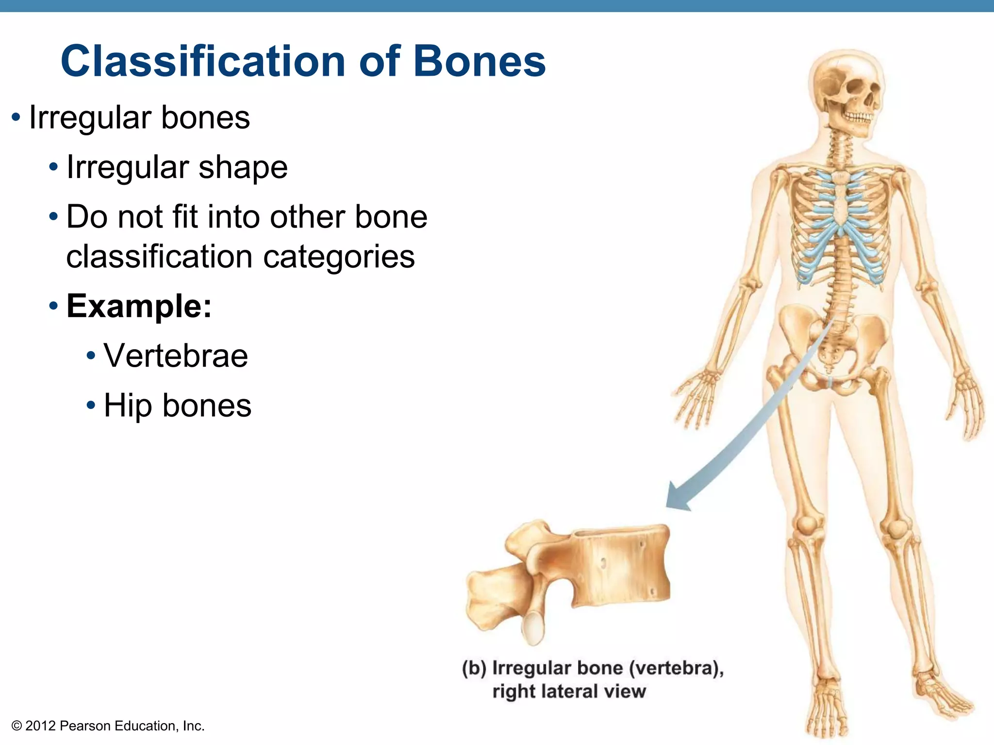
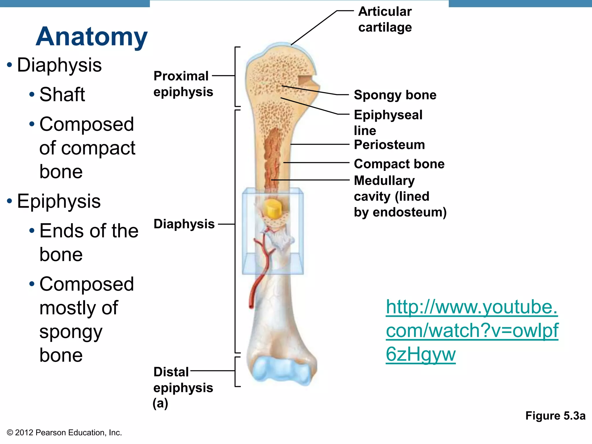
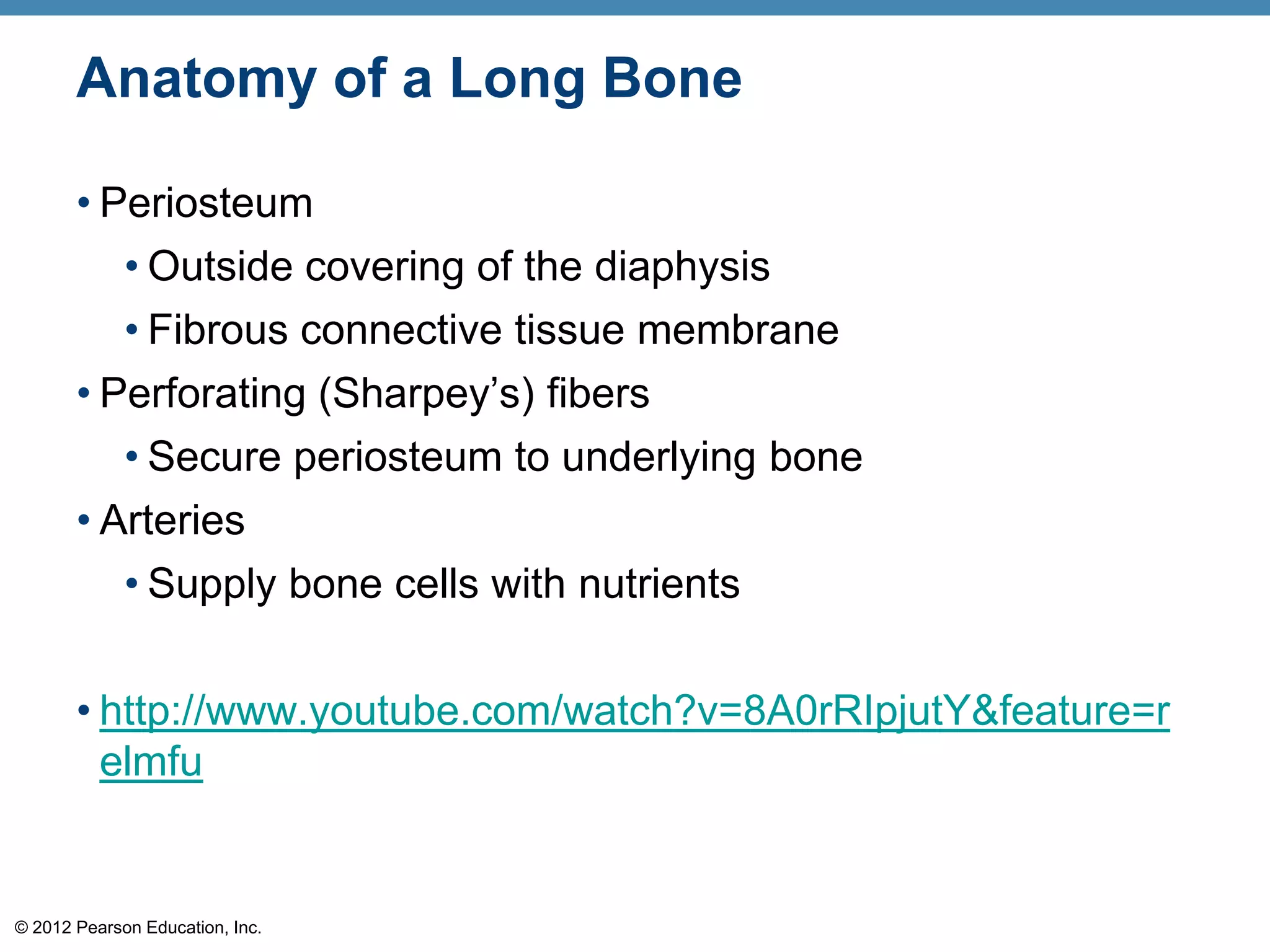
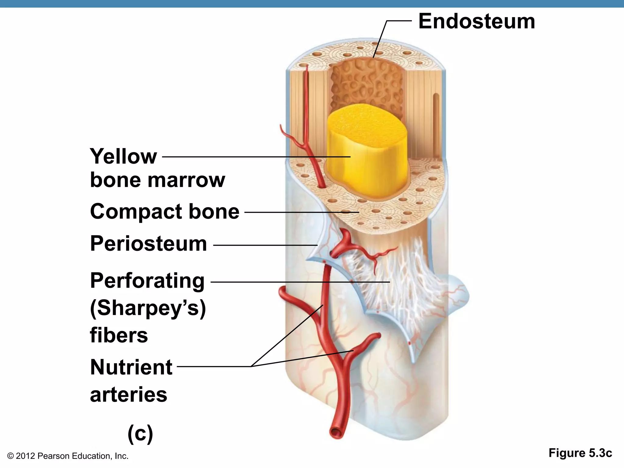
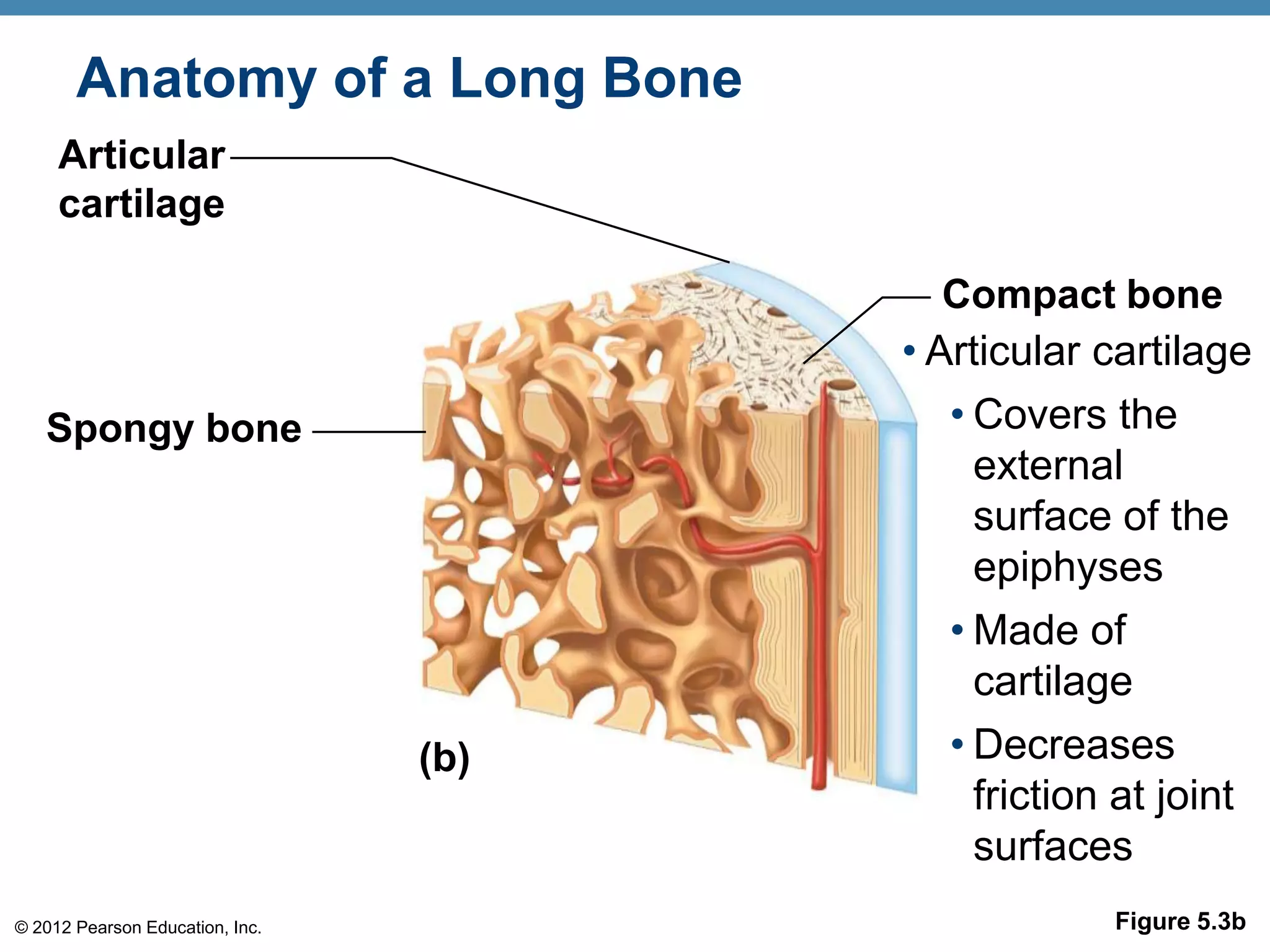
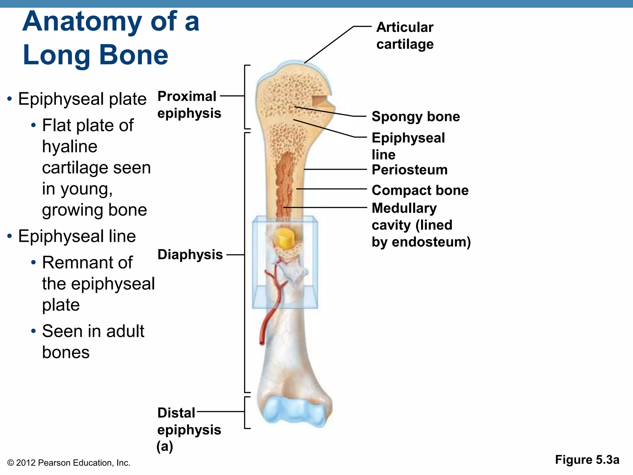
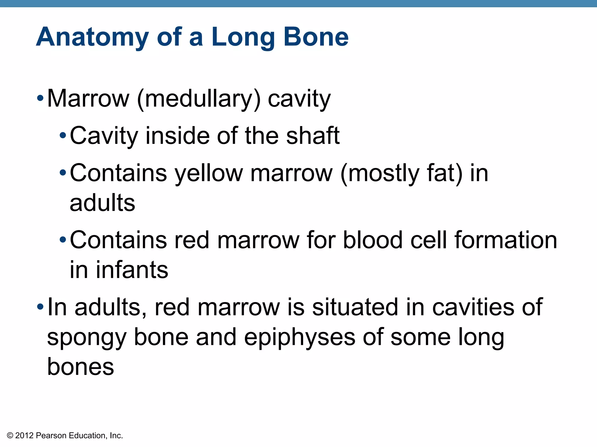
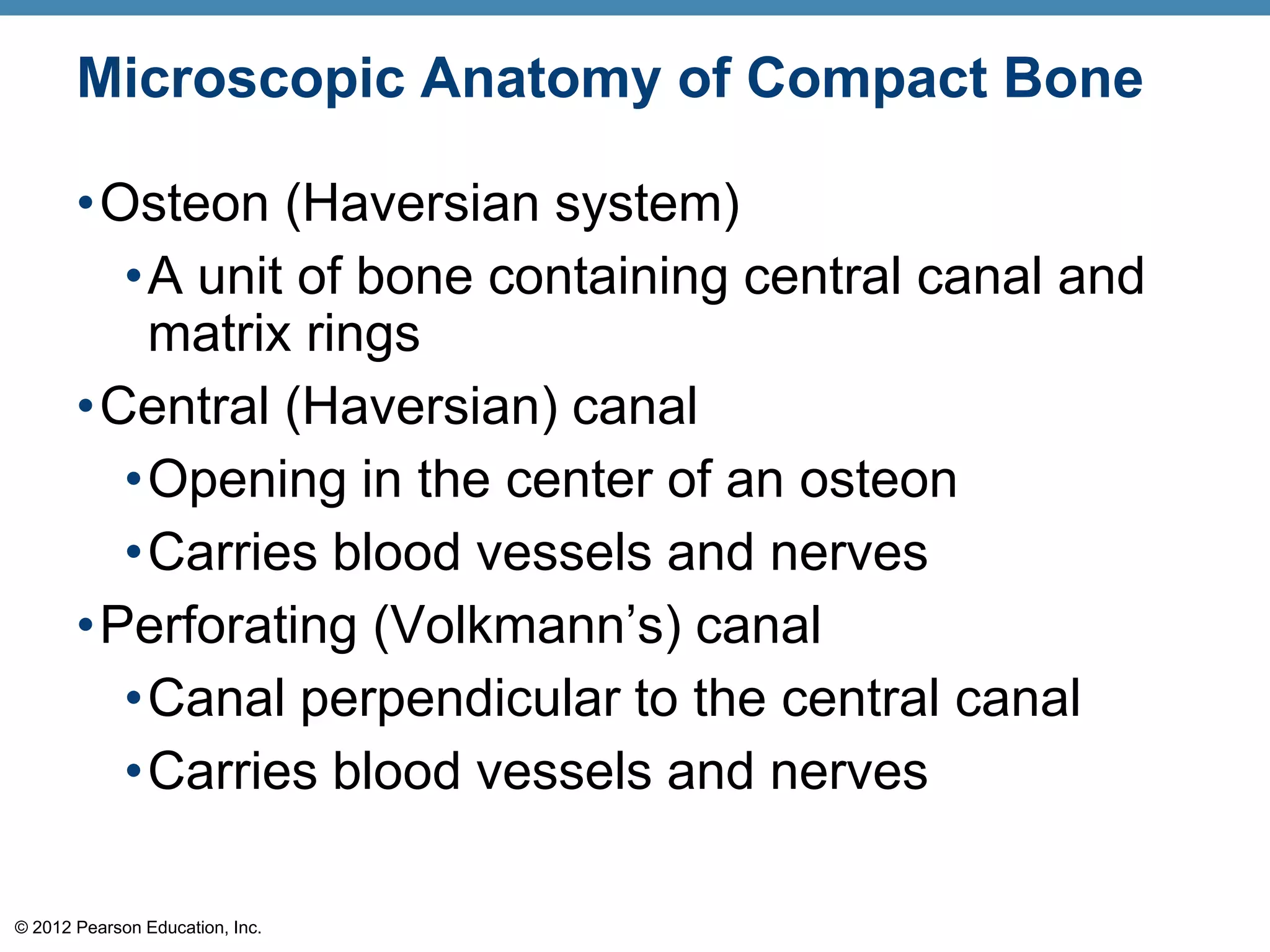
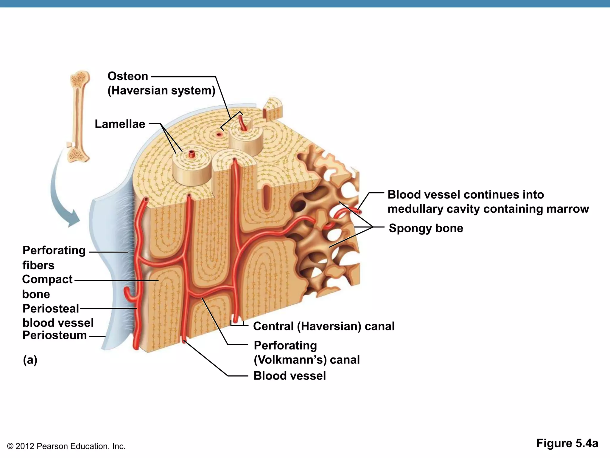
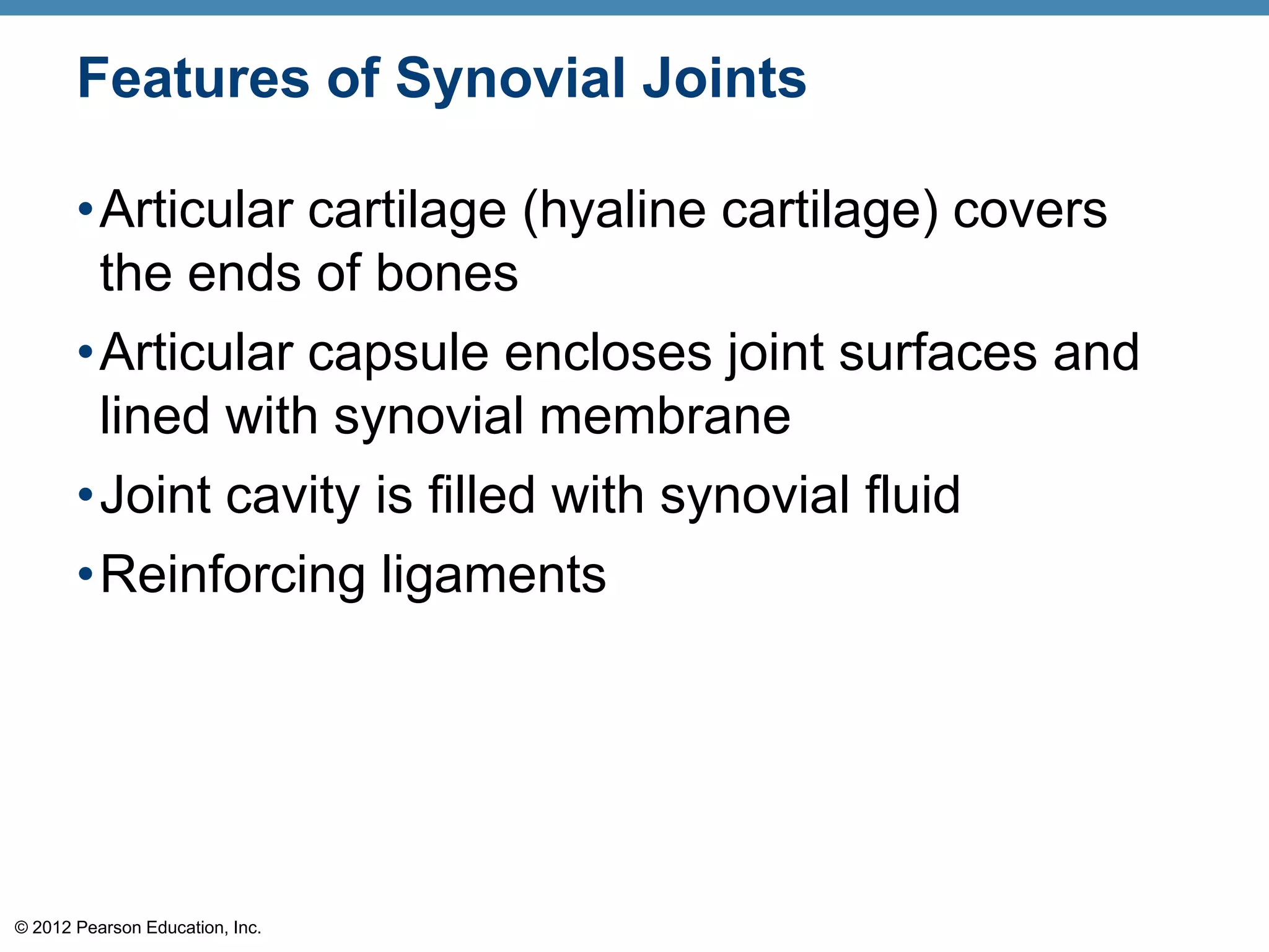
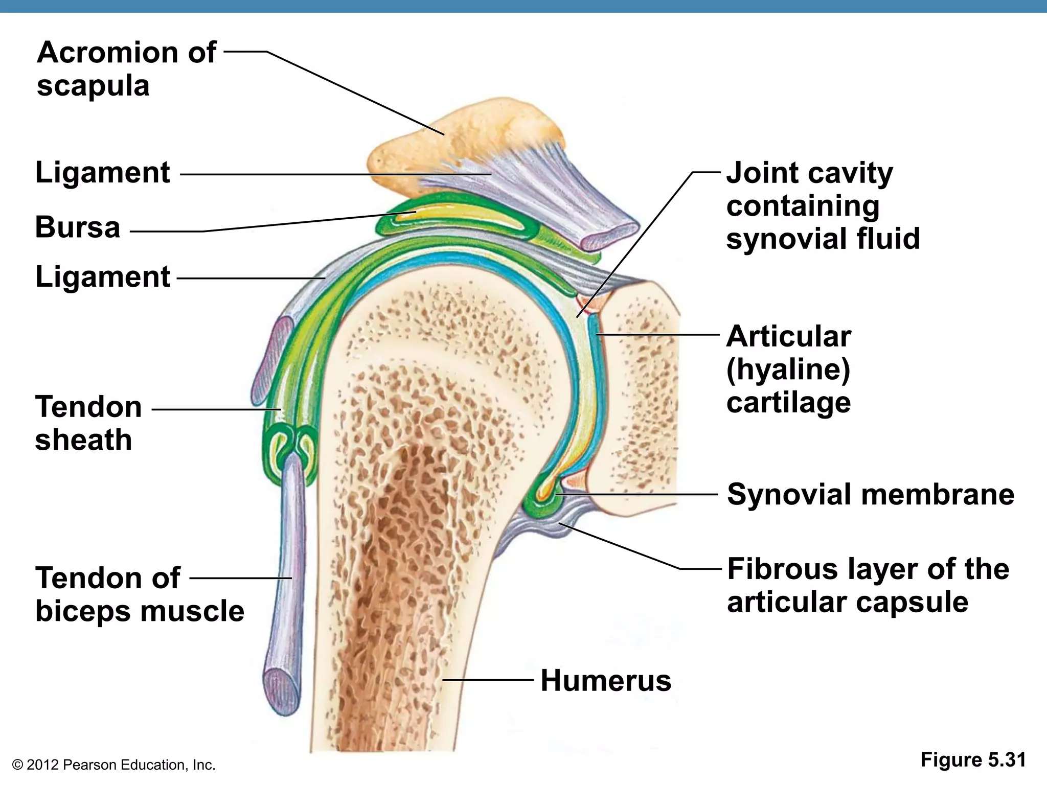
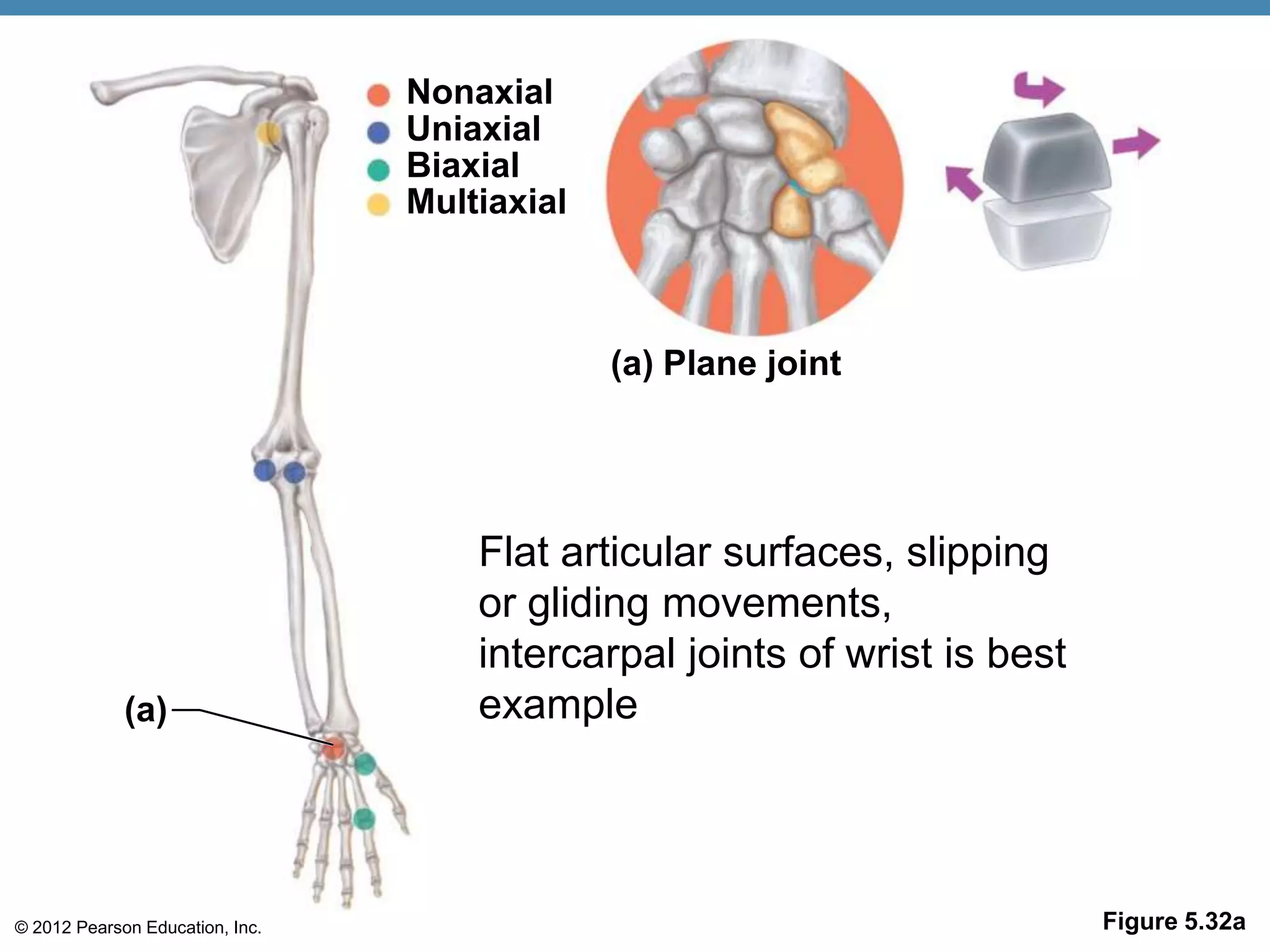
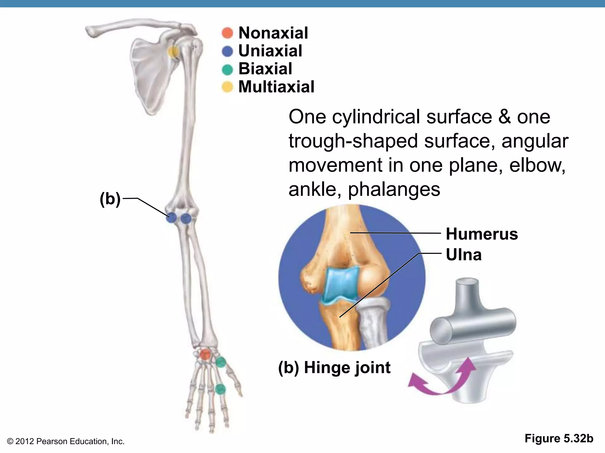
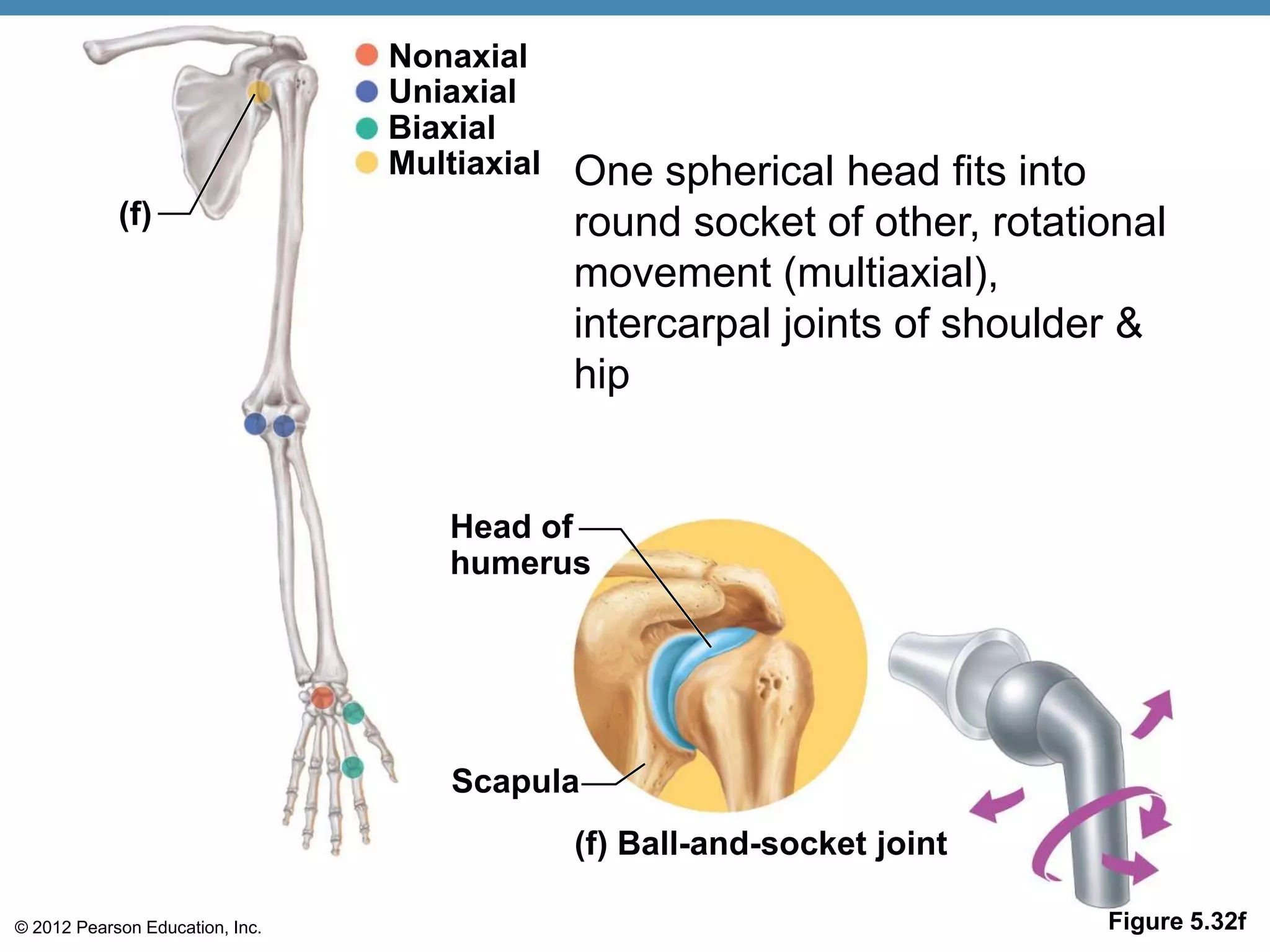
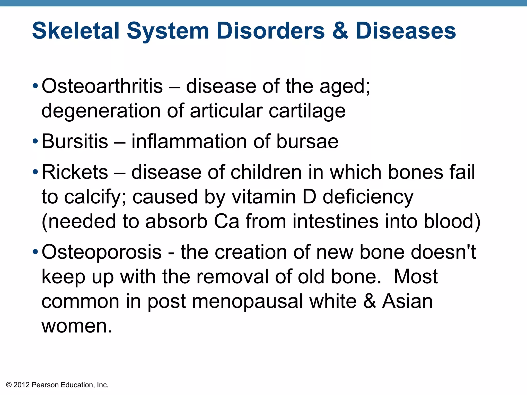
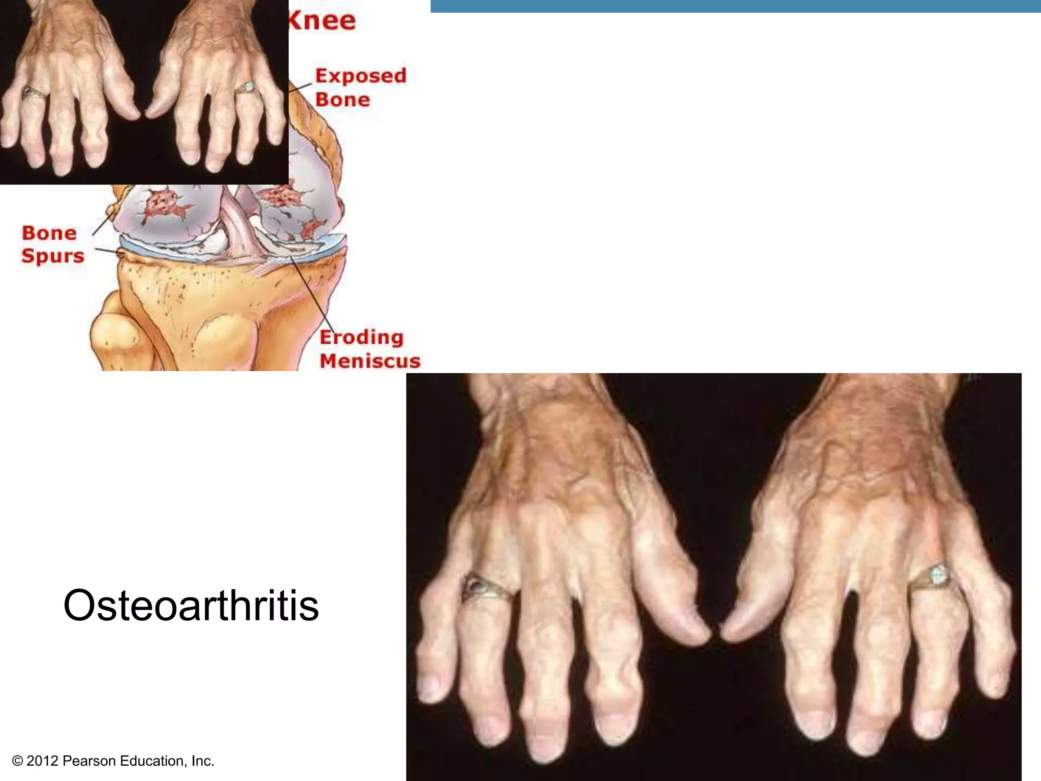
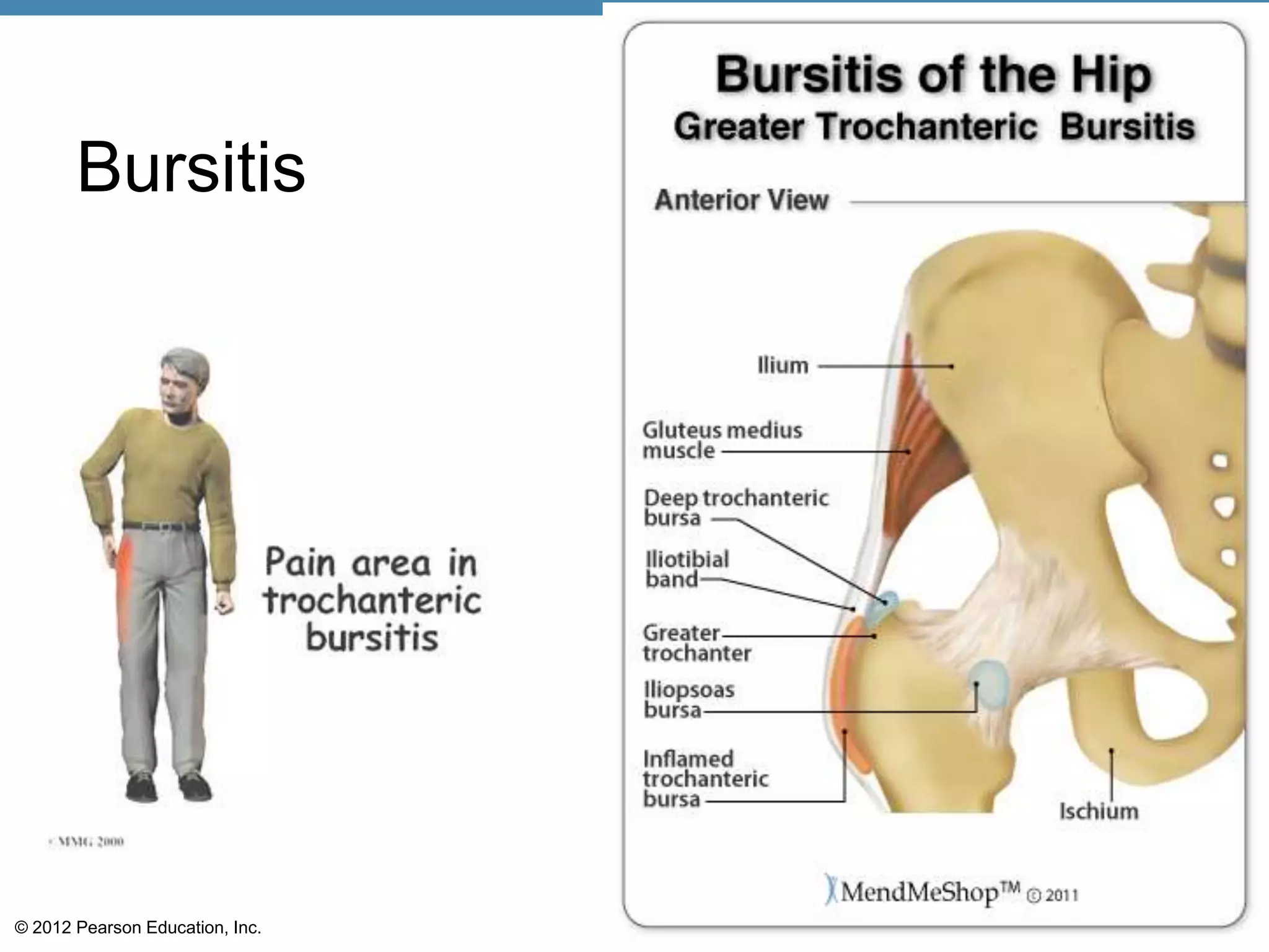
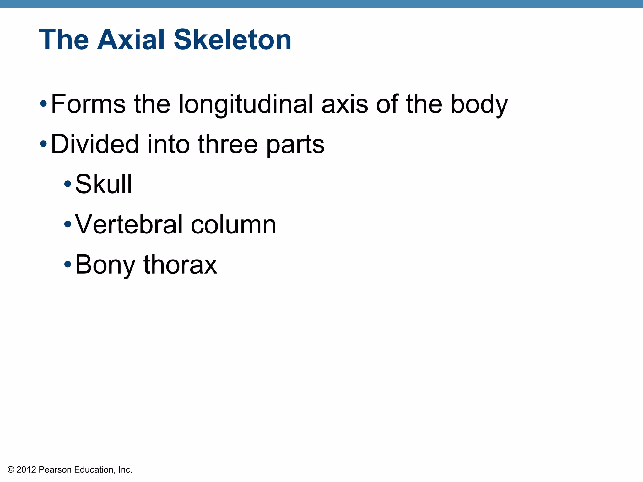
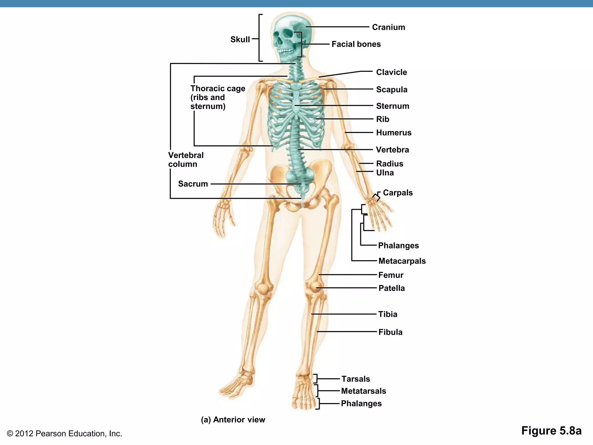
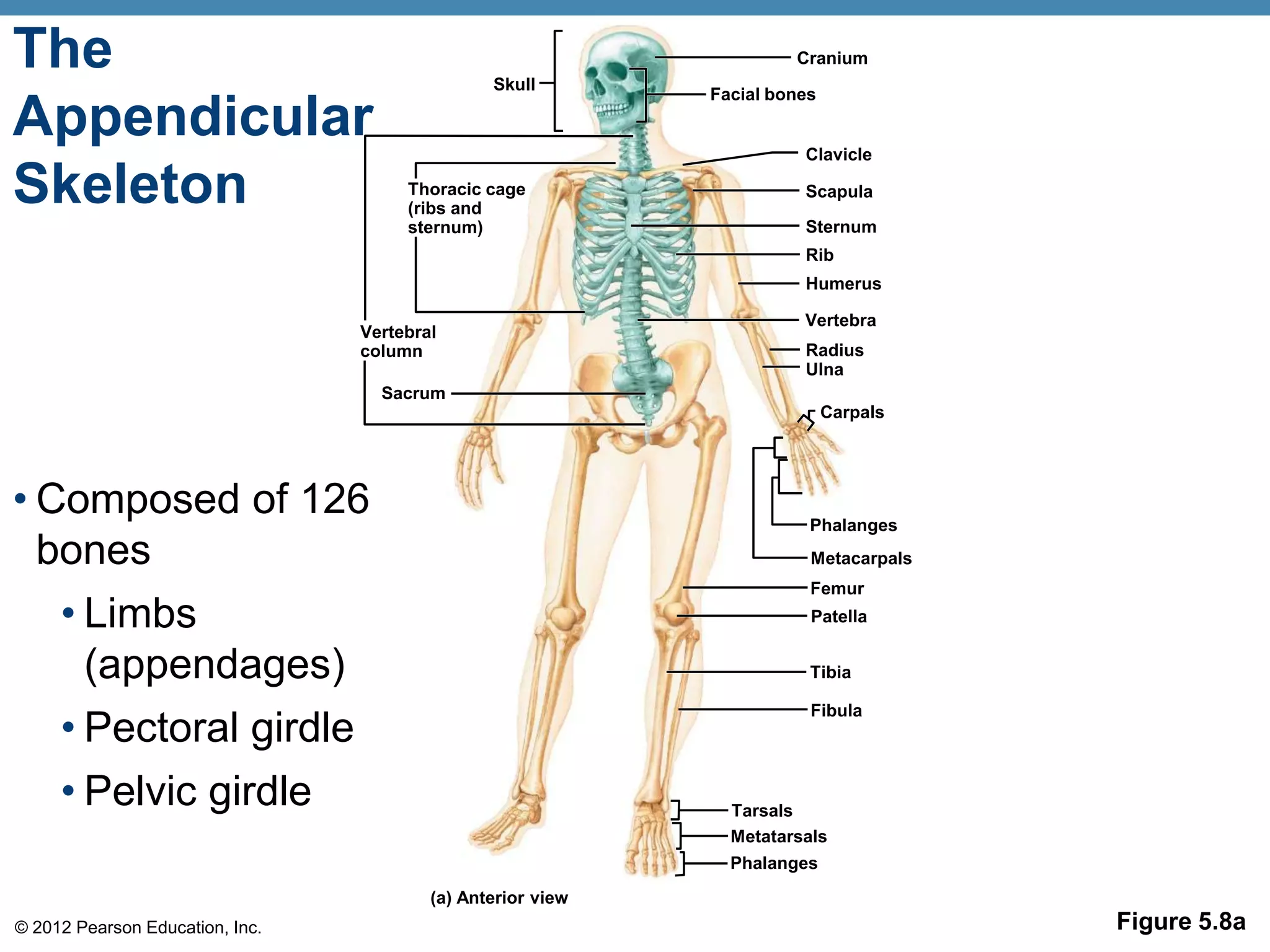
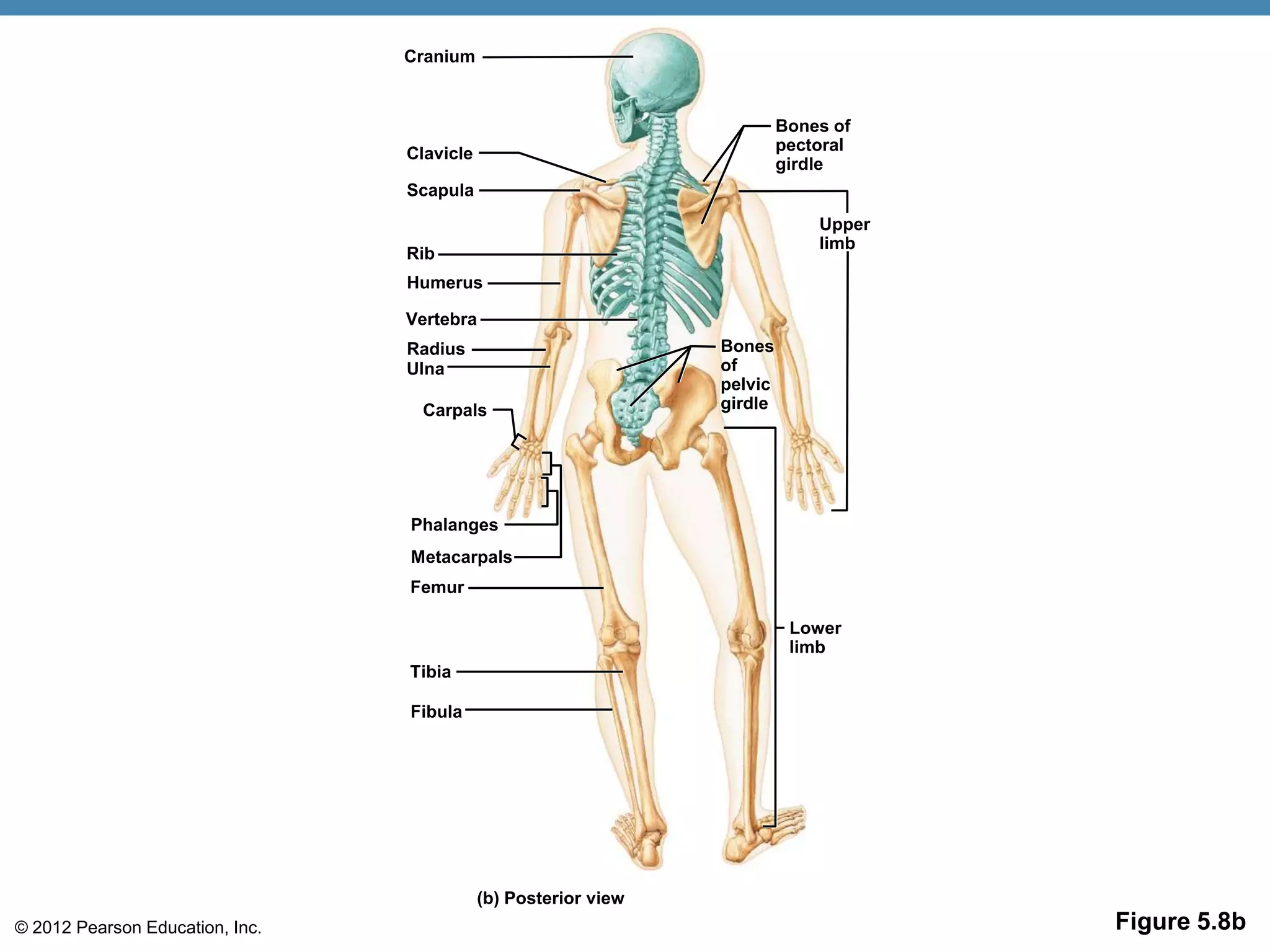
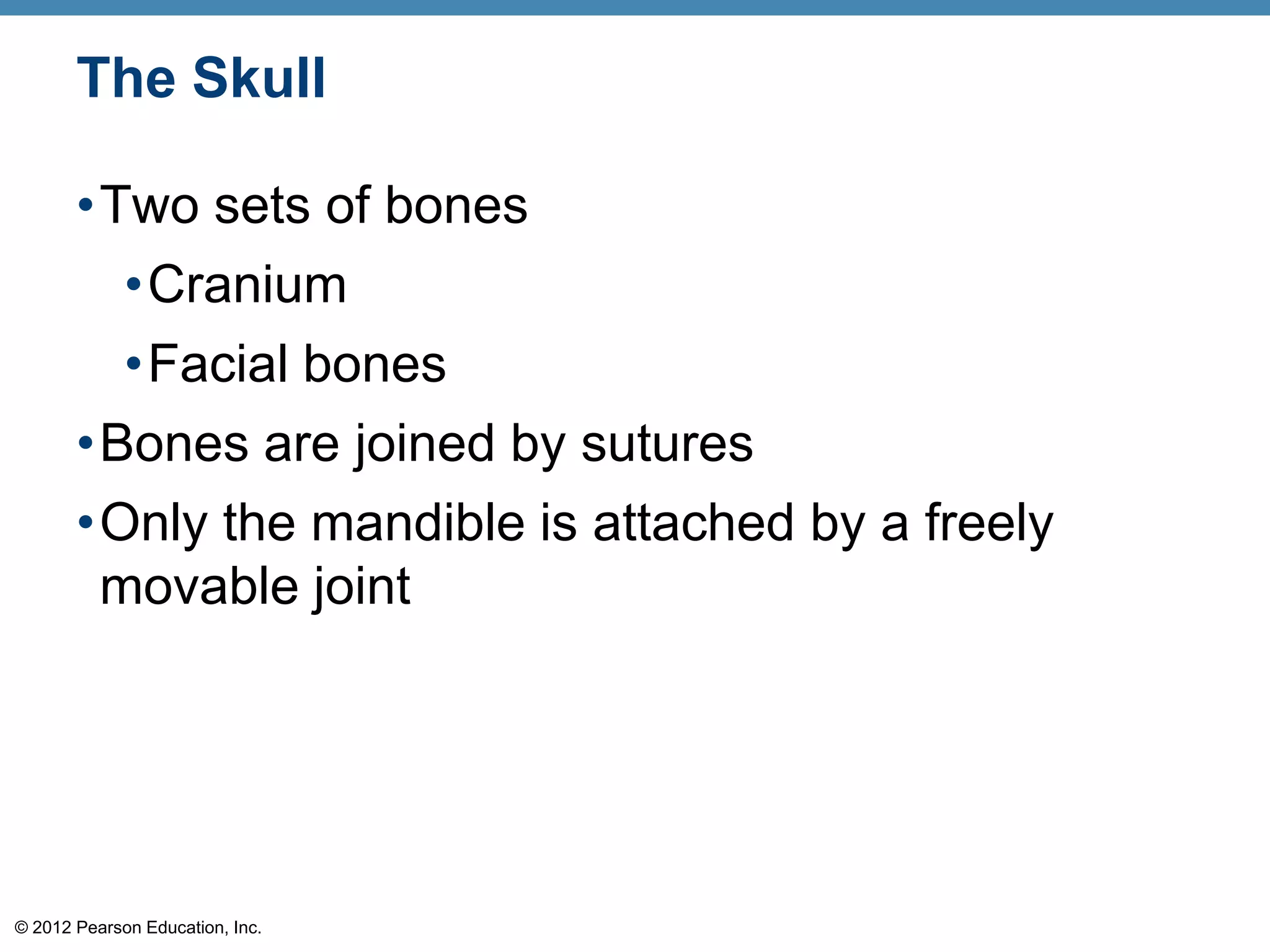
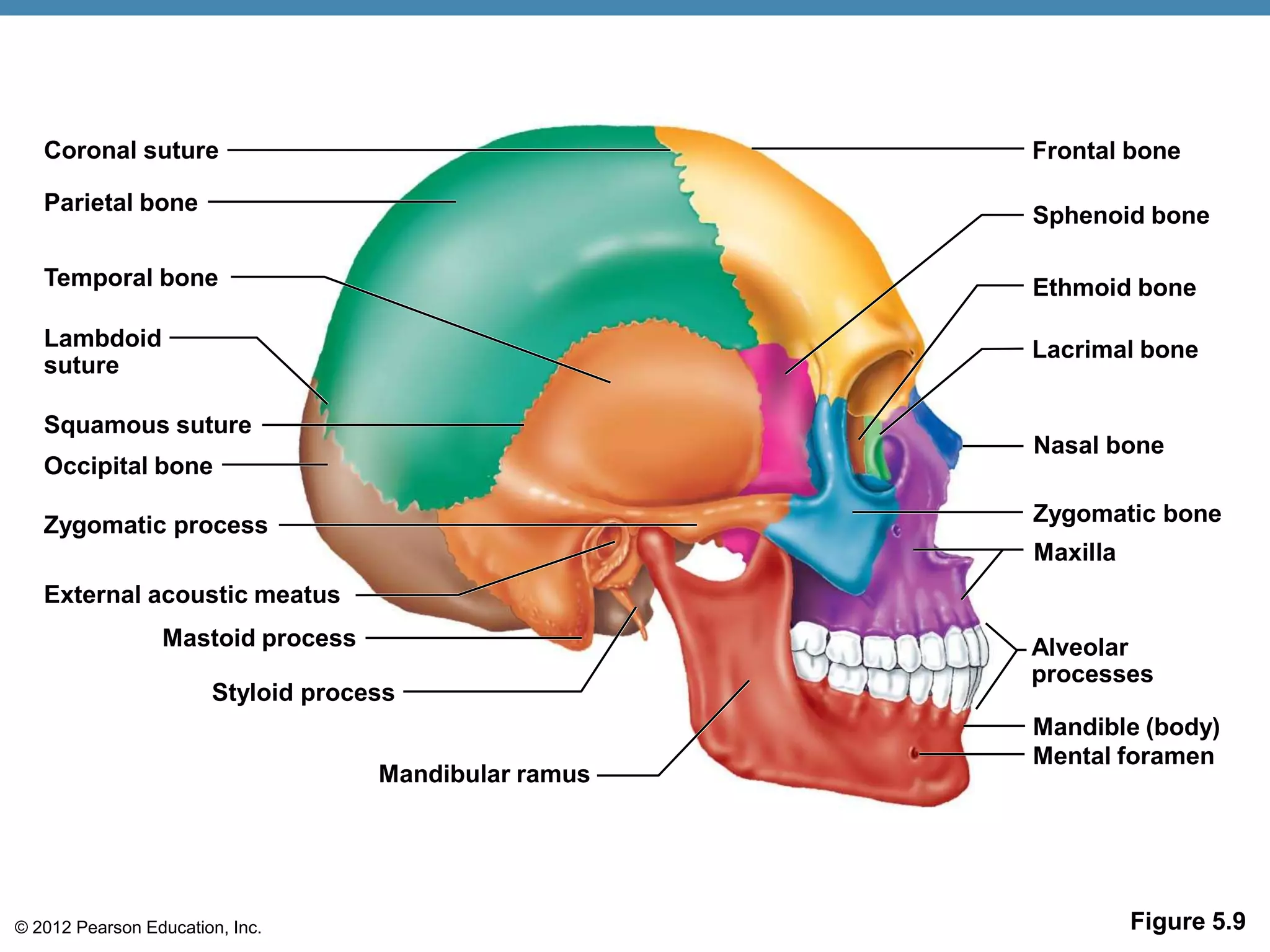
The skeletal system consists of bones, joints, and cartilages that make up the endoskeleton of the human body. It has several functions including support, protection, movement, mineral storage, and blood cell formation. The skeletal system is divided into the axial skeleton which includes the skull, vertebral column, and chest, and the appendicular skeleton which connects to the axial skeleton and includes the upper and lower limbs. Bones can be classified based on their shape as long, short, flat, or irregular. The anatomy of long bones includes diaphyses, epiphyses, periosteum, marrow cavity, and growth plates. Joints allow movement and come in several types including ball-and-socket and
The skeletal system document provides information about the skeletal system in 3 paragraphs. It describes the parts of the skeletal system including bones, joints, cartilages and ligaments. It then explains the two subdivisions of the skeleton: the axial skeleton and appendicular skeleton. Finally, it lists the functions of bones which include support, protection, movement, mineral storage, and blood cell formation.
The document describes the process of bone formation through intramembranous and endochondral ossification. Intramembranous ossification involves bone matrix secretion within a fibrous membrane to form woven bone and periosteum. Endochondral ossification uses hyaline cartilage models and involves the breakdown of cartilage and formation of bone collar, spongy bone, and medullary cavity through several stages. Bone growth and remodeling occur through the epiphyseal plate in long bones and the coordinated action of osteoblasts and osteoclasts.
Dr. Ali Selk Ghafari: Biomechanics of Musculoskeletal System: Components of t...a_selkghafari
in this lecture students learn about components of the musculoskeletal system in three sections:
• Section I – Skeletal tissue and skeletal system
• Section II – Muscular tissues
• Section III – Joints
This document provides an overview of bone physiology. It begins with definitions of bone and discusses its various functions. It then covers the classification, structure, development and types of bone cells. Key points include that bone is composed of inorganic minerals deposited in an organic collagen matrix. It undergoes two types of ossification - intramembranous and endochondral. Bone growth involves the coordinated activities of osteoblasts, osteoclasts and osteocytes. The document provides detailed descriptions of bone anatomy and histology.
In my presentation, I have described about the anatomy and physiology of bone and also did a elaboration on the pathways which is responsible for bone cells formation. For more details related to bone you can contact me on my email : sheershopramanik24@gmail.com
The Indian Dental Academy is the Leader in continuing dental education , training dentists in all aspects of dentistry and
offering a wide range of dental certified courses in different formats.for more details please visit
www.indiandentalacademy.com
This document discusses the anatomy of bone, including its structure, function, classification, development, and blood supply. It describes bone as a composite connective tissue made up of organic components like bone cells and collagen and inorganic components like calcium crystals. It classifies bone tissue into woven bone, lamellar bone, cortical bone, and cancellous bone. Bone development occurs through either intramembranous or endochondral ossification. Finally, it outlines the dynamic process of bone remodeling carried out by osteoblasts, osteoclasts, and the blood supply to bone.
The document provides an overview of the skeletal system, including its structure, functions, and common diseases. It discusses the main parts of the skeletal system including bones, joints, and cartilage. It describes the different types of bones, joints, and bone cells. Common diseases like arthritis, osteoporosis, fractures, and cancers that affect the skeletal system are summarized. The roles of the axial and appendicular skeleton are outlined.
The Indian Dental Academy is the Leader in continuing dental education , training dentists in all aspects of dentistry and
offering a wide range of dental certified courses in different formats.for more details please visit
www.indiandentalacademy.com
The skeletal system has several important functions:
1. It provides structure and support for the body, protects internal organs, and allows for movement through muscle attachment points.
2. Bones store minerals like calcium and aid in mineral homeostasis. Certain bones also produce blood cells.
3. The skeletal system is composed of bones, cartilage, ligaments, and tendons. Bones are living tissues with osteogenic cells, osteoblasts, osteocytes, and osteoclasts. They have an extracellular matrix containing collagen and minerals like hydroxyapatite.
The Indian Dental Academy is the Leader in continuing dental education , training dentists in all aspects of dentistry and
offering a wide range of dental certified courses in different formats.for more details please visit
www.indiandentalacademy.com
BONE – AN INTRODUCTION
A bone is a rigid organ that constitutes part of the vertebrate skeleton.
There are around 270 to 300+ bones in Infants which gets reduced to 206 bones in adults.
Bones are dynamic structures that are undergoing constant change and remodelling in
response to the ever-changing environment.
Bones support and protect the various organs of the body, produce red and white blood cells,
store minerals, provide structure and support for the body, and enable mobility.
It has a honeycomb-like matrix internally, which helps to give the bone rigidity.
The largest bone in the body is the femur or thigh-bone, and the smallest is the stapes in
the middle ear.
This document provides an overview of alveolar bone, including its development, histology, cellular components, and remodeling. It begins with a brief introduction to bone classification and composition. Key points include that alveolar bone forms via intramembranous ossification, and is composed of inorganic minerals and organic collagen fibers. It contains two main cell types - osteoblasts, which build bone, and osteoclasts, which resorb bone. Alveolar bone is continually modeled and remodeled throughout life to adapt to forces.
Alveolar bone forms the sockets that hold teeth and is composed of alveolar bone proper surrounding tooth roots and supporting alveolar bone. It develops during tooth eruption through both intramembranous and endochondral ossification. Alveolar bone is maintained through remodeling where bone resorption by osteoclasts is followed by bone formation by osteoblasts, regulated by hormones and growth factors to maintain calcium homeostasis.
Bone & Its Importance to ProsthodontistSelf employed
Bone is a hard connective tissue that forms the skeleton. It has an inorganic mineral component made up of calcium salts and an organic collagen matrix. Bone comes in two types - dense cortical bone forming the hard outer shell and spongy cancellous bone on the inside. Bone is constantly remodeled through the actions of bone-forming osteoblasts and bone-resorbing osteoclasts. Diseases like osteoporosis weaken the bones through decreased bone density while Paget's disease involves abnormal bone deposition and resorption.
The document discusses the anatomy and physiology of bone. It covers the functions, classification, microscopic structure, development, growth and common disorders of bone. Key points include that bone provides structure, protection and movement; is made of collagen fibers and hydroxyapatite; develops through intramembranous or endochondral ossification; and can be affected by conditions like fractures, osteoporosis or cancer.
The document discusses the structure and types of bone, including alveolar bone. It notes that bone is composed of osteoprogenitor cells, bone cells, and intercellular substance. There are two types of bone: compact bone and spongy bone. Compact bone is dense and found in shafts, while spongy bone is porous and found in flat bones. Alveolar bone lines the tooth socket and is composed of bundle bone, with Sharpey's fibers inserting into it. It includes the alveolar bone proper and supporting alveolar bone made of cortical and trabecular plates.
- Bone is composed of compact cortical bone and spongy trabecular bone. Compact bone contains osteons arranged in concentric layers. Trabecular bone is made of spicules or plates with many cells on the surfaces.
- Bone is strengthened by deposits of calcium salts in an organic matrix composed primarily of collagen fibers. Hydroxyapatite crystals precipitate on collagen fibers during bone calcification.
- Bone remodeling involves bone resorption by osteoclasts and bone formation by osteoblasts to continually renew and reshape bone in response to stress and repair microdamage.
The document discusses alveolar bone and its development, structure, and functions. It covers the following key points:
- Alveolar bone develops through intramembranous ossification and surrounds the teeth to support them and absorb forces. It has a layer of compact bone and spongy bone interior.
- The alveolar bone receives blood supply from the alveolar arteries and innervations from the inferior alveolar nerve. Its functions include supporting teeth and absorbing chewing forces.
- Orthodontic tooth movement relies on optimal force application and tissue response to forces. Drug effects and biological considerations also impact alveolar bone remodeling during orthodontic treatment.
The document provides an overview of alveolar bone, including its classification, histology, composition, development, parts, functions, remodeling, blood and nerve supply, and age-related changes. Key points include that alveolar bone develops during tooth eruption, consists of alveolar bone proper and supporting alveolar bone (cortical plates and spongy bone), and its morphology is determined by the shape and location of teeth. Alveolar bone anchors teeth, distributes occlusal forces, and is remodeled through the coupling of bone resorption and formation.
Bone is a living tissue that provides structure and support. It can be classified based on shape, development, histology, and composition. The alveolar process forms with tooth development and eruption to support teeth in the jaw. It consists of cortical and cancellous bone layers surrounded by osteoblasts and osteoclasts, which build and resorb bone through various signaling pathways and enzymes.
This document provides information on alveolar bone structure and composition. It discusses the different types of bone cells that make up alveolar bone, including osteoprogenitor cells, osteoblasts, osteocytes, and osteoclasts. It also describes the composition and structural elements of alveolar bone, such as Sharpey's fibers, vascular supply, gross morphology, and the periosteum and endosteum layers.
The document summarizes key aspects of the skeletal system. It describes the two types of bone tissue as compact bone and spongy bone. It explains the structure of long bones as having an epiphysis, diaphysis and medullary cavity. There are two types of ossification - intramembranous which forms flat bones, and endochondral which forms most other bones from hyaline cartilage models. Postnatal bone growth occurs through longitudinal growth at the epiphyseal plate and appositional growth increasing bone width.
This document provides an overview of bone histology and development. It discusses that bone is a specialized mineralized connective tissue that can be divided into compact and cancellous bone microscopically. Bone develops through either endochondral or intramembranous ossification. The key cells involved are osteoblasts, which form new bone, and osteoclasts, which resorb bone. Bone is remodeled throughout life by the balanced actions of these cells.
The document summarizes key aspects of the skeletal system, including:
1. Bones develop from cartilage early in development and continue changing in structure throughout life.
2. There are 206 bones in the human body classified as either axial or appendicular based on location and role.
3. Bones are living tissues composed of both organic and inorganic materials and have distinct structures depending on their shape and role.
4. Bones continuously remodel through the processes of deposition and resorption to maintain strength and mineral homeostasis.
Bone is a living connective tissue that makes up the skeleton. The alveolar process is the tooth-supporting portion of the jaws that forms the sockets housing the teeth. It develops from the dental follicle and surrounding bone as the teeth erupt. The alveolar process consists of inner and outer cortical plates surrounding a core of cancellous bone. It is continually remodeled through the balanced actions of bone-forming osteoblasts and bone-resorbing osteoclasts to maintain healthy teeth and absorb forces from chewing. Fenestrations and dehiscences are defects where the alveolar bone no longer fully surrounds the tooth roots.
The document discusses the structure and function of bones. It describes that bones are composed of cells embedded in a calcified matrix. The main cell types are osteoprogenitor cells, osteoblasts, osteoclasts and osteocytes. Bone has two types of osseous tissue - compact bone and spongy bone. Compact bone has concentric lamellae surrounding Haversian canals, while spongy bone has a more porous structure. Bones provide structure, protection, enable movement and store minerals. Remodeling and modeling allow bones to adapt throughout life.
The skeletal system includes bones and cartilage that provide structure, allow for movement, and protect organs. The medical specialty of orthopedics focuses on treating the skeleton and joints. Skeletal tissue includes cartilage and bone, which are types of dense connective tissue. Bones have important functions like support, protection, movement, mineral storage, and blood cell production. Bone is made up of osteogenic cells, osteoblasts that form bone, osteocytes embedded in bone matrix, and osteoclasts that resorb bone. There are different types of bones like long bones with a diaphysis, epiphyses, and metaphysis. Bone formation occurs through intramembranous ossification or endochondral ossification. Common bone
The document discusses the axial skeleton, which includes the bones of the skull, vertebral column, and thoracic cage. It provides details on the individual bones that make up these regions, including the skull bones (cranium and facial bones), vertebrae, ribs, and sternum. Descriptions include bone landmarks and structures, as well as their anatomical relationships and functional roles in supporting organs and allowing muscle attachment. Diagrams are included to illustrate the skeletal features and their positions within the body.
a brief ppt description about cartilage which may be usefull for teaching for first year mbbs, bds and paramedical students, hope it is helpfull to everyone
The Indian Dental Academy is the Leader in continuing dental education , training dentists in all aspects of dentistry and
offering a wide range of dental certified courses in different formats.for more details please visit
www.indiandentalacademy.com
The skeletal system has several important functions:
1. It provides structure and support for the body, protects internal organs, and allows for movement through muscle attachment points.
2. Bones store minerals like calcium and aid in mineral homeostasis. Certain bones also produce blood cells.
3. The skeletal system is composed of bones, cartilage, ligaments, and tendons. Bones are living tissues with osteogenic cells, osteoblasts, osteocytes, and osteoclasts. They have an extracellular matrix containing collagen and minerals like hydroxyapatite.
The Indian Dental Academy is the Leader in continuing dental education , training dentists in all aspects of dentistry and
offering a wide range of dental certified courses in different formats.for more details please visit
www.indiandentalacademy.com
BONE – AN INTRODUCTION
A bone is a rigid organ that constitutes part of the vertebrate skeleton.
There are around 270 to 300+ bones in Infants which gets reduced to 206 bones in adults.
Bones are dynamic structures that are undergoing constant change and remodelling in
response to the ever-changing environment.
Bones support and protect the various organs of the body, produce red and white blood cells,
store minerals, provide structure and support for the body, and enable mobility.
It has a honeycomb-like matrix internally, which helps to give the bone rigidity.
The largest bone in the body is the femur or thigh-bone, and the smallest is the stapes in
the middle ear.
This document provides an overview of alveolar bone, including its development, histology, cellular components, and remodeling. It begins with a brief introduction to bone classification and composition. Key points include that alveolar bone forms via intramembranous ossification, and is composed of inorganic minerals and organic collagen fibers. It contains two main cell types - osteoblasts, which build bone, and osteoclasts, which resorb bone. Alveolar bone is continually modeled and remodeled throughout life to adapt to forces.
Alveolar bone forms the sockets that hold teeth and is composed of alveolar bone proper surrounding tooth roots and supporting alveolar bone. It develops during tooth eruption through both intramembranous and endochondral ossification. Alveolar bone is maintained through remodeling where bone resorption by osteoclasts is followed by bone formation by osteoblasts, regulated by hormones and growth factors to maintain calcium homeostasis.
Bone & Its Importance to ProsthodontistSelf employed
Bone is a hard connective tissue that forms the skeleton. It has an inorganic mineral component made up of calcium salts and an organic collagen matrix. Bone comes in two types - dense cortical bone forming the hard outer shell and spongy cancellous bone on the inside. Bone is constantly remodeled through the actions of bone-forming osteoblasts and bone-resorbing osteoclasts. Diseases like osteoporosis weaken the bones through decreased bone density while Paget's disease involves abnormal bone deposition and resorption.
The document discusses the anatomy and physiology of bone. It covers the functions, classification, microscopic structure, development, growth and common disorders of bone. Key points include that bone provides structure, protection and movement; is made of collagen fibers and hydroxyapatite; develops through intramembranous or endochondral ossification; and can be affected by conditions like fractures, osteoporosis or cancer.
The document discusses the structure and types of bone, including alveolar bone. It notes that bone is composed of osteoprogenitor cells, bone cells, and intercellular substance. There are two types of bone: compact bone and spongy bone. Compact bone is dense and found in shafts, while spongy bone is porous and found in flat bones. Alveolar bone lines the tooth socket and is composed of bundle bone, with Sharpey's fibers inserting into it. It includes the alveolar bone proper and supporting alveolar bone made of cortical and trabecular plates.
- Bone is composed of compact cortical bone and spongy trabecular bone. Compact bone contains osteons arranged in concentric layers. Trabecular bone is made of spicules or plates with many cells on the surfaces.
- Bone is strengthened by deposits of calcium salts in an organic matrix composed primarily of collagen fibers. Hydroxyapatite crystals precipitate on collagen fibers during bone calcification.
- Bone remodeling involves bone resorption by osteoclasts and bone formation by osteoblasts to continually renew and reshape bone in response to stress and repair microdamage.
The document discusses alveolar bone and its development, structure, and functions. It covers the following key points:
- Alveolar bone develops through intramembranous ossification and surrounds the teeth to support them and absorb forces. It has a layer of compact bone and spongy bone interior.
- The alveolar bone receives blood supply from the alveolar arteries and innervations from the inferior alveolar nerve. Its functions include supporting teeth and absorbing chewing forces.
- Orthodontic tooth movement relies on optimal force application and tissue response to forces. Drug effects and biological considerations also impact alveolar bone remodeling during orthodontic treatment.
The document provides an overview of alveolar bone, including its classification, histology, composition, development, parts, functions, remodeling, blood and nerve supply, and age-related changes. Key points include that alveolar bone develops during tooth eruption, consists of alveolar bone proper and supporting alveolar bone (cortical plates and spongy bone), and its morphology is determined by the shape and location of teeth. Alveolar bone anchors teeth, distributes occlusal forces, and is remodeled through the coupling of bone resorption and formation.
Bone is a living tissue that provides structure and support. It can be classified based on shape, development, histology, and composition. The alveolar process forms with tooth development and eruption to support teeth in the jaw. It consists of cortical and cancellous bone layers surrounded by osteoblasts and osteoclasts, which build and resorb bone through various signaling pathways and enzymes.
This document provides information on alveolar bone structure and composition. It discusses the different types of bone cells that make up alveolar bone, including osteoprogenitor cells, osteoblasts, osteocytes, and osteoclasts. It also describes the composition and structural elements of alveolar bone, such as Sharpey's fibers, vascular supply, gross morphology, and the periosteum and endosteum layers.
The document summarizes key aspects of the skeletal system. It describes the two types of bone tissue as compact bone and spongy bone. It explains the structure of long bones as having an epiphysis, diaphysis and medullary cavity. There are two types of ossification - intramembranous which forms flat bones, and endochondral which forms most other bones from hyaline cartilage models. Postnatal bone growth occurs through longitudinal growth at the epiphyseal plate and appositional growth increasing bone width.
This document provides an overview of bone histology and development. It discusses that bone is a specialized mineralized connective tissue that can be divided into compact and cancellous bone microscopically. Bone develops through either endochondral or intramembranous ossification. The key cells involved are osteoblasts, which form new bone, and osteoclasts, which resorb bone. Bone is remodeled throughout life by the balanced actions of these cells.
The document summarizes key aspects of the skeletal system, including:
1. Bones develop from cartilage early in development and continue changing in structure throughout life.
2. There are 206 bones in the human body classified as either axial or appendicular based on location and role.
3. Bones are living tissues composed of both organic and inorganic materials and have distinct structures depending on their shape and role.
4. Bones continuously remodel through the processes of deposition and resorption to maintain strength and mineral homeostasis.
Bone is a living connective tissue that makes up the skeleton. The alveolar process is the tooth-supporting portion of the jaws that forms the sockets housing the teeth. It develops from the dental follicle and surrounding bone as the teeth erupt. The alveolar process consists of inner and outer cortical plates surrounding a core of cancellous bone. It is continually remodeled through the balanced actions of bone-forming osteoblasts and bone-resorbing osteoclasts to maintain healthy teeth and absorb forces from chewing. Fenestrations and dehiscences are defects where the alveolar bone no longer fully surrounds the tooth roots.
The document discusses the structure and function of bones. It describes that bones are composed of cells embedded in a calcified matrix. The main cell types are osteoprogenitor cells, osteoblasts, osteoclasts and osteocytes. Bone has two types of osseous tissue - compact bone and spongy bone. Compact bone has concentric lamellae surrounding Haversian canals, while spongy bone has a more porous structure. Bones provide structure, protection, enable movement and store minerals. Remodeling and modeling allow bones to adapt throughout life.
The skeletal system includes bones and cartilage that provide structure, allow for movement, and protect organs. The medical specialty of orthopedics focuses on treating the skeleton and joints. Skeletal tissue includes cartilage and bone, which are types of dense connective tissue. Bones have important functions like support, protection, movement, mineral storage, and blood cell production. Bone is made up of osteogenic cells, osteoblasts that form bone, osteocytes embedded in bone matrix, and osteoclasts that resorb bone. There are different types of bones like long bones with a diaphysis, epiphyses, and metaphysis. Bone formation occurs through intramembranous ossification or endochondral ossification. Common bone
The document discusses the axial skeleton, which includes the bones of the skull, vertebral column, and thoracic cage. It provides details on the individual bones that make up these regions, including the skull bones (cranium and facial bones), vertebrae, ribs, and sternum. Descriptions include bone landmarks and structures, as well as their anatomical relationships and functional roles in supporting organs and allowing muscle attachment. Diagrams are included to illustrate the skeletal features and their positions within the body.
a brief ppt description about cartilage which may be usefull for teaching for first year mbbs, bds and paramedical students, hope it is helpfull to everyone
This document provides an overview of bone structure and formation. It discusses the two types of ossification - intramembranous and endochondral. Intramembranous ossification forms flat bones of the skull directly from mesenchymal tissue, while endochondral ossification involves cartilage models that are later replaced by bone. Bone cells, blood supply, composition and fracture healing are also summarized. The document classifications bones by shape, developmental origin, and microscopic and macroscopic structure.
This document provides an overview of the anatomy of the temporal bone as visualized on HRCT scans. It describes the 3 main planes of scanning and their utility. It then details the individual bones that make up the temporal bone and the external, middle, and inner ear structures. Numerous axial, coronal, and sagittal HRCT images are presented to illustrate key anatomic landmarks and relationships. Structures like the ossicles, facial nerve canal, internal auditory canal, labyrinthine and cochlear anatomy are specifically called out.
The document discusses the musculoskeletal system and provides an overview of bone structure and classification. It identifies the main functions of bones as support, protection, movement, storage of minerals/fats, and blood cell formation. Bones are classified based on location in the body, shape, and internal structure as either compact or spongy bone.
Bone is a mineralized connective tissue that forms the endoskeleton of vertebrates. It has both compact and spongy structures and develops through either membranous or endochondral ossification. Bone is made up of osteoprogenitor cells, osteoblasts, osteocytes, and osteoclasts. Osteoblasts form new bone tissue while osteoclasts resorb old or damaged bone. The intricate blood supply through nutrient arteries is essential for bone growth and healing. Fractures heal through the formation of a hematoma, fibrocartilaginous callus, bony callus, and remodeling into secondary bone.
This document discusses the structure and function of skeletal muscle. It begins with an introduction to skeletal muscle and then covers topics like muscle fiber structure, development of muscle cells, muscle proteins, the sarcomere, sarcoplasmic reticulum, and excitation-contraction coupling. Diagrams are provided to illustrate muscle fiber anatomy, the arrangement of actin and myosin filaments in the sarcomere, and the relationship between the sarcoplasmic reticulum and t-tubules. The document provides definitions of key muscle terms and describes the roles of various muscle proteins.
The document describes the structure and function of bones and bone tissue. It discusses the different types of bone tissue (compact and spongy bone), the cells found in bone tissue, and the organization of bones. Compact bone forms the dense outer walls and is made up of concentric rings called osteons. Spongy bone forms a porous inner structure of branching plates called trabeculae. Bones provide structure, protect organs, allow movement, and store minerals. The periosteum and endosteum are membranes that line the outer and inner surfaces of bones.
The skeleton is composed of bones that provide structure, protect organs, allow movement, and store minerals. Bones are classified by their shape as long, flat, irregular, short, or sesamoid. Long bones act as levers pulled by muscles and have a hollow shaft (diaphysis) of compact bone surrounded by spongy bone in the ends (epiphyses). Bones grow in childhood as the epiphyseal plate ossifies. The periosteum and endosteum lining bones are involved in growth, repair, and marrow production of red and white blood cells.
This document discusses the skeletal system and bone structure. It describes the types of bone tissue as compact bone and spongy bone. Compact bone forms the dense outer shell and is made up of microscopic bone units called osteons. Spongy or trabecular bone forms a lattice within bones and is composed of thin struts called trabeculae. The document outlines the cells involved in bone formation and resorption, including osteoblasts, osteocytes, osteoclasts, and osteoprogenitor cells. It also discusses bone development through intramembranous and endochondral ossification.
This document discusses the skeletal system and bone structure. It describes the key components of bone, including compact and spongy bone. Compact bone consists of osteons, which have concentric lamellae and central canals. Spongy bone is made up of trabeculae that form an open, porous network. Bones also contain bone cells like osteoblasts, osteocytes, and osteoclasts. The document outlines bone development through intramembranous and endochondral ossification, and the roles of calcification, periosteum and endosteum in bone formation and growth.
2401 lecture05 ch 6 bones and bone tissue for class and webAbhishek Sharma
This document provides an overview of bones and skeletal tissues. It discusses the three types of skeletal cartilages, their locations in the body, and that they are surrounded by a vasculature-rich perichondrium. It also classifies bones into two regions - axial and appendicular - and four shapes - long, short, flat, and irregular. The structures of long bones and other bone shapes are described. The functions, microscopic structure, chemical composition, types of fractures and their repair, and homeostatic imbalances of bone are summarized as well.
Bones can be classified in several ways based on their shape, development, location in the body, and microscopic structure. The main types of bones by shape are long bones found in the limbs, short bones in the wrists and ankles, flat bones that make up surfaces like the skull, and irregularly shaped bones like vertebrae. Bones also develop differently as membrane bones, cartilaginous bones, or a combination. Key functions of bones include support, protection, movement, mineral storage, and blood cell formation. The basic structure of bones includes compact cortical bone on the outside and spongy cancellous bone on the inside, with bone cells and matrix throughout.
The skeletal system consists of bones, joints, cartilage, and ligaments that make up the framework of the body. It has several functions including support, protection, movement, storage, and blood cell formation. There are 206 bones in the adult human body that are classified based on their shape as long, short, flat, irregular, or sesamoid bones. Bones are living tissues that undergo remodeling throughout life. The skeletal system is divided into the axial skeleton, which includes the skull, vertebral column, and thoracic cage, and the appendicular skeleton, consisting of the upper and lower limbs.
Bones, Joints, and the Architecture of Movement: Exploring the Skeletal SystemNursing Mastery
Title: Bones, Joints, and the Architecture of Movement: Exploring the Skeletal System
Introduction:
Welcome to our engaging SlideShare presentation on the Skeletal System & Joints, where we embark on a fascinating exploration of the framework that supports our bodies and facilitates movement. Join us as we delve into the intricate world of bones, joints, and the dynamic interplay that shapes our mobility and stability.
The skeletal system serves as the foundation of our anatomy, providing structural support, protection for vital organs, and a framework for locomotion. Comprised of bones, cartilage, ligaments, and tendons, this complex system forms the structural scaffold upon which our bodies are built.
In this presentation, we'll take a closer look at the anatomy of bones, from their composition and classification to their roles in mineral storage and blood cell production. Through detailed illustrations and interactive diagrams, we'll explore the dynamic nature of bone remodeling and the factors that influence bone health and density.
But bones alone do not account for the versatility of human movement. Joints, the articulations where two or more bones meet, play a crucial role in facilitating motion and absorbing impact. From synovial joints like the knee and shoulder to fibrous and cartilaginous joints, we'll unravel the mechanics of joint structure and function.
Together, the skeletal system and joints form a complex network that enables us to walk, run, jump, and perform a myriad of daily activities. Whether you're a student studying anatomy, a fitness enthusiast seeking to optimize performance, or simply curious about the mechanics of the human body, our presentation offers valuable insights into the wonders of the skeletal system and joints.
Join us as we embark on a journey through the bony landmarks and articulations that define our physical form and discover the intricate architecture of movement hidden beneath our skin.
This document discusses the anatomy of bone and fracture healing. It begins by introducing bone as the basic unit of the human skeleton and describing its composition and regenerative abilities. It then classifies bones based on their position, size, shape and structure. Key parts of long bones are defined, including the diaphysis, epiphysis and metaphysis. The microscopic structure and composition of compact and cancellous bone are outlined. In summary, the document provides an overview of bone anatomy and classification, with a focus on the structure of long bones and their constituent parts.
The skeletal system is comprised of bones, joints, and cartilages. It has two divisions: the axial skeleton which includes the skull, vertebral column, and rib cage, and the appendicular skeleton which includes the limbs and girdles. Bones provide structure, protection, movement, storage, and blood cell formation. There are over 200 bones in the human body which are classified by shape. Joints connect bones and allow movement. Common diseases of the skeletal system include arthritis, inflammation of joints and tendons, fractures, osteoporosis, scoliosis, kyphosis, and lordosis. Tuberculosis can also affect the spine.
The document discusses the skeletal system and bones. It covers the structure and function of bones, classification of bones, bone tissues, bone cells, bone growth and healing, joints, and common bone diseases. The skeletal system includes bones, cartilage, ligaments, and joints. It is divided into the axial skeleton which includes the skull, vertebral column, ribs, and thoracic cage, and the appendicular skeleton which includes the limbs and their attachments.
Description :
The Indian Dental Academy is the Leader in continuing dental education , training dentists in all aspects of dentistry and
offering a wide range of dental certified courses in different formats.for more details please visit
www.indiandentalacademy.com
The skeletal system is divided into the axial skeleton and appendicular skeleton. The axial skeleton forms the body's central axis and includes the skull, vertebral column, and bony thorax. The skull is composed of two sets of bones - the cranium and facial bones. The cranium is made up of eight flat bones: the frontal, parietal, temporal, occipital, sphenoid, and ethmoid bones.
This document provides an overview of bone and skeletal development. It discusses [1] the initiation of cartilage formation through mesenchymal cell condensation and signaling, [2] the maturation of chondrocytes within the cartilage template, and [3] the replacement of cartilage by bone through endochondral ossification. It also covers [4] the factors controlling bone formation by osteoblasts and bone resorption by osteoclasts, which maintain bone homeostasis. Additional topics include [5] bone repair during fracture healing and [6] joint formation through segmentation of skeletal elements.
This document defines bone and describes its classification, structure, and functions. It notes that bone is living connective tissue composed of cells and an inorganic calcium phosphate matrix. It classifies bones based on location in the body, shape, and ossification process. The key types are long bones of the limbs, flat bones that make up surfaces, and irregularly shaped bones. Bone structure and development is also summarized, including how compact cortical and spongy cancellous bone differ and how bones ossify from cartilage models. The key functions of providing structure and protecting organs are also covered.
There are two types of bone tissue: compact bone and spongy bone. Compact bone contains concentric rings surrounding haversian canals, while spongy bone is less dense and contains thin bone columns. Bones come in long, short, flat, and irregular shapes. Long bones have a diaphysis made of compact bone, epiphyses containing spongy bone, articular cartilage at the ends, a periosteum outer layer, and medullary cavity containing marrow. Key bone markings include heads, condyles, epicondyles, and trochanters for muscle attachment and articulation.
The document summarizes the key components and functions of the skeletal system. It describes how bones provide structure, protection, movement, mineral storage, and blood cell formation. It outlines the different bone types and internal bone structure. Key bone groups like the vertebral column, thoracic cage, and extremities are depicted. Bone growth and remodeling over the lifespan is summarized. Common bone diseases like osteoporosis, rickets, and arthritis are also briefly outlined.
Bone tissue is a highly vascular, mineralized connective tissue consisting of cells and extracellular matrix. There are three main cell types - osteoblasts which form bone, osteocytes embedded in bone matrix, and osteoclasts which resorb bone. Bone has a gross structure of either compact or cancellous bone and a microstructure of lamellar or woven bone. Bone forms through either intramembraneous or endochondral ossification and is remodeled throughout life by the balanced activities of osteoblasts and osteoclasts. Bones provide structure, protection, calcium storage and leverage for movement. Bone diseases result from issues with formation, mineralization or remodeling.
The document provides an overview of alveolar bone, including its development, structure, blood and nerve supply, functions, and clinical considerations. Alveolar bone develops with the formation and eruption of teeth and supports the teeth by forming the bony sockets within the maxilla and mandible. It has two parts - the alveolar bone proper that directly surrounds the tooth root and the supporting alveolar bone that provides structural support. Age-related changes such as loss of bone with tooth loss are discussed. Therapeutic options for treating alveolar bone defects through grafting and regeneration techniques are also summarized.
Similar to A & p ch 5 skeleton student version (20)
This document contains a quiz about the muscular system organized into multiple levels and rounds of questions. It covers topics such as the three types of muscle tissue, muscle fiber structure, muscle contraction mechanisms, roles of calcium and acetylcholine, and functions of connective tissues like the endomysium. The final question indicates that muscle contraction occurs through actin fibers sliding past myosin fibers, due to the cross-bridge cycling action of the myosin heads.
Ch 6 Lab quiz study practice anterior body muscleszernwoman
La Unión Europea ha acordado un embargo petrolero contra Rusia en respuesta a la invasión de Ucrania. El embargo prohibirá las importaciones marítimas de petróleo ruso a la UE y pondrá fin a las entregas a través de oleoductos dentro de seis meses. Esta medida forma parte de un sexto paquete de sanciones de la UE destinadas a aumentar la presión económica sobre Rusia y privar al gobierno de Vladimir Putin de fondos para financiar la guerra.
Ch 6 Muscular System Lab quiz study practice connective tissue wrappings of m...zernwoman
The document discusses the benefits of exercise for mental health. Regular physical activity can help reduce anxiety and depression and improve mood and cognitive functioning. Exercise causes chemical changes in the brain that may help boost feelings of calmness, happiness and focus.
A & P Ch 6 Muscular System Lab quiz study practice facial muscleszernwoman
The document discusses the benefits of exercise for mental health. Regular physical activity can help reduce anxiety and depression and improve mood and cognitive functioning. Exercise causes chemical changes in the brain that may help boost feelings of calmness, happiness and focus.
A & P Ch 6 Muscular System Lab Quiz Practice - Posterior Muscleszernwoman
The document discusses the benefits of exercise for mental health. Regular physical activity can help reduce anxiety and depression and improve mood and cognitive functioning. Exercise causes chemical changes in the brain that may help protect against mental illness and improve symptoms.
Muscular system chapter overview:
- 3 types of muscle tissue: skeletal, smooth, and cardiac
- Muscles have 4 main functions and are organized in a muscular system
- Skeletal muscles are striated and attached via tendons or aponeuroses
- Contraction occurs when myosin cross-bridges bind actin fibers, sliding them past each other
- Nerve impulses trigger calcium release and muscle contraction via sliding filament theory
- Exercise improves muscle endurance, size and strength through aerobic and resistance training
Ch 6 Muscle Lab Quiz Study Practice Anterior Muscleszernwoman
The document discusses the benefits of exercise for mental health. Regular physical activity can help reduce anxiety and depression and improve mood and cognitive function. Exercise causes chemical changes in the brain that may help protect against mental illness and improve symptoms for those who already suffer from conditions like anxiety and depression.
The document outlines the goals and key concepts to be covered in a chapter on photosynthesis, including distinguishing between autotrophic and heterotrophic nutrition, describing the structure and function of chloroplasts, explaining the light and dark reactions of photosynthesis including the Calvin cycle, and summarizing alternative carbon fixation pathways such as C4 and CAM photosynthesis.
The document provides information about the skin and its layers. It discusses the three main layers of the skin - the epidermis, dermis and subcutaneous tissue. It describes the layers of the epidermis in detail, including the stratum basale, stratum spinosum, stratum granulosum and stratum corneum. It also discusses melanin production, skin color, skin functions, and skin appendages such as sweat glands, oil glands and hair.
This document summarizes key concepts about membrane structure and function, transport mechanisms, and the cell cycle. It describes the fluid mosaic model of the membrane and discusses different types of transport like passive diffusion, osmosis, and active transport. It also explains endocytosis and exocytosis. Regarding the cell cycle, it outlines the phases of interphase when the cell grows and duplicates its DNA, and the stages of mitosis and cytokinesis when the cell divides. Control mechanisms ensure the cell cycle proceeds at the proper times.
This document provides an overview of cells and cellular structures. It begins with an introduction to cells and explains that cells are the basic units of life. It then describes three types of microscopes used to view cells - light microscopes, transmission electron microscopes, and scanning electron microscopes - and notes one benefit of light microscopes. The document outlines the key differences between prokaryotic and eukaryotic cells and explains the importance of compartmentalization in eukaryotic cells. It provides descriptions and functions of major cellular structures and organelles found in plant and animal cells.
Ap bio ch 3 Functional Groups & Macromoleculeszernwoman
1. Organic molecules like carbohydrates, lipids, proteins, and nucleic acids are made up of monomers linked together through covalent bonds.
2. Carbon is a versatile building block due to its ability to form four covalent bonds (tetravalency). This allows it to link to other carbon atoms to form chains, branches, and rings.
3. Organic molecules contain functional groups that influence their chemical properties. Common functional groups include hydroxyl, carbonyl, carboxyl, amino, and phosphate groups.
4. The structure and bonding of organic molecules contribute to isomerism, including structural, geometric, and enantiomer isomers. Spatial arrangement of atoms and groups affects molecular properties.
This document discusses the four primary types of tissues in the body: epithelial, connective, muscle, and nervous tissue. It focuses on epithelial tissues, which are grouped into glands and coverings. Epithelial tissues are classified based on the number of cell layers (simple or stratified) and the shape of cells (squamous, cuboidal, columnar). Simple epithelia include squamous, cuboidal and columnar types which line various organs. Stratified epithelia provide additional protection with multiple layers including squamous, cuboidal and columnar cell types. Glandular epithelia are responsible for secreting products into openings.
Ap bio ch 2 ppt The Chemistry of Life and Waterzernwoman
The document discusses the importance of water for life, including its unique properties like polarity and hydrogen bonding that allow it to moderate temperatures, dissolve many substances, and enable processes like diffusion. Water's high specific heat also means it can absorb or release large amounts of heat without much change in temperature. These properties are crucial for biological functions and maintaining Earth's habitability.
This document discusses key concepts in population ecology including:
1. It defines population characteristics like density and dispersion and explains how demography studies population growth and decline through factors like birth rates, death rates, immigration and emigration.
2. Population age structure, generation time, and sex ratio are important in determining growth and decline as they impact reproductive rates. Survivorship curves show mortality rates at different ages.
3. Organisms must make life history trade-offs between energy invested in reproduction versus survival. Their life history is determined by number of reproductive events, offspring per event, and age of first reproduction.
4. Population growth can be exponential or logistic, with the latter constrained by environmental carrying
This chapter discusses animal behavior and behavioral biology. It defines key concepts like behavioral ecology, instinct, learning, and communication. It provides examples of innate behaviors like fixed action patterns and imprinting. It also covers different types of learning such as habituation, associative learning, and insight. The chapter discusses social behaviors including aggression, dominance hierarchies, territoriality, and courtship. It addresses migration, kin selection, and altruism. The overall goals are to explain different kinds of animal behavior and how they help organisms survive and reproduce.
Integrated Science Unit 1 nature of sciencezernwoman
This document provides an overview of key concepts in the scientific method including observation, hypotheses, variables, experiments, data collection and analysis, inferences, conclusions, and reliability and validity. It uses a hypothetical example experiment testing how color affects the dissolving rate of M&Ms in water to illustrate these concepts. Key steps discussed are developing testable questions, controlling variables, collecting quantitative data, analyzing results using graphs, and drawing conclusions supported by evidence.
This document lists 4 sources for memory and study techniques. The sources include a US Air Force Academy webpage on memory techniques, a New York Times article on improving memory, a Middle Tennessee State University webpage on memory principles including using interest, and a design share webpage on brain-based learning principles from 1998.
This presentation was provided by Rebecca Benner, Ph.D., of the American Society of Anesthesiologists, for the second session of NISO's 2024 Training Series "DEIA in the Scholarly Landscape." Session Two: 'Expanding Pathways to Publishing Careers,' was held June 13, 2024.
🔥🔥🔥🔥🔥🔥🔥🔥🔥
إضغ بين إيديكم من أقوى الملازم التي صممتها
ملزمة تشريح الجهاز الهيكلي (نظري 3)
💀💀💀💀💀💀💀💀💀💀
تتميز هذهِ الملزمة بعِدة مُميزات :
1- مُترجمة ترجمة تُناسب جميع المستويات
2- تحتوي على 78 رسم توضيحي لكل كلمة موجودة بالملزمة (لكل كلمة !!!!)
#فهم_ماكو_درخ
3- دقة الكتابة والصور عالية جداً جداً جداً
4- هُنالك بعض المعلومات تم توضيحها بشكل تفصيلي جداً (تُعتبر لدى الطالب أو الطالبة بإنها معلومات مُبهمة ومع ذلك تم توضيح هذهِ المعلومات المُبهمة بشكل تفصيلي جداً
5- الملزمة تشرح نفسها ب نفسها بس تكلك تعال اقراني
6- تحتوي الملزمة في اول سلايد على خارطة تتضمن جميع تفرُعات معلومات الجهاز الهيكلي المذكورة في هذهِ الملزمة
واخيراً هذهِ الملزمة حلالٌ عليكم وإتمنى منكم إن تدعولي بالخير والصحة والعافية فقط
كل التوفيق زملائي وزميلاتي ، زميلكم محمد الذهبي 💊💊
🔥🔥🔥🔥🔥🔥🔥🔥🔥
Walmart Business+ and Spark Good for Nonprofits.pdfTechSoup
"Learn about all the ways Walmart supports nonprofit organizations.
You will hear from Liz Willett, the Head of Nonprofits, and hear about what Walmart is doing to help nonprofits, including Walmart Business and Spark Good. Walmart Business+ is a new offer for nonprofits that offers discounts and also streamlines nonprofits order and expense tracking, saving time and money.
The webinar may also give some examples on how nonprofits can best leverage Walmart Business+.
The event will cover the following::
Walmart Business + (https://business.walmart.com/plus) is a new shopping experience for nonprofits, schools, and local business customers that connects an exclusive online shopping experience to stores. Benefits include free delivery and shipping, a 'Spend Analytics” feature, special discounts, deals and tax-exempt shopping.
Special TechSoup offer for a free 180 days membership, and up to $150 in discounts on eligible orders.
Spark Good (walmart.com/sparkgood) is a charitable platform that enables nonprofits to receive donations directly from customers and associates.
Answers about how you can do more with Walmart!"
Gender and Mental Health - Counselling and Family Therapy Applications and In...PsychoTech Services
A proprietary approach developed by bringing together the best of learning theories from Psychology, design principles from the world of visualization, and pedagogical methods from over a decade of training experience, that enables you to: Learn better, faster!
Beyond Degrees - Empowering the Workforce in the Context of Skills-First.pptxEduSkills OECD
Iván Bornacelly, Policy Analyst at the OECD Centre for Skills, OECD, presents at the webinar 'Tackling job market gaps with a skills-first approach' on 12 June 2024
Temple of Asclepius in Thrace. Excavation resultsKrassimira Luka
The temple and the sanctuary around were dedicated to Asklepios Zmidrenus. This name has been known since 1875 when an inscription dedicated to him was discovered in Rome. The inscription is dated in 227 AD and was left by soldiers originating from the city of Philippopolis (modern Plovdiv).
Elevate Your Nonprofit's Online Presence_ A Guide to Effective SEO Strategies...TechSoup
Whether you're new to SEO or looking to refine your existing strategies, this webinar will provide you with actionable insights and practical tips to elevate your nonprofit's online presence.