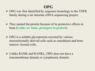
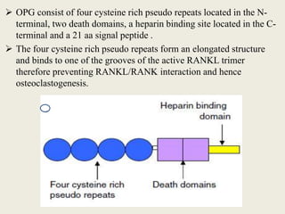
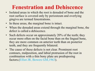
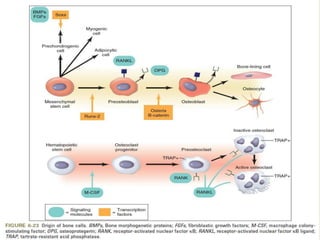
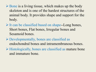
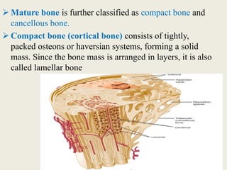
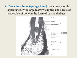
Bone is a living tissue that provides structure and support. It can be classified based on shape, development, histology, and composition. The alveolar process forms with tooth development and eruption to support teeth in the jaw. It consists of cortical and cancellous bone layers surrounded by osteoblasts and osteoclasts, which build and resorb bone through various signaling pathways and enzymes.
Coronal and radicular pulp
Apical foramen
Accessory canal
Functions of dental pulp
Components of dental pulp
Functions of pulpal extracellular matrix
Organization of cells in the pulp
The principle cells of the pulp
The pathways of collagen synthesis
Matrix and ground substances
Vasculature and lymphatic supply
Innervation of Dentin- pulp complex
Disorders of the dental pulp
Advances in pulp vitality testing
alveolar bone in health with microscopic features and details about bone formation, resorption also includes bone remodelling and changes after extraction
I prepared this presentation during the first year of my MDS. This will give you a basic idea and necessary information about the pulp of the teeth and its histology. Hope you guys find it useful.
Indian Dental Academy: will be one of the most relevant and exciting training center with best faculty and flexible training programs for dental professionals who wish to advance in their dental practice,Offers certified courses in Dental implants,Orthodontics,Endodontics,Cosmetic Dentistry, Prosthetic Dentistry, Periodontics and General Dentistry.
Development of bone
Microstructure of bone
Composition of bone
Formation of osteoblasts
Mineralisation of bone
Formation of osteoclasts
Resorption of bone
Macrostructure of bone
Volume changes in bone
Bone healing
Acute scrotum is a general term referring to an emergency condition affecting the contents or the wall of the scrotum.
There are a number of conditions that present acutely, predominantly with pain and/or swelling
A careful and detailed history and examination, and in some cases, investigations allow differentiation between these diagnoses. A prompt diagnosis is essential as the patient may require urgent surgical intervention
Testicular torsion refers to twisting of the spermatic cord, causing ischaemia of the testicle.
Testicular torsion results from inadequate fixation of the testis to the tunica vaginalis producing ischemia from reduced arterial inflow and venous outflow obstruction.
The prevalence of testicular torsion in adult patients hospitalized with acute scrotal pain is approximately 25 to 50 percent
HOT NEW PRODUCT! BIG SALES FAST SHIPPING NOW FROM CHINA!! EU KU DB BK substit...GL Anaacs
Contact us if you are interested:
Email / Skype : kefaya1771@gmail.com
Threema: PXHY5PDH
New BATCH Ku !!! MUCH IN DEMAND FAST SALE EVERY BATCH HAPPY GOOD EFFECT BIG BATCH !
Contact me on Threema or skype to start big business!!
Hot-sale products:
NEW HOT EUTYLONE WHITE CRYSTAL!!
5cl-adba precursor (semi finished )
5cl-adba raw materials
ADBB precursor (semi finished )
ADBB raw materials
APVP powder
5fadb/4f-adb
Jwh018 / Jwh210
Eutylone crystal
Protonitazene (hydrochloride) CAS: 119276-01-6
Flubrotizolam CAS: 57801-95-3
Metonitazene CAS: 14680-51-4
Payment terms: Western Union,MoneyGram,Bitcoin or USDT.
Deliver Time: Usually 7-15days
Shipping method: FedEx, TNT, DHL,UPS etc.Our deliveries are 100% safe, fast, reliable and discreet.
Samples will be sent for your evaluation!If you are interested in, please contact me, let's talk details.
We specializes in exporting high quality Research chemical, medical intermediate, Pharmaceutical chemicals and so on. Products are exported to USA, Canada, France, Korea, Japan,Russia, Southeast Asia and other countries.
Prix Galien International 2024 Forum ProgramLevi Shapiro
June 20, 2024, Prix Galien International and Jerusalem Ethics Forum in ROME. Detailed agenda including panels:
- ADVANCES IN CARDIOLOGY: A NEW PARADIGM IS COMING
- WOMEN’S HEALTH: FERTILITY PRESERVATION
- WHAT’S NEW IN THE TREATMENT OF INFECTIOUS,
ONCOLOGICAL AND INFLAMMATORY SKIN DISEASES?
- ARTIFICIAL INTELLIGENCE AND ETHICS
- GENE THERAPY
- BEYOND BORDERS: GLOBAL INITIATIVES FOR DEMOCRATIZING LIFE SCIENCE TECHNOLOGIES AND PROMOTING ACCESS TO HEALTHCARE
- ETHICAL CHALLENGES IN LIFE SCIENCES
- Prix Galien International Awards Ceremony
Title: Sense of Smell
Presenter: Dr. Faiza, Assistant Professor of Physiology
Qualifications:
MBBS (Best Graduate, AIMC Lahore)
FCPS Physiology
ICMT, CHPE, DHPE (STMU)
MPH (GC University, Faisalabad)
MBA (Virtual University of Pakistan)
Learning Objectives:
Describe the primary categories of smells and the concept of odor blindness.
Explain the structure and location of the olfactory membrane and mucosa, including the types and roles of cells involved in olfaction.
Describe the pathway and mechanisms of olfactory signal transmission from the olfactory receptors to the brain.
Illustrate the biochemical cascade triggered by odorant binding to olfactory receptors, including the role of G-proteins and second messengers in generating an action potential.
Identify different types of olfactory disorders such as anosmia, hyposmia, hyperosmia, and dysosmia, including their potential causes.
Key Topics:
Olfactory Genes:
3% of the human genome accounts for olfactory genes.
400 genes for odorant receptors.
Olfactory Membrane:
Located in the superior part of the nasal cavity.
Medially: Folds downward along the superior septum.
Laterally: Folds over the superior turbinate and upper surface of the middle turbinate.
Total surface area: 5-10 square centimeters.
Olfactory Mucosa:
Olfactory Cells: Bipolar nerve cells derived from the CNS (100 million), with 4-25 olfactory cilia per cell.
Sustentacular Cells: Produce mucus and maintain ionic and molecular environment.
Basal Cells: Replace worn-out olfactory cells with an average lifespan of 1-2 months.
Bowman’s Gland: Secretes mucus.
Stimulation of Olfactory Cells:
Odorant dissolves in mucus and attaches to receptors on olfactory cilia.
Involves a cascade effect through G-proteins and second messengers, leading to depolarization and action potential generation in the olfactory nerve.
Quality of a Good Odorant:
Small (3-20 Carbon atoms), volatile, water-soluble, and lipid-soluble.
Facilitated by odorant-binding proteins in mucus.
Membrane Potential and Action Potential:
Resting membrane potential: -55mV.
Action potential frequency in the olfactory nerve increases with odorant strength.
Adaptation Towards the Sense of Smell:
Rapid adaptation within the first second, with further slow adaptation.
Psychological adaptation greater than receptor adaptation, involving feedback inhibition from the central nervous system.
Primary Sensations of Smell:
Camphoraceous, Musky, Floral, Pepperminty, Ethereal, Pungent, Putrid.
Odor Detection Threshold:
Examples: Hydrogen sulfide (0.0005 ppm), Methyl-mercaptan (0.002 ppm).
Some toxic substances are odorless at lethal concentrations.
Characteristics of Smell:
Odor blindness for single substances due to lack of appropriate receptor protein.
Behavioral and emotional influences of smell.
Transmission of Olfactory Signals:
From olfactory cells to glomeruli in the olfactory bulb, involving lateral inhibition.
Primitive, less old, and new olfactory systems with different path
TEST BANK for Operations Management, 14th Edition by William J. Stevenson, Ve...kevinkariuki227
TEST BANK for Operations Management, 14th Edition by William J. Stevenson, Verified Chapters 1 - 19, Complete Newest Version.pdf
ARTIFICIAL INTELLIGENCE IN HEALTHCARE.pdfAnujkumaranit
Artificial intelligence (AI) refers to the simulation of human intelligence processes by machines, especially computer systems. It encompasses tasks such as learning, reasoning, problem-solving, perception, and language understanding. AI technologies are revolutionizing various fields, from healthcare to finance, by enabling machines to perform tasks that typically require human intelligence.
New Directions in Targeted Therapeutic Approaches for Older Adults With Mantl...i3 Health
i3 Health is pleased to make the speaker slides from this activity available for use as a non-accredited self-study or teaching resource.
This slide deck presented by Dr. Kami Maddocks, Professor-Clinical in the Division of Hematology and
Associate Division Director for Ambulatory Operations
The Ohio State University Comprehensive Cancer Center, will provide insight into new directions in targeted therapeutic approaches for older adults with mantle cell lymphoma.
STATEMENT OF NEED
Mantle cell lymphoma (MCL) is a rare, aggressive B-cell non-Hodgkin lymphoma (NHL) accounting for 5% to 7% of all lymphomas. Its prognosis ranges from indolent disease that does not require treatment for years to very aggressive disease, which is associated with poor survival (Silkenstedt et al, 2021). Typically, MCL is diagnosed at advanced stage and in older patients who cannot tolerate intensive therapy (NCCN, 2022). Although recent advances have slightly increased remission rates, recurrence and relapse remain very common, leading to a median overall survival between 3 and 6 years (LLS, 2021). Though there are several effective options, progress is still needed towards establishing an accepted frontline approach for MCL (Castellino et al, 2022). Treatment selection and management of MCL are complicated by the heterogeneity of prognosis, advanced age and comorbidities of patients, and lack of an established standard approach for treatment, making it vital that clinicians be familiar with the latest research and advances in this area. In this activity chaired by Michael Wang, MD, Professor in the Department of Lymphoma & Myeloma at MD Anderson Cancer Center, expert faculty will discuss prognostic factors informing treatment, the promising results of recent trials in new therapeutic approaches, and the implications of treatment resistance in therapeutic selection for MCL.
Target Audience
Hematology/oncology fellows, attending faculty, and other health care professionals involved in the treatment of patients with mantle cell lymphoma (MCL).
Learning Objectives
1.) Identify clinical and biological prognostic factors that can guide treatment decision making for older adults with MCL
2.) Evaluate emerging data on targeted therapeutic approaches for treatment-naive and relapsed/refractory MCL and their applicability to older adults
3.) Assess mechanisms of resistance to targeted therapies for MCL and their implications for treatment selection
NVBDCP.pptx Nation vector borne disease control programSapna Thakur
NVBDCP was launched in 2003-2004 . Vector-Borne Disease: Disease that results from an infection transmitted to humans and other animals by blood-feeding arthropods, such as mosquitoes, ticks, and fleas. Examples of vector-borne diseases include Dengue fever, West Nile Virus, Lyme disease, and malaria.
micro teaching on communication m.sc nursing.pdfAnurag Sharma
Microteaching is a unique model of practice teaching. It is a viable instrument for the. desired change in the teaching behavior or the behavior potential which, in specified types of real. classroom situations, tends to facilitate the achievement of specified types of objectives.
These lecture slides, by Dr Sidra Arshad, offer a quick overview of physiological basis of a normal electrocardiogram.
Learning objectives:
1. Define an electrocardiogram (ECG) and electrocardiography
2. Describe how dipoles generated by the heart produce the waveforms of the ECG
3. Describe the components of a normal electrocardiogram of a typical bipolar leads (limb II)
4. Differentiate between intervals and segments
5. Enlist some common indications for obtaining an ECG
Study Resources:
1. Chapter 11, Guyton and Hall Textbook of Medical Physiology, 14th edition
2. Chapter 9, Human Physiology - From Cells to Systems, Lauralee Sherwood, 9th edition
3. Chapter 29, Ganong’s Review of Medical Physiology, 26th edition
4. Electrocardiogram, StatPearls - https://www.ncbi.nlm.nih.gov/books/NBK549803/
5. ECG in Medical Practice by ABM Abdullah, 4th edition
6. ECG Basics, http://www.nataliescasebook.com/tag/e-c-g-basics
Lung Cancer: Artificial Intelligence, Synergetics, Complex System Analysis, S...Oleg Kshivets
RESULTS: Overall life span (LS) was 2252.1±1742.5 days and cumulative 5-year survival (5YS) reached 73.2%, 10 years – 64.8%, 20 years – 42.5%. 513 LCP lived more than 5 years (LS=3124.6±1525.6 days), 148 LCP – more than 10 years (LS=5054.4±1504.1 days).199 LCP died because of LC (LS=562.7±374.5 days). 5YS of LCP after bi/lobectomies was significantly superior in comparison with LCP after pneumonectomies (78.1% vs.63.7%, P=0.00001 by log-rank test). AT significantly improved 5YS (66.3% vs. 34.8%) (P=0.00000 by log-rank test) only for LCP with N1-2. Cox modeling displayed that 5YS of LCP significantly depended on: phase transition (PT) early-invasive LC in terms of synergetics, PT N0—N12, cell ratio factors (ratio between cancer cells- CC and blood cells subpopulations), G1-3, histology, glucose, AT, blood cell circuit, prothrombin index, heparin tolerance, recalcification time (P=0.000-0.038). Neural networks, genetic algorithm selection and bootstrap simulation revealed relationships between 5YS and PT early-invasive LC (rank=1), PT N0—N12 (rank=2), thrombocytes/CC (3), erythrocytes/CC (4), eosinophils/CC (5), healthy cells/CC (6), lymphocytes/CC (7), segmented neutrophils/CC (8), stick neutrophils/CC (9), monocytes/CC (10); leucocytes/CC (11). Correct prediction of 5YS was 100% by neural networks computing (area under ROC curve=1.0; error=0.0).
CONCLUSIONS: 5YS of LCP after radical procedures significantly depended on: 1) PT early-invasive cancer; 2) PT N0--N12; 3) cell ratio factors; 4) blood cell circuit; 5) biochemical factors; 6) hemostasis system; 7) AT; 8) LC characteristics; 9) LC cell dynamics; 10) surgery type: lobectomy/pneumonectomy; 11) anthropometric data. Optimal diagnosis and treatment strategies for LC are: 1) screening and early detection of LC; 2) availability of experienced thoracic surgeons because of complexity of radical procedures; 3) aggressive en block surgery and adequate lymph node dissection for completeness; 4) precise prediction; 5) adjuvant chemoimmunoradiotherapy for LCP with unfavorable prognosis.
2. Bone is a living tissue, which makes up the body
skeleton and is one of the hardest structures of the
animal body. It provides shape and support for the
body.
It can be classified based on shape--Long bones,
Short bones, Flat bones, Irregular bones and
Sesamoid bones.
Developmentally, bones are classified as
endochondral bones and intramembranous bones.
Histologically, bones are classified as mature bone
and immature bone.
3. Mature bone is further classified as compact bone and
cancellous bone.
Compact bone (cortical bone) consists of tightly,
packed osteons or haversian systems, forming a solid
mass. Since the bone mass is arranged in layers, it is also
called lamellar bone
4. Cancellous bone (spongy bone) has a honeycomb
appearance, with large marrow cavities and sheets of
trabeculae of bone in the form of bars and plates.
5. Woven or immature bone is the first formed bone
with irregularly oriented collagen fibers of varying
diameters. This type of bone is not usually seen after
birth. However it is seen in the alveolar bone and
during healing of fractures. Since this bone forms
rapidly, it incorporates many osteocytes
6. The alveolar process is defined as that part of the
maxilla and the mandible that forms and supports the
sockets of the teeth.
7. DEVELOPMENT OF ALVEOLAR
PROCESS
• Near the end of the second month of fetal life, the maxilla as
well as the mandible form a groove that is open towards the
surface of the oral cavity.
• Tooth germs develop within the bony structures at late bell
stage. Bony septa and bony bridge begin to form and separate
the individual tooth germs from one another, keeping
individual tooth germs in clearly outlined bony compartments.
• The major changes in the alveolar process begin to occur with
development of roots and tooth eruption.
• As roots develop, the alveolar process increases in height.
8. • At the same time, some cells in the dental follicle differentiate
into osteoblasts and form alveolar bone proper
• An alveolar process develops only during the eruption of the
teeth. It is important to realize that, during growth, part of the
alveolar process is gradually incorporated into the maxillary or
mandibular body while it grows at a fairly rapid rate at its free
borders
• The alveolar process forms with the development and the
eruption of teeth, and conversely, it gradually diminishes in
height after the loss of teeth.
9. • The alveolar process consists of the following:
An external plate of cortical bone is formed by haversian
bone and compacted bone lamellae.
The inner socket wall of thin, compact bone called the
alveolar bone proper is seen as the lamina dura in
radiographs.
Cancellous trabeculae between these two compact
layers act as supporting alveolar bone.
Most of the facial and lingual portions of the sockets are
formed by compact bone alone; cancellous bone
surrounds the lamina dura in apical, apicolingual, and
interradicular areas.
10. • The alveolar process is divisible into separate
areas on an anatomic basis, but it functions as
a unit, with all parts interrelated in the
support of the teeth.
11. COMPOSITION OF BONE
• Bone is a connective tissue composed of cells, fibers
and ground substance.
• The inorganic part of bone is made of bone minerals.
• The mineral component is composed of
hydroxyapatite crystals(Ca10[PO4]6[OH]2).
• Bone crystals are packed closely with long axis
nearly parallel to collagen fibrils axis.
• The ions present are calcium phosphate, hydroxyl and
carbonate. Citrate, magnesium, sodium, potassium,
fluoride, iron, zinc, copper, aluminum, lead,
strontium, silicon and boron are present in small
quantities.
12. • Type I collagen (> 95%) is the principal collagen in
mineralized bone and, together with type V collagen (< 5%),
forms heterotypic fiber bundles that provide the basic
structural integrity of connective tissues.
• The elasticity of collagen imparts resiliency to the tissue and
helps to resist fractures.
• Type XII collagen is also present in alveolar bone and has
been found to be expressed under conditions of mechanical
strain.
• Types I, V and XII collagens are expressed by osteoblasts.
13. Noncollagenous proteins
• These proteins are released from bone by demineralization,
reflecting the predominant association with the mineral
phase.
• Osteocalcin and Bone sialoprotein, are essentially unique
to mineralized tissues, whereas others, such as
Osteonectin/SPARC (secreted protein, acidic, rich in
cysteine) and Osteopontin have a more general
distribution.
• In addition to those proteins produced by bone-forming
cells, certain proteins derived from blood and tissue fluids
are concentrated in bone, largely due to their affinity for the
mineral crystals.These include albumin, α2HS
glycoprotein, immunoglobulins and matrix gla protein.
( Delmas PD et al. 1993)
14. Osteocalcin
It is a small, highly conserved, 5.8-kDa acidic protein
that is characteristically modified by vitamin K–
dependent carboxylating enzymes that convert two to
three glutamic acids into γ-carboxyglutamic acids
(gla groups).
Represents 15% of the noncollagenous proteins and
was the first noncollagenous bone protein to be
characterized. Its presence in alveolar bone has been
demonstrated immunohistochemically.
15. • The gla groups formed on the pro-osteocalcin prior to
secretion, bind calcium ions strongly and increase the
affinity of osteocalcin for bone mineral.
The small osteocalcin molecule forms two α helical
sections; the gla helix containing the Ƴ-
carboxyglutamyl groups interacts with mineral crystals
(HA).
16. Bone sialoprotein and osteopontin
• Osteopontin and bone sialoprotein, originally
characterized as bone sialoproteins I and II , are
expressed in alveolar bone and have been localized
using immunohistochemistry —(chen j et al,1993,
Helder MN et al,1993, Mckee MD et al,1996)
• Bone sialoprotein is essentially restricted to
mineralizing tissues, osteopontin has a more general
distribution that reflects a broader biological role.
Similar to blood clotting factors, osteopontin is also
susceptible to thrombin, indicative of an origin in the
blood or blood-forming organs.
17. • Bone sialoprotein is expressed coincident with the
first appearance of mineral crystals in cementum and
bone and is also able to nucleate hydroxyapatite
crystal formation in vitro through the polyglutamate
sequence , it is thought to function in the initiation of
mineral crystal formation in vivo.(Chen j et al,1992,
Hunter GK, Goldberg HA , 1993)
• In contrast, osteopontin is a potent inhibitor of
hydroxyapatite crystal growth and is enriched at all
cell-matrix interfaces where it can mediate the
attachment of bone cells, including
osteoclasts.(Hunter GK, Goldberg HA , 1995, Mckee
MD, Nanci A, 1996)
18. Bone sialoprotein and osteopontin are chemically similar molecules with an
open flexible structure. Both are highly glycosylated and phosphorylated, with
mineral (HA) and cell-binding (RGD) sites. In bone sialoprotein, a number of
sulfated tyrosines surround the RGD while osteopontin has a thrombin (Thr)
sensitive site (arrow) near the RGD.
19. Osteonectin/SPARC( secreted protein,
acidic , rich in cysteine)
• SPARC/osteonectin, a 40-kDa glycoprotein that is
predominantly bound to hydroxyapatite.
• SPARC has both a high-affinity EF-hand calcium-binding
site and a number of low-affinity calcium-binding sites
concentrated towards the amino-terminus.
• SPARC can comprise as much as 25% of the non-
collagen proteins.
• Four domain present in SPARC.
• Domain I, which is the least conserved, appears to be
responsible for mineral binding, resulting in its presence
in mineralized tissues. (Romberg RW et al, 1986).
20. • The second domain (domain II) of SPARC is rich in
cysteines, which stabilize the protein structure
through disulfide bridges.
• Domain III is a-helical and is susceptible to
proteolysis.
• Domain IV contains the EF-hand high-affinity
calcium binding site.
21. Cellular components
Osteoblast
Osteoblasts arise from pluripotent stem cells, which are of
mesenchymal origin in the axial and appendicular
skeleton and of ectomesenchymal origin (neural crest
cells that migrate in mesenchyme) in the head.
The secretory products of osteoblasts include type I
collagen, the dominant component of the organic matrix,
small amounts of type V collagen and proteoglycans, and
several noncollagenous proteins
Osteoblasts produce a range of growth factors under a
variety of stimuli including the insulin-like growth factors
(IGF),platelet-derived growth factor (PDGF), basic
fibroblast growth factor (bFGF), transforming growth
factor-beta (TGF-β) and the bone morphogenetic proteins
(BMP).
22. • Recent gene deletion studies have shown that absence of
Runt related transcription factor 2 (Runx2) or of a
downstream factor, Osterix, is critical for osteoblast
differentiation(DUCY, P. et al.1997) .
• Deletion of Runx2 leads to a complete lack of bone
formation in mice and to craniofacial dysostosis in
heterozygous humans (Ducy P et al 1997, Otto F et al ,
1997, Mundols S et al 1997)
• Deletion of osterix leads to an arrest along the osteoblast
differentiation pathway and several skeletal
abnormalities in mice (Nakashima k et al 2002)
23. Wnt Signaling: Canonical Pathway
• The Wnt canonical signaling pathway relies mainly on the
stabilization of cytosolic β-catenin.
• In the absence of Wnt proteins, β-catenin is
phosphorylated by several kinases,mainly glycogen
synthase kinase 3 (GSK-3) , casein kinase 1, and
targeted to ubiquitination and degradation by the
proteasomal machinery.
• Wnt binding to its receptor complex results in the
inhibition of GSK-3 activity.
• This inhibition mediates the prevention of β-catenin
degradation,leading to an accumulation of β-catenin
in the cytoplasm.
24. • β-catenin is essential in determining whether
mesenchymal progenitors become osteoblasts or
chondrocytes (Hill TP et al,2006, Day TF et al 2005).
Thus Wnt signaling can determine the cell fate of
mesenchymal precursors.
• In addition to regulating osteoblast commitment, Wnt/β-
catenin signaling is also suspected of affecting osteoblast
proliferation.
• Wnt regulates the expression of osteoprotegerin, that
inhibits osteoclast differentiation by interacting with
RANKL.
• Thus Wnt signaling indirectly controls osteoclast
differentiation via its effect on osteoblasts.
25. A, In the absence of Wnt ligand, β-catenin is phosphorylated by GSK-3
leading to its degra- dation and pathway signaling inactivation. B, After Wnt
binding to its LRP5/6 and Fz coreceptors, GSK-3 is inactivated. β-Catenin is
then stabilized and accumulates in the cytoplasm. β-Catenin will consequently
translocate into the nucleus where it affects gene expression.
26. Osteocytes
• Osteoblasts that have been trapped in the osteoid are
called osteocytes.
• Osteocytes have numerous long cell processes rich in
microfilaments that are organized during the
formation of the matrix and before its calcification.
They form a network of thin canaliculi permeating
the entire bone matrix.
27. • A young osteocyte has most of the structural characteristics of
the osteoblast but decreased cell volume and capacity of
protein synthesis.
• An older osteocyte, located deeper within the calcified bone,
presents with a further decrease in cell volume and an
accumulation of glycogen in the cytoplasm.
• The osteocytes are finally phagocytosed and digested during
osteoclastic bone resorption.(ELMARDI et al 1990.)
• It has been suggested that dying or dead osteocytes send
signals of resorption (VERBORGT et al,2002) and recently it
has been shown that a protein highly expressed in osteocytes
called sclerostin can target osteoblasts to inhibit bone
formation (VAN BEZOOIJEN et al 2005).
28. Osteoclast
• Osteoclasts are members of the
monocyte/macrophage lineage and are formed by
multiple cellular fusions from their mononuclear
precursors.
• Fully differentiated human osteoclasts are large cells,
approximately with five to eight nuclei.
• Their differentiation is regulated by a number of other
cells and their products, especially by RANKL and
M-CSF.
29. • When osteoclasts are resorbing the bone,four distinct
membrane domains can be detected both using morphological
criteria as well as molecular markers (J. Salo et al,1996).
• Sealing zone membrane attaches the resorbing cell to the
mineralized extracellular matrix and circulates the ruffled
border membrane, which is the actual resorbing organ.
• Basolateral or non-bone facing plasma membrane is also
divided into two distinct domains since a specific membrane
domain, called functional secretory domain (FSD), is formed
in the center of the basolateral membrane (J. Salo et al,1997).
30.
31. Osteoclasts secrete cathepsin K to degrade
organic bone matrix
• Two group of enzymes, namely matrix metalloproteinases
(MMPs) and lysosomal cathepsins have a major role in the
extracellular degradation of bone matrix.
• Cathepsin K is a key enzyme in the degradation of bone matrix
in the resorption lacuna. Catepsin K is very highly expressed
in osteoclasts and is secreted into the resorption lacuna (V.
Everts, et al,2006. B.R. Troen , et al 2006).
• It can split insoluble type I collagen at acidic pH and inhibition
of its enzymatic activity in various in vitro and in vivo models
prevents effectively matrix degradation M.J. Bossard et al
1996).
• Osteoclasts are also rich in acid phosphatases and tartrate
resistant acid phosphatase (TRACP) is used as a cellular
marker for osteoclasts.
32. • Serum levels of osteoclast-specific isoform, TRACP5b,
correlates with resorption activity and can be used as a clinical
marker of resorption activity in various clinical situations(J.M.
Halleen, et al, 2006).
• Cathepsin K activates TRACP
• TRACP has two distinct enzymatic activities. It functions as a
phosphatase and is also able to generate reactive oxygen
species via Fenton reaction.
• ROS-generating activity of TRACP has been shown to
facilitate collagen degradation and may thus have a role in the
final degradation of resorption products (J.M. Halleen, et al,
2003).
• Since TRACP is also highly expressed in dendritic cells. It is
interesting to speculate that there it may have a role in antigen
processing and its function in osteoclasts is more to reduce the
risk of autoimmunity against bone proteins than to have a
functional role in bone resorption as such.
33. • In contrast to cathepsin K, a role of MMPs in osteoclast
function is still much debated.
• MMP 9, MMP14 are produced by osteoclasts(T.L. Andersen ,
et al 2004, B. Linsuwanont-Santiwong et al, 2006).
• The present data suggest that organic matrix degradation is
initiated by MMPs and continued by cysteine proteases and
only the latter event is regulated by estrogen(V. Parikka et al,
2001).
• Osteoclasts express semaphorin 4D (Sema4D), previously
shown to be an axon guidance molecule, which potently
inhibits bone formation.
• The binding of Sema4D to its receptor Plexin-B1 on
osteoblasts resulted in the activation of the small GTPase
RhoA, which inhibits bone formation by suppressing insulin-
like growth factor-1 (IGF-1) signaling and by modulating
osteoblast motility.
34.
35. Alveolar bone proper
• Forms the inner wall of the socket is perforated by many openings
that carry branches of the interalveolar nerves and blood vessels
into the periodontal ligament, and it is therefore called the
cribriform plate.
• Bone between the teeth is called interdental septum and is
composed entirely of cribriform plate.
• The interdental and interradicular septa contain the perforating
canals of Zuckerkandl and Hirschfeld (nutrient canals) which
house the interdental and interradicular arteries, veins, lymph
vessels and nerves .
36.
37. Interdental Septum
• The interdental septum consists of cancellous bone that is
bordered by the socket wall cribriform plates (i.e., lamina dura
or alveolar bone proper) of approximating teeth and the facial
and lingual cortical plates.
• If the interdental space is narrow, the septum may consist of
only the cribriform plate.
• If the roots are too close together, an irregular “window”can
appear in the bone between adjacent roots.
38. • Between maxillary molars, the septum consisted of cribriform
plate and cancellous bone in 66.6% of cases; it was composed
of only cribriform plate in 20.8%, and it had a fenestration in
12.5%.
• The mesiodistal angulation of the crest of the interdental
septum usually parallels a line drawn between the
cementoenamel junctions of the approximating teeth.(Ritchey
B, Orban B. 1953)
• The distance between the crest of the alveolar bone and the
cementoenamel junction in young adults varies between 0.75
and 1.49 mm (average, 1.08 mm). This distance increases with
age to an average of 2.81 mm.(Gargiulo AW et al 1961)
• The mesiodistal and faciolingual dimensions and shape of the
interdental septum are governed by the size and convexity of
the crowns of the two approximating teeth as well as by the
position of the teeth in the jaw and their degree of eruption.
39. Bone Marrow
• In the embryo and the newborn, the cavities of all bones
are occupied by red hematopoietic marrow. The red
marrow gradually undergoes a physiologic change to the
fatty or yellow inactive type of marrow.
• In the adult, the marrow of the jaw is normally of the
latter type.
• However, foci of the red bone marrow are occasionally
seen in the jaws, often accompanied by the resorption of
bony trabeculae.
• Common locations are the maxillary tuberosity, the
maxillary and mandibular molar and premolar areas, and
the mandibular symphysis and ramus angle, which may
be visible radiographically as zones of radiolucency.
40. Periosteum and Endosteum
• The tissue that covers the outer surface of bone is termed
periosteum, whereas the tissue that lines the internal bone
cavities is called endosteum.
• The periosteum consists of an inner layer composed of
osteoblasts surrounded by osteoprogenitor cells, which have
the potential to differentiate into osteoblasts, and an outer layer
rich in blood vessels and nerves and composed of collagen
fibers and fibroblasts.
• The endosteum is composed of a single layer of osteoblasts
and sometimes a small amount of connective tissue. The inner
layer is the osteogenic layer, and the outer layer is the fibrous
layer.
41. RANK, RANKL & OPG
RANK
RANK was discovered by Anderson et al by direct sequencing
cDNA from a human bone marrow-derived myeloid dendritic
cell.
Sequencing of the RANK gene showed it to be a type I
transmembrane glycoprotein and further gene mapping
showed this newly discovered protein to be located on
chromosome 18q22.1 and also a member of the tumour
necrosis factor receptor (TNFR) family.
The expression of RANK has since been found on the surface
of a wide variety of cells such as; osteoclast precursors
(circulating monocytes) ,mature osteoclasts ,dendritic cells
,mammary gland epithelial cells ,breast cancer cells and
prostate cancer cells .
42. RANKL
• RANKL is a tumour necrosis factor (TNF)- related cytokine
expressed by various bone cells including osteoblasts and their
immature precursors , T lymphocytes , B lymphocytes and
megakaryocytes.
• Membrane bound RANKL ensures cell–cell contact with
osteoclasts and their precursors ,whereas sRANKL{The third
isoforms of RANK having both the transmembrane and
cytoplasmic domains and acts as a soluble ligand} allows for
diffusion to activate target cells.
• In the first case, bone resorption will be tightly localised to the
cells expressing RANKL.
• The second case, resorption will be more generalized .
43. OPG
OPG was first identified by sequence homology to the TNFR
family during a rat intestine cDNA sequencing project.
They named the protein because of its protective effects in
bone (Latin: os- bone, protegere to protect).
OPG is a soluble glycoprotein secreted by various
mesenchymally derived cells such as osteoblasts and bone
marrow stromal cells.
Unlike RANK and RANKL, OPG does not have a
transmembrane domain or cytoplasmic domain .
44. OPG consist of four cysteine rich pseudo repeats located in the N-
terminal, two death domains, a heparin binding site located in the C-
terminal and a 21 aa signal peptide .
The four cysteine rich pseudo repeats form an elongated structure
and binds to one of the grooves of the active RANKL trimer
therefore preventing RANKL/RANK interaction and hence
osteoclastogenesis.
45. Bone remodeling
• Remodeling takes place at a microscopic locus known as the
bone (or basic) multicellular unit (BMU), which consists of a
unit of coupled osteoblast and osteoclast activity on the bone
surface.
• An orderly sequence of osteoclast attachment, resorption,
osteoblast attachment and proliferation and, finally, matrix
synthesis proceeds at the BMU.
• In cortical bone the BMU forms a cylindrical canal about
2,000µm long and 150–200µm wide and gradually burrows
through the bone with a speed of 20–40 µm/day.
• The events at the bone multicellular unit are regulated by cell-
cell interactions and cytokines.
46. 2% to 5% of cortical bone is being remodeled each year.
The trabecular bone is more actively remodeled than cortical
bone due to the much larger surface to volume ratio.
Osteoclasts travel across the trabecular surface with a speed of
approximately 25 µm/day, digging a trench with a depth of
40–60 µm.
The remodeling cycle consists of three consecutive phases:
resorption, reversal,and formation.
Resorption probably continues for about 2 weeks, the reversal
phase may last up to 4 or 5 weeks, while formation can
continue for 4 months until the new bone structural unit is
completely created.
47. • pathway involves three factors:
The transmembrane receptor RANK (receptor activator for NF-
κB), which is expressed on osteoclast precursors.
RANK ligand, (RANKL) which is expressed on osteoblasts and
marrow stromal cells.
osteoprotegerin (OPG), a secreted “decoy” receptor(a soluble
receptor acting as antagonist) made by osteoblasts and several
other types of cells that can bind RANKL and thus prevent its
interaction with RANK.
• When stimulated by RANKL, RANK signaling activates the
transcription factor NF-κB, which is essential for the generation
and survival of osteoclasts.
48.
49. • A second important pathway involves Macrophage colony
stimulating factor (M-CSF) produced by osteoblasts.
• Activation of the M-CSF receptor on osteoclast precursors
stimulates a tyrosine kinase cascade that is also crucial for the
generation of osteoclasts.
• WNT proteins produced by osteoprogenitor cells bind to the
LRP5 and LRP6 receptors on osteoblasts and thereby trigger the
activation of β-catenin and the production of OPG.
• Conversely, sclerostin, which is produced by osteocytes, inhibits
the WNT/β-catenin pathway.
• The balance between net bone formation and resorption is
modulated by the signals that connect to the RANK and WNT
signaling pathways.
• because OPG and RANKL oppose one another, either bone
resorption or bone formation can be favored by tipping the
RANK-to-OPG ratio.
50.
51. • Systemic factors that affect this balance include hormones
(parathyroid hormone, estrogen, testosterone, and
glucocorticoids), vitamin D, inflammatory cytokines (e.g., IL-
1), and growth factors (e.g., bone morphogenetic factors).
• Each of the above presumably acts by altering the levels of
RANK and WNT/β-catenin signaling in osteoblasts.
• Parathyroid hormone, IL-1 and glucocorticoids promote
osteoclast differentiation .
• Bone morphogenic proteins and sex hormones generally block
osteoclast differentiation or activity by favoring OPG
expression.
52. • Another level of control involves paracrine crosstalk between
osteoblasts and osteoclasts. Breakdown of matrix by
osteoclasts liberates and activates matrix proteins, growth
factors, cytokines, and enzymes (e.g., collagenase),including
some that stimulate osteoblasts.
• Thus, as bone is broken down to its elemental units, substances
are released into the microenvironment that initiate its renewal.
53. The development of bone is controlled by a
number of local and systemic factors:
Growth hormone (GH) is secreted by the anterior pituitary.It
acts on resting chondrocytes to induce and maintain
proliferation.
Thyroid hormone (T3) is secreted by the thyroid gland, and
acts on proliferating chondrocytes to induce hypertrophy.
Indian hedgehog (Ihh) is a locally secreted regulator, made by
prehypertrophic chondrocytes, that coordinates chondrocyte
proliferation and differentiation and osteoblasts proliferation.
Parathyroid hormone related protein (PTHrP) is a local
factor, expressed by perichondrial stromal cells and early
proliferating chondrocytes, that activates the PTH receptor and
maintains proliferation of chondrocytes.
54. Wnt is a family of secreted factors that are expressed at highest
levels in the proliferating zone and bind to the receptors Frizzled
and LRP5/6 to activate β-catenin signaling. They can promote both
proliferation and maturation of chondrocytes.
SOX9 is a transcription factor expressed by proliferating but not
hypertrophic chondrocytes, that is essential for differentiation of
precursor cells into chondrocytes.
RUNX2 is a transcription factor involved in chondrocyte and
osteoblast differentiation. It is expressed in early hypertrophic
chondrocytes and immature mesenchymal cells and controls
terminal chondrocyte and osteoblast differentiation respectively.
Fibroblast growth factors (FGFs) are secreted by a variety of
mesenchymal cells. FGF (most notably FGF3) acts on hypertrophic
chondrocytes to inhibit proliferation and promote differentiation.
55. • Bone morphogenic proteins (BMPs) are members of the
TGF-β family. They are expressed at various stages of
chondrocyte development in the growth plate and have diverse
effects on chondrocyte proliferation and hypertophy.
56. Functions of alveolar bone are:
Houses the roots of teeth.
Anchors the roots of teeth to the alveoli, which is achieved by
the insertion of Sharpey’s fibers into the alveolar bone proper.
Helps to move the teeth for better occlusion.
Helps to absorb and distribute occlusal forces generated during
tooth contact.
Supplies vessels to periodontal ligament.
Houses and protects developing permanent teeth, while
supporting primary teeth.
Organizes eruption of primary and permanent teeth.
57. Osseous Topography
• Alveolar bone anatomy varies among patients and has
important clinical implications.
• The height and thickness of the facial and lingual
bony plates are affected by the alignment of the teeth,
the angulation of the root to the bone, and occlusal
forces.
• On teeth in labial version, the margin of the labial
bone is located farther apically than it is on teeth that
are in proper alignment. The bone margin is thinned
to a knife edge.
58. • On teeth in lingual version, the facial bony plate is thicker than
normal. The margin is blunt, rounded, and horizontal rather
than arcuate.
• The effect of the root-to-bone angulation on the height of
alveolar bone is most noticeable on the palatal roots of the
maxillary molars. The bone margin is located farther apically
on the roots, and it forms relatively acute angles with the
palatal bone.
• The cervical portion of the alveolar plate is sometimes
considerably thickened on the facial surface, apparently as
reinforcement against occlusal forces.
59. Fenestration and Dehiscence
• Isolated areas in which the root is denuded of bone and the
root surface is covered only by periosteum and overlying
gingiva are termed fenestrations.
• In these areas, the marginal bone is intact.
• When the denuded areas extend through the marginal bone, the
defect is called a dehiscence.
• Such defects occur on approximately 20% of the teeth; they
occur more often on the facial bone than on the lingual bone,
they are more common on anterior teeth than on posterior
teeth, and they are frequently bilateral.
• The cause of these defects is not clear. Prominent root
contours, malposition, and labial protrusion of the root in
combination with a thin bony plate are predisposing
factors.(Elliot JR, Bowers GM,1963).
60.
61. AGE CHANGES
• Alveolar sockets appear jagged and uneven.
• The marrow spaces have fatty infiltration.
• The alveolar process in edentulous jaws decreases in size.
• Loss of maxillary bone is accompanied by increase in size of
the maxillary sinus.
• Internal trabecular arrangement is more open, which indicates
bone loss.
• The distance between the crest of the alveolar bone and CEJ
increases with age—approximately by 2.81 mm.