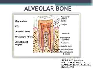
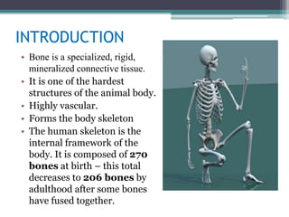
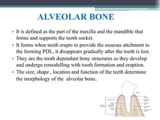
alveolar bone in health with microscopic features and details about bone formation, resorption also includes bone remodelling and changes after extraction
Coronal and radicular pulp
Apical foramen
Accessory canal
Functions of dental pulp
Components of dental pulp
Functions of pulpal extracellular matrix
Organization of cells in the pulp
The principle cells of the pulp
The pathways of collagen synthesis
Matrix and ground substances
Vasculature and lymphatic supply
Innervation of Dentin- pulp complex
Disorders of the dental pulp
Advances in pulp vitality testing
The bone of the skeleton is a mineralized vascular type of connective tissue with a solid matrix. The alveolar process is the bony extension of the mandible and maxilla that provides the necessary support for the teeth and serves as a site of attachment for the periodontal ligament fibers. By its resorption and deposition, it also compensates for tooth movement.
This ppt gives information about alveolar bone . I am a post graduate student from the department of periodontics and implantology , I've tried to make the topic as easy as possible. Hope it is useful for the ug students.
• Osseous tissue, a specialised form of dense connective tissue consisting of bone cells (osteocytes)• Embedded in a matrix of calcified intercelluarsubstance• Bone matrix contains collagen fibres and the minerals calcium phosphate and calcium carbonate
Seminar of U.V. Spectroscopy by SAMIR PANDASAMIR PANDA
Spectroscopy is a branch of science dealing the study of interaction of electromagnetic radiation with matter.
Ultraviolet-visible spectroscopy refers to absorption spectroscopy or reflect spectroscopy in the UV-VIS spectral region.
Ultraviolet-visible spectroscopy is an analytical method that can measure the amount of light received by the analyte.
Cancer cell metabolism: special Reference to Lactate PathwayAADYARAJPANDEY1
Normal Cell Metabolism:
Cellular respiration describes the series of steps that cells use to break down sugar and other chemicals to get the energy we need to function.
Energy is stored in the bonds of glucose and when glucose is broken down, much of that energy is released.
Cell utilize energy in the form of ATP.
The first step of respiration is called glycolysis. In a series of steps, glycolysis breaks glucose into two smaller molecules - a chemical called pyruvate. A small amount of ATP is formed during this process.
Most healthy cells continue the breakdown in a second process, called the Kreb's cycle. The Kreb's cycle allows cells to “burn” the pyruvates made in glycolysis to get more ATP.
The last step in the breakdown of glucose is called oxidative phosphorylation (Ox-Phos).
It takes place in specialized cell structures called mitochondria. This process produces a large amount of ATP. Importantly, cells need oxygen to complete oxidative phosphorylation.
If a cell completes only glycolysis, only 2 molecules of ATP are made per glucose. However, if the cell completes the entire respiration process (glycolysis - Kreb's - oxidative phosphorylation), about 36 molecules of ATP are created, giving it much more energy to use.
IN CANCER CELL:
Unlike healthy cells that "burn" the entire molecule of sugar to capture a large amount of energy as ATP, cancer cells are wasteful.
Cancer cells only partially break down sugar molecules. They overuse the first step of respiration, glycolysis. They frequently do not complete the second step, oxidative phosphorylation.
This results in only 2 molecules of ATP per each glucose molecule instead of the 36 or so ATPs healthy cells gain. As a result, cancer cells need to use a lot more sugar molecules to get enough energy to survive.
introduction to WARBERG PHENOMENA:
WARBURG EFFECT Usually, cancer cells are highly glycolytic (glucose addiction) and take up more glucose than do normal cells from outside.
Otto Heinrich Warburg (; 8 October 1883 – 1 August 1970) In 1931 was awarded the Nobel Prize in Physiology for his "discovery of the nature and mode of action of the respiratory enzyme.
WARNBURG EFFECT : cancer cells under aerobic (well-oxygenated) conditions to metabolize glucose to lactate (aerobic glycolysis) is known as the Warburg effect. Warburg made the observation that tumor slices consume glucose and secrete lactate at a higher rate than normal tissues.
Comparing Evolved Extractive Text Summary Scores of Bidirectional Encoder Rep...University of Maribor
Slides from:
11th International Conference on Electrical, Electronics and Computer Engineering (IcETRAN), Niš, 3-6 June 2024
Track: Artificial Intelligence
https://www.etran.rs/2024/en/home-english/
THE IMPORTANCE OF MARTIAN ATMOSPHERE SAMPLE RETURN.Sérgio Sacani
The return of a sample of near-surface atmosphere from Mars would facilitate answers to several first-order science questions surrounding the formation and evolution of the planet. One of the important aspects of terrestrial planet formation in general is the role that primary atmospheres played in influencing the chemistry and structure of the planets and their antecedents. Studies of the martian atmosphere can be used to investigate the role of a primary atmosphere in its history. Atmosphere samples would also inform our understanding of the near-surface chemistry of the planet, and ultimately the prospects for life. High-precision isotopic analyses of constituent gases are needed to address these questions, requiring that the analyses are made on returned samples rather than in situ.
Observation of Io’s Resurfacing via Plume Deposition Using Ground-based Adapt...Sérgio Sacani
Since volcanic activity was first discovered on Io from Voyager images in 1979, changes
on Io’s surface have been monitored from both spacecraft and ground-based telescopes.
Here, we present the highest spatial resolution images of Io ever obtained from a groundbased telescope. These images, acquired by the SHARK-VIS instrument on the Large
Binocular Telescope, show evidence of a major resurfacing event on Io’s trailing hemisphere. When compared to the most recent spacecraft images, the SHARK-VIS images
show that a plume deposit from a powerful eruption at Pillan Patera has covered part
of the long-lived Pele plume deposit. Although this type of resurfacing event may be common on Io, few have been detected due to the rarity of spacecraft visits and the previously low spatial resolution available from Earth-based telescopes. The SHARK-VIS instrument ushers in a new era of high resolution imaging of Io’s surface using adaptive
optics at visible wavelengths.
Multi-source connectivity as the driver of solar wind variability in the heli...Sérgio Sacani
The ambient solar wind that flls the heliosphere originates from multiple
sources in the solar corona and is highly structured. It is often described
as high-speed, relatively homogeneous, plasma streams from coronal
holes and slow-speed, highly variable, streams whose source regions are
under debate. A key goal of ESA/NASA’s Solar Orbiter mission is to identify
solar wind sources and understand what drives the complexity seen in the
heliosphere. By combining magnetic feld modelling and spectroscopic
techniques with high-resolution observations and measurements, we show
that the solar wind variability detected in situ by Solar Orbiter in March
2022 is driven by spatio-temporal changes in the magnetic connectivity to
multiple sources in the solar atmosphere. The magnetic feld footpoints
connected to the spacecraft moved from the boundaries of a coronal hole
to one active region (12961) and then across to another region (12957). This
is refected in the in situ measurements, which show the transition from fast
to highly Alfvénic then to slow solar wind that is disrupted by the arrival of
a coronal mass ejection. Our results describe solar wind variability at 0.5 au
but are applicable to near-Earth observatories.
This presentation explores a brief idea about the structural and functional attributes of nucleotides, the structure and function of genetic materials along with the impact of UV rays and pH upon them.
What is greenhouse gasses and how many gasses are there to affect the Earth.moosaasad1975
What are greenhouse gasses how they affect the earth and its environment what is the future of the environment and earth how the weather and the climate effects.
Nutraceutical market, scope and growth: Herbal drug technologyLokesh Patil
As consumer awareness of health and wellness rises, the nutraceutical market—which includes goods like functional meals, drinks, and dietary supplements that provide health advantages beyond basic nutrition—is growing significantly. As healthcare expenses rise, the population ages, and people want natural and preventative health solutions more and more, this industry is increasing quickly. Further driving market expansion are product formulation innovations and the use of cutting-edge technology for customized nutrition. With its worldwide reach, the nutraceutical industry is expected to keep growing and provide significant chances for research and investment in a number of categories, including vitamins, minerals, probiotics, and herbal supplements.
Richard's entangled aventures in wonderlandRichard Gill
Since the loophole-free Bell experiments of 2020 and the Nobel prizes in physics of 2022, critics of Bell's work have retreated to the fortress of super-determinism. Now, super-determinism is a derogatory word - it just means "determinism". Palmer, Hance and Hossenfelder argue that quantum mechanics and determinism are not incompatible, using a sophisticated mathematical construction based on a subtle thinning of allowed states and measurements in quantum mechanics, such that what is left appears to make Bell's argument fail, without altering the empirical predictions of quantum mechanics. I think however that it is a smoke screen, and the slogan "lost in math" comes to my mind. I will discuss some other recent disproofs of Bell's theorem using the language of causality based on causal graphs. Causal thinking is also central to law and justice. I will mention surprising connections to my work on serial killer nurse cases, in particular the Dutch case of Lucia de Berk and the current UK case of Lucy Letby.
2. CONTENTS
• Introduction;
• Functions
• Classification;
• Composition;
• Alveolar bone
• Histology;
• Bone formation;
• Bone resorption;
• Bone remodelling;
• Vascular supply;
• Lymphatic
drainage
• Nerve supply;
• Clinical
application.
3. INTRODUCTION
• Bone is a specialized, rigid,
mineralized connective tissue.
• It is one of the hardest
structures of the animal body.
• Highly vascular.
• Forms the body skeleton
• The human skeleton is the
internal framework of the
body. It is composed of 270
bones at birth – this total
decreases to 206 bones by
adulthood after some bones
have fused together.
4. FUNCTIONS
• It provides shape and support to the body.
• Protects the vital organs of the body.
• Gives attachment to muscles and tendons,which are essential
for locomotion.
• Acts as a storage site for minerals such as calcium and
phosphate.
• It provides the medium, the marrow for the development and
storage of blood cells.
• It possesses a certain degree of toughness and elasticity to the
body.
6. LONG BONES:
Longer than they are wider.
They have a tubular diaphysis
or shaft,made up of compact
bone surrounding the central
marrow cavity which contains
yellow marrow.
The two bulky ends are called
epiphysis. They are made up of
compact bone at the periphery
and central spongy bone.
7. Epiphyseal line is present
between the epiphysis and
diaphysis.
This is a remnant of
epiphyseal plate.
Eg:arm:humerus,radius,ulna
leg: femur, tibia, fibula,
fingers and toes-each
phalanx
metacarpals and
metatarsals
8. SHORT BONES
These bones are usually cube
shaped of nearly equal length
and width.
They consist of spongy bone
with a thin layer of compact
bone.
Eg; bones of wrist and ankle.
9. FLAT BONES
Flat, thin, curved with no
marrow cavity.
Spongy bone is present
between upper and lower
layer of compact bone.
Eg;sternum,ribs,scapula,cla
vicle and bones that form
roof of skull.
10. IRREGULAR BONES
Complex shapes
Notched or with ridges
Spongy bone covered with
thin layer of compact bone
Eg; bones of
vertebrae,ethmoid.sphenoid,
pelvic bones,calcareous and
mandible.
12. IMMATURE OR WOVEN
BONE
MATURE OR LAMELLAR BONE
Intertwined collagen fibers, oriented in
many directions.
Orderly arrangement of collagen. Fibers
in one lamella are at right angles to
collagen in other lamella
Great amount of interfibrillar space less amount of interfibrillar space.
in H&E sections ,the matrix appears
blue due to higher proteoglycan content
Comparatively uniform acidophilic
staining of the matrix.
Faster rate of deposition and
mineralization.
Comparatively slower rate of deposition
and mineralization.
Woven bone is enriched in BAG -75
and bone sialoprotein.
Lamellar bone is enriched in osteocalcin.
13. D
Mineral density is lower Mineral density is higher
Higher water content Lesser water content.
Can entirely be removed by osteoclasts Only a portion of lamellar matrix is
resorbed at once.
14. COMPOSITI
ON
ORGANIC
TYPE 1
COLLAGEN(95%
)
TYPE 5 (5%)
NON COLLAGENOUS
PROTEINS
Osteocalcin
Osteopontin
Osteonectin
Bone sialoprotein
proteoglycans
INORGANIC
calcium, phosphate,
hydroxyl ,
Carbonate ,citrate
and trace amount
of other ions.
The mineral salts
are in the form of
Hydroxyapatite
crystals.
15. OSTOCALCIN First non collagenous protein to be
recognised.
Also known as bone Gla protein as it
contains γ-carboxy glutamic acid.
Secreted by osteoblasts.
Acts as a marker of new bone formation as it
is secreted by osteoblasts.
The carboxy terminal of it is a
chemoattractant to osteoclast precursors
suggesting its role is bone resorption.
OSTEOPONTIN AND
BONE SIALOPROTEIN
Though they have similar structure,they differ in
their functions.Bone sialoprotein is involved in the
initiation of mineral crystal formation.
Osteopontin is a potent inhibitor of hydroxyapatite
crystal growth.
OSTEONECTIN It is a secreted calcium binding glycoprotein.
Plays a role in regulation of cell adhesion,
proliferation, and modulation of cytokine activity.
PROTEOGLYCANS Regulate fibrillogenesis
16. ALVEOLAR BONE
• It is defined as the part of the maxilla and the mandible that
forms and supports the tooth socket.
• It forms when tooth erupts to provide the osseous attchment to
the forming PDL, it disappears gradually after the tooth is lost.
• They are the tooth dependant bony structures as they develop
and undergo remodelling with tooth formation and eruption.
• The size, shape , location and function of the teeth determine
the morphology of the alveolar bone.
17. FUNCTIONS
• Houses the roots of teeth
• Anchors roots of teeth to alveoli with the help of sharpey’s
fibers.
• Helps to move the teeth for better occlusion.
• Helps to absorb and distribute occlusal forces
• Supplies vessels to Periodontal ligament
• Houses and protects developing permanent teeth , while
supporting primary teeth.
• Organizes eruption of teeth
18. STRUCTURE OF ALVEOLAR PROCESS
• Alveolar process has two parts alveolar bone proper and the
supporting alveolar bone
1.Alveolar bone proper consists partly of lamellated and partly
of bundle bone.
• Bundle bone: Is that bone in which principal fibers of pdl are
anchored. Radiographically, it is also referred to as lamina
dura, because of increased radiopacity,which is due to
presence of thick bone without trabeculations.
• Alveolar bone proper which forms the inner wall of the socket
is perforated by many openings that carry branches of the
interalveolar nerves and vessels into the periodontal ligament,
and it is therefore called the cribriform plate.
19.
20. 2. Supporting alveolar bone consists of two parts: cortical
plates and spongy bone
• Cortical plates consist of compact bone and form the outer and
inner plates of alveolar processes.
• They are much thinner in maxilla than in mandible
• They are thickest in the premolar and molar region of the
lower jaw, especially on the buccal side.
• Spongy bone fills the area between the cortical plates and
alveolar bone proper.
21. • In the region of the anterior teeth of both jaws , the
supporting bone is very thin. No spongy is found here.
22. • Cortical bone and alveolar bone proper meet at the
alveolar crest usually 0.75-1.49mm below the
cementoenamel junction on the tooth it surrounds.
24. • Osteoid is an
unmineralized bone matrix
on the surface, where active
bone formation is taking
place.
• All mature bones have
outer sheet of dense
compact bone and central
medullary cavity. The cavity
shows network of
trabeculae.
• Outer aspect of compact
bone consists of dense
fibrocollagen layer the
periosteum.
25. • Periosteum has two layers:
outer fibrous layer and inner
osteogenic layer
• The inner surfaces of
compact and cancellous
bones are covered by a thin
cellular layer called
endosteum.
26. • CIRCUMFERENTIAL LAMELLAE: They are present at
the periosteal and endosteal surface arranged parallelly.
• HAVERSIAN SYSTEM / OSTEON : Deep to the
circumferential lamellae ,the concentric lamellae are arranged
concentrically around HAVERSIAN CANAL together
known as the haversian system.
• The adult bones, in between the osteons contain interstitial
lamellae.
• Adjacent haversian canals are interconnected by
Volkmann’s canals.
27. • REVERSAL LINES/
CEMENTING LINES
A cement line of
mineralized matrix
delineates the haversian
system. It marks the limit of
bone erosion prior to the
formation of osteon and is
therefore also known as
reversal line. It is highly
irregular.
• RESTING LINES:
This lines has more
regular appearance which
denotes period of rest
during bone formation.
28. • Like other connective tissues, bone tissue contains an
abundant matrix surrounding the cells. The matrix is about
25% water, 25% protein fibres and 50% mineral salts.
• There are 4 types of cells in bone tissue.
1. OSTEOPROGENITOR cells
2. OSTEOBLASTS
3. OSTEOCYTES
4. OSTEOCLASTS
29. 1. OSTEOPROGENITOR CELLS
• The osteoprogenitor cells are divided into two types
determined and inducible.
• The DOPC are present in the bone marrow,endosteum,and
periosteum and differentiate into osteoblasts under the
influence of systemic and bone derived growth factors.
• The IOPC represent mesenchymal cells present in other organs
and tissues and differentiate into bone forming cells when
stimulated.
• They express transcription factors which are essential for
osteoblast differentiation.
30. .
2.OSTEOBLASTS
• Osteoblasts are mononucleated cells
responsible for the synthesis and secretion
organic constituents of bone matrix,
remodelling and bone mineralization.
• These are derived from osteoprogenitor cells
of mesenchymal origin.
• Osteoblasts exhibit high level of alkaline
phosphatase which have been recognized as
a reliable indicator of osteoblastic activity.
• The osteoblasts recognize resorptive signal
and transmit it to the osteoclast.
31. REGULATION OF OSTEOBLAST ACTIVITY
PARTHORMONE REGULATES SERUM CALCIUM LEVEL BY
STIMULATION OF BONE RESORPTION
VITAMIN D3 ENHANCES BONE FORMATION -LOW CONCENTRATION
AND RESORPTION -HIGH CONCENTRATION
GROWTH HORMONE REQUIRED FOR ATTAINING NORMAL BONE MASS.
INSULIN STIMULATES BONE MATRIX FORMATION AND
MINERALIZATION
BONE
MORPHOGENIC
PROTEIN
BMPs 2,4,6 DIRECT PLURIPOTENT CELLS TO COMMIT
TO AN OSTEOBLAST LINEAGE.
ALSO INCREASE DIFFERENTIATION OF COMMITTED
CELLS TO OSTEOBLAST LINEAGE.
INSULIN LIKE
GROWTH FACTOR
INCREASES PROLIFERATION AND PLAY A MAJOR
ROLE IN STIMULATING MATURE OSTEOBLAST
FUNCTION.
32. FIBROBLAST
GROWTH
FACTOR
EXERT EFFECT ON BONE
FORMATION,PRIMARILY THROUGH
INCREASED PROLIFERATION OF
OSTEOPROGENITOR CELLS AND PROMTION
OF OSTEOGENIC DIFFERENTIATION.
GLUCOCORTICOID
S
PROMOTE DIFFERENTIATION OF OSTEOBLSATS
AND STIMULATE BONE MATRIX FORMATION.
PROLONGED TREATMENT WITH IT RESULTS IN
BONE LOSS.
PLATELET
DERIVED
GROWTH FACTOR
PROMOTES OSTEOGENESIS.
BUT MAY ALSO HAVE EFFECT ON BONE
RESORPTION BY THE UPREGULATION OF
COLLAGENASE TRANSCRIPTION AND INCREASE IN
IL-6 EXPRESSION IN OSTEOBLASTS.
VASCULAR
ENDOTHELIAL
GROWTH FACTOR
ACTS DIRECTLY ON OSTEOBLATS TO PROMOTE
OSTEOBLSAT MIGRATION,PROLIFERATION AND
DIFFERENTIATION.
33. 3.OSTEOCYTES
• As the osteoblasts form the bone
matrix, they get entrapped within the
matrix they secrete. They are known
as osteocytes.
• Within the bone matrix , the
osteocyte reduces in size, creating a
space around it, called osteocytic
lacuna.
• Narrow extensions of these lacunae
form channels called canaliculi.
• Osteocytic processes are present
within these canaliculi.
• The main function of these canaliculi
is to bring oxygen and nutrients to
the osteocytes through the blood
vessels and to remove the metabolic
waste products
34. 4. OSTEOCLAST
• Multinucleated giant cells
• Removes bone tissue by removing
mineralized matrix of bone.
• They are large cells approximately
40-100 μm diameter with 15-20
closely packed nuclei.
• The presence of acid phosphatase
distinguishes the osteoclast from
other multinucleated giant cells.
• FORMATION: They are derived
from hemopoietic cells of monocyte
macrophage lineage.
35. REGULATION OF OSTEOCLAST ACIVITY
ESTROGEN SUPPRESSES PRODUCTION OF BONE
RESORBING CYTOKINES INCLUDING IL-1
AND IL-6
VITAMIN D3 AND
PARATHYROID
HORMONE
PROMOTES DIFFERENTIATION OF OSTEOCLASTS
FROM MONOCYTE MACROPHAGE STEM CELL
PRECURSORS
CALCITONIN INHIBITOR OF OSTEOCLAST ACTIVITY.
IL-1,IL-6.IL-8,IL-11 ENHANCE OSTEOCLASTOGENESIS
IL-4,IL-10,IL-12,IL-13,IL-
18
LIMIT OSTEOCLAST FORMATION.
36. TNF-ά STIMULATES DIFFERENTIATION OF
OSTEOCLAST PROGENITORS INTO
OSTEOCLASTS.
OSTEOCLAST
INHIBITORY
LECTIN(OCIL)
INHIBITS OSTEOCLAST FORMATION
TGF-β AND
INTERFERON -γ
INHIBIT PROLIFERATION AND DIFFERENTIATION OF
COMMITTED PRECURSORS INTO MATURE
OSTEOCLASTS
BISPHOSPHONATES SUPPRESS BONE RESORPTION BY CAUSING
OSTEOCLAST APOPTOSIS.
PROSTAGLANDINS POWERFUL MEDIATOR OF BONE RESORPTION AND
ALSO STIMULATES BONE FORMATION.
37. HAEMOPOIETIC TISSUE IN BONE
In newborn infants, the
medullary cavity and all
areas of spongy bone
contain red bone marrow.
In adults , red marrow is
found only in
ribs,sternum,vertebrae,
skull and humerus.
Red marrow contains stem
cells of both
fibroblast/mesenchymal
type and blood cell lineage.
Yellow marrow:
Yellow marrow is seen in
epiphysis of long bones.
In old bones, the marrow is
yellow , with loss of hemopoietic
potential and increased
accumulation of fat cells.
Yellow marrow of the medullary
cavity can revert to red marrow ,
if a person is anemic and needs
increased red blood cell
production.
•,
38. BONE FORMATION:
• The process by which bone forms is called OSSIFICATION.
• Ossification begins around the 6th or 7th week of embryonic
life and continues throughout adulthood
• Bone formation follows one of 2 patterns;
• Intramembranous ossification- refers to the formation of bone
directly on or within the fibrous connective tissue membranes.
• Endochondral ossification- refers to the formation of bone in
hyaline cartilage
39. • Maxilla forms by intramembranous ossification.
• Mandible forms partly by intramembranous and partly by
intra-cartilaginous ossification. Greater part of body, ramus,
condyloid and coronoid process are intra-membranous in
origin. Only the tip of condyloid and coronoid process are of
endochondral origin.
40.
41. • At te site of bone formation:loose mesenchyme
capillaries enter the mesenchyme
center cells differentiate
into osteoblasts
▫
▫ lay down bone matrix
42. • The first small mass of newly formed bone matrix is
an irregular shaped spicule
these spicules gradually lengthen to form
anastomosing structures called trabecullae
this early bone is termed as woven bone.
The new bone formation occurs on the pre existing
bone.This is known as appositional growth
43. • As layers of bone build up by apposition,the
trabecullae thicken and soft tissue space gets
narrowed .This process converts cancellous bone to
compact bone..
• The mechanism of intramembranous bone formation
involves bone morphogenic protein.
BMP
Activates
transcription factor called cbfa1
• Mesenchymal cells osteoblasts
44. ENDOCHONDRAL BONE FORMATION
1.FORMATION OF CARTILAGE
MODEL.
2.GROWTH OF CARTILAGE
MODEL.
3.FORMATION OF BONE
COLLAR.
4.FORMATION OF
PERIOSTEAL BUD.
5.FORMATION OF PRIMARY
OSSIFICATION CENTER.
6.FORMATION OF
MEDULLARY CAVITY.
7.FORMATION OF
SECONDARY OSSIFICATION
CENTER.
45. BONE RESORPTION
• Removal of the mineral and organic components of
extracellular matrix of bone under the action of osteolytic cells
especially osteoclasts .
Sequence of bone resorption:
• 1st phase :
Formation of osteoclast progenitors in hematopoietic tissues.
• Their vascular dissemination.
• Generation of resting preosteoclasts and osteoclast in bone.
46. • 2nd phase
Activation of osteoclasts at the surface of bone.
• 3rd phase
Activated osteoclasts resorbing bone.
47. Alterations in osteoclasts
Osteoclasts undergo changes just
before resorption :
1. Development of ruffled border
Many infoldings of cell membrane
resulting in fingerlike projections of
the cytoplasm creating an extensive
surface suited for resorption.
2. Sealing zone of the plasma
membrane
At the periphery of the ruffled border
plasma membrane is smooth and
closely apposed to bone surface.
48. • Cytoplasm contains contractile actin microfilaments
surrounded by 2 vinculin rings .
• This region is called sealing zone or clear zone.
• Facilitates attachment of osteoclast to resorption sites.
• Creates an isolated microenvironment for resorption.
• Osteoclasts binds to bone by Integrin and
Vitronectin.
49. Removal of hydroxyapatite
The initial phase involves
the dissolution of mineral
component.
Protons are released across
the ruffled border into
resorption zone by proton
pump.
This leads to fall in pH to
2.5-3 in the osteoclast
resorption space leading to
resorption of mineral
content.
50. Degradation of organic matrix
• After dissolution of minerals, organic matrix is is resorbed.
• Proteolytic enzymes suck as cathepsin-K and MMP( 9 &13)
are involved in this process.
• Cathepsin-K degrades major amount of type 1 collagen and
non collagenous proteins.
• MMP is required for osteoclast migration ,bone resorption and
osteoclast differentiation.
51. Removal of degradation products from
lacunae.
• Once liberated from bone, the organic and inorganic particles of
bone matrix are taken in by osteoclast through the ruffled
border.
• They are packed into vesicles.
• These vesicles are then releassed by exocytosis.
• This indicates that the matrix components are released away
from the bone.
52. TRAP- Tartrate resistant acid
phosphatase
• Active enzyme which plays an important role in bone
resorption both inside and outside osteoclast cell.
53. • TENCATE DESCRIBED THE SEQUENCE OF EVENTS
IN THE RESORPTIVE PROCESS:
Attachment of osteoclasts to the
mineralized surface of bone.
creation of sealed acidic environment
through the action of proton pump
which demineralizes bone and exposes
the organic matrix
Degradation of the exposed organic
matrix to its constituent amino
acids via the action of released
enzymes.
Sequestering of mineral ions
and amino acids within the
osteoclast.
55. 2.Resorption phase :
Once differentiated, osteoclasts adhere to the bone surface
and begin to dissolve bone.
They resorb the haversian lamellae and a part of the
circumferential lamellae and form a resorption tunnel or
cutting cone.
3. Reversal phase :
• After removal of debris produced during matrix degradation,
osteoclasts are replaced by osteoblasts
56. 4.Formation phase:
• Bone matrix resorption leads to the release of several growth
factors:
• Bone morphogenetic proteins (BMPs)
• Fibroblast growth factors (FGFs)
• Transforming growth factor β (TGF β)
• Recruitment of the osteoblasts in the reabsorbed area
• Osteoblasts produce the new bone matrix
57.
58. MEDIATORS OF BONE REMODELING
HORMONES
• Parathyroid hormone
• Vitamin D metabolites
• Estrogen
• Growth hormone
• Glucocorticoids
59. LOCAL FACTORS
• IL-1
• TNF- α and TNF- β
• Prostaglandins
• IGF-1 and II
• Bone morphogenic protein
• Bacterial products
MECHANICAL FACTOR
• Under muscular action, tension is transmitted to the
bone ,which is detected by osteocyte network. These
osteocytes produce prostaglandins and IGF-1 which
stimulate osteoblast activity leading to increased
bone formation.
60. MARKERS OF BONE FORMATION
• Alkaline phosphatase
• Osteocalcin
• Procollagen 1 extension peptide
MARKERS OF BONE RESORPTION
• Urine calcium
• Urine hydroxyproline
• Collagen crosslink fragments
• Urine N-telopeptide
• Urine C-telopeptide
• Urine total pyridinoline
• Urine free deoxypyridinoline
65. Conditions involving loss of alveolar bone
■ The various causes of alveolar bone loss are:
I. Extension of gingival inflammation
II. Trauma from occlusion
III. Systemic factors
Other factors :
I. Periodontitis
II. Periodontal abscess
III. Food impaction
IV. Overhanging restoration
V. Adjacent tooth extraction
VI. Ill-fitting prosthesis
66. BONE DESTRUCTION CAUSED BY
EXTENTION OF GINGIVAL INFLAMMATION
• Most common cause of bone loss in periodontal disease is
extension of inflammation from marginal gingiva into
supporting periodontal tissues.
• The transition from gingivitis to periodontitis is associated
with changes in compostion of bacterial plaque.
• In advanced stages number of motile organisms and
spirochetes increases.
• The cellular composition of the infiltrated connective tissue
also changes with increasing severity of the lesion
67. Radius of action of plaque
■ Garant &Cho suggest that bacterial plaque can induce
bone loss within range of 1.5 to 2.5 mm.
■ Page and Schroeder on the basis of waerhaug’s
measurements made on human autopsy specimens,
postulated that there is range of effectiveness of about
1.5 to 2.5mm within which bacterial plaque can induce
loss of bone. This is known as radius of action.
68. BONE DESTRUCTION PATTERN IN
PERIODONTAL DISEASE
1)Horizontal bone loss
2)Vertical bone loss
Horizontal bone loss:
■ Most common pattern of bone
loss in periodontal disease.
■ Bone is reduced in height but
margin remains approximately
perpendicular to tooth surface.
69. VERTICAL BONE LOSS
Vertical/angular bone loss
occurs in an oblique
direction.
The base of the defect is
apical to the surrounding
bone.
It occurs when the pathway
of inflammation travels
directly into the pdl space.
This type of bone loss
produce infrabony pocket.
70. • Goldman and Cohen classified angular defects on
the bases of number of osseous walls.
•
•
• three walled
•
• two walled
• one
• walled combined defect
71. • REVERSAL OF ARCHITECTURE:
Produced by loss of interdental bone, including
facial and lingual plates without concomitant
loss of radicular bone, there by reversing the
architecture.
72. CRATERS
• These are specific type of two-walled defects;the
present as concavities in the crest of interdental
bone that is confined within the facial and
lingual walls.
75. TRAUMA FROM OCCLUSION
When occlusal forces exceeds the adaptive capacity of the
tissue , tissue injury results Trauma from occlusion
1) ACUTE TRAUMA FROM OCCLUSION
2) CHRONIC TRAUMA FROM OCCLUSION
PRIMARY TRAUMA FROM OCCLUSION:
Alteration in occlusal forces with normal periodontium with
normal height of bone.
SECONDARY TRAUMA FROM OCCLUSION:
Due to reduced ability of tissues to resist forces .
76. FENESTRATION
• Isolated areas in which root is denuded of bone and the root
surface is covered only by periosteum and underlying gingiva
are termed as fenestration. Here the marginal bone is intact.
DEHISCENCE
• When the denuded areas extend through the marginal bone,
the defect is called as dehiscence.
78. DISEASES
OSTEOPOROSIS
• Osteoporosis is a common condition characterized by both
alterations in the macro and micro architecture of the bone.
• There are multiple etiologies of this systemic disease,including
post menopausal,age associated, glucocorticoid induced,
secondary to cancer, androgen ablation, and aromatase
inhibitors (Kanis 2002).
• All these forms result in decrease in strength and cause
fractures.
• Post menopausal osteoporosis is the most common cause.
• There is a decrease in bone mineral density.
79. OSTEOPETROSIS
Osteopetrosis is a group of related diseases in which there is a
pronounced increase in BMD due to abnormal bone turnover.
They are due to a variety of defects in osteoclastic bone
resorption.
80. VITAMIN D DEFICIENCY
When inadequate vitamin D is available, mineralization of the bones
is impaired, resulting in a condition referred to as osteomalacia.
When the disease occurs in children, it is referred to as rickets.
The key features of osteomalacia are bones that contain a normal
collagen matrix and osteoid structure, but lack proper
mineralization, resulting in the softening of bones(Russell2010).
81. DIABETES MELLITUS
• Chronic hyperglycemia adversely affects the synthesis,
maturation and maintenance of collagen and extracellular
matrix.
• Hyperglycemia results in formation of accumulated glycation
end products .
• Collagen is crosslinked with AGE formation , which makes
the collagen less soluble and less likely to be normally
repaired or replaced resulting in bone loss
82. OSTEONECROSIS
When ischemia occurs in bone for an extended
period of time, often due to an interruption in blood supply,
cell death occurs.
Osteonecrosis has multiple etiologies including radiation,
bisphosphonate use, steroid use, hypertension, and in some
cases arthritis or lupus.
Bisphosphonate‐related osteonecrosis of the jaw (ONJ) is of
growing concern in the dental field.
83. HYPERPARATHYROIDISM
Hyperparathyroidism is an overproduction of PTH, which
promotes resorption of calcium and phosphorus from bone
to increase serum calcium to normal levels (Unnanuntana et
al. 2011)
Primary hyperparathyroidism is most commonly caused by
a parathyroid gland adenoma, whereas secondary
hyperparathyroidism occurs when PTH production is
overstimulated in response to low serum calcium.
Tooth mobility, malocclusion, osteoporosis, widening of pdl
space and loss of lamina dura are the oral manifestations.
84. PAGETS DISEASE
• Paget’s disease is a condition where bone metabolism is
significantly higher than normal, with bone formation
exceeding that of resorption (Noor & Shoback2000). This
results in excessive bone formation and may affect one or
multiple bones.
• The affected bones, despite having increased bone formation,
are weak and deformed.
• This is due to irregular collagen fiber formation within the
bones.
85. FIBROUS DYSPLASIA
• Fibrous dysplasia is a skeletal developmental anomaly
of the bone-forming mesenchyme that manifests as a
defect in osteoblastic differentiation and maturation
• Symptoms of this condition include fractures and bone pain.
• Notably, this condition has other craniofacial symptoms,
including craniofacial bone deformities, exophthalmos, and
dental abnormalities.
87. CONCLUSION
• It is a rigid ,specialized connective tissue.
• Its formation and function is dependant on the
dentition.
• It is of utmost importance for the healthy
periodontium.
88. REFERENCES:
• Carranza’s Clinical Periodontology, 13th edition.
• Clinical Periodontology & Implant Dentistry, 6th
edition, Jan Lindhe
• Orban’s oral histology and embryology 13th th
edition
• Marie PJ Bone remodeling: a social network of
cells . Medicographia. 2012;34:149-154
• Bartold, P. M., Cantley, M. D., & Haynes, D. R.
(2010). Mechanisms and control of pathologic
bone loss in periodontitis. Periodontology 2000,
53(1), 55–69